. 2019 Oct 30;9(1):15678.
doi: 10.1038/s41598-019-51680-1.

Mechanisms of soft tissue and protein preservation in Tyrannosaurus rex

Affiliations

Mechanisms of soft tissue and protein preservation in Tyrannosaurus rex

Elizabeth M Boatman et al. Sci Rep. .

Abstract

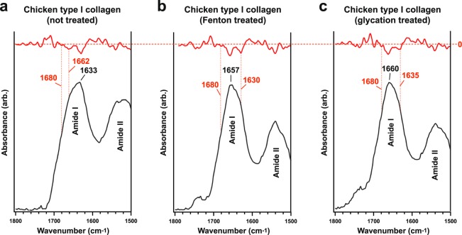
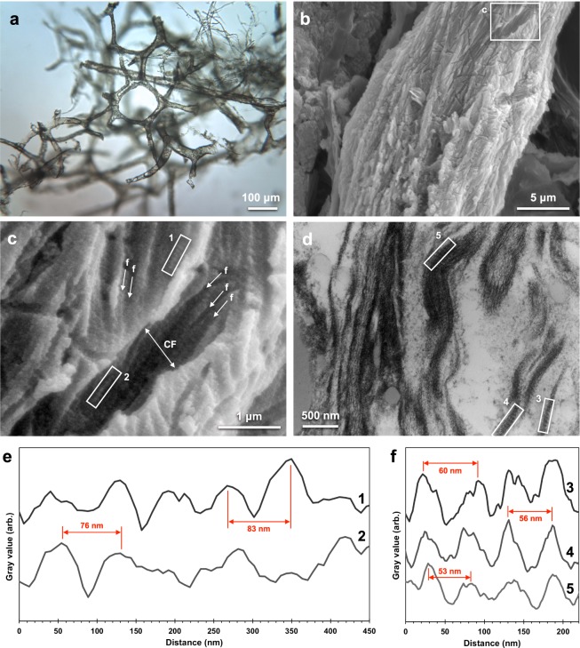
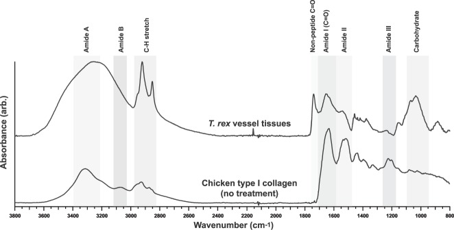
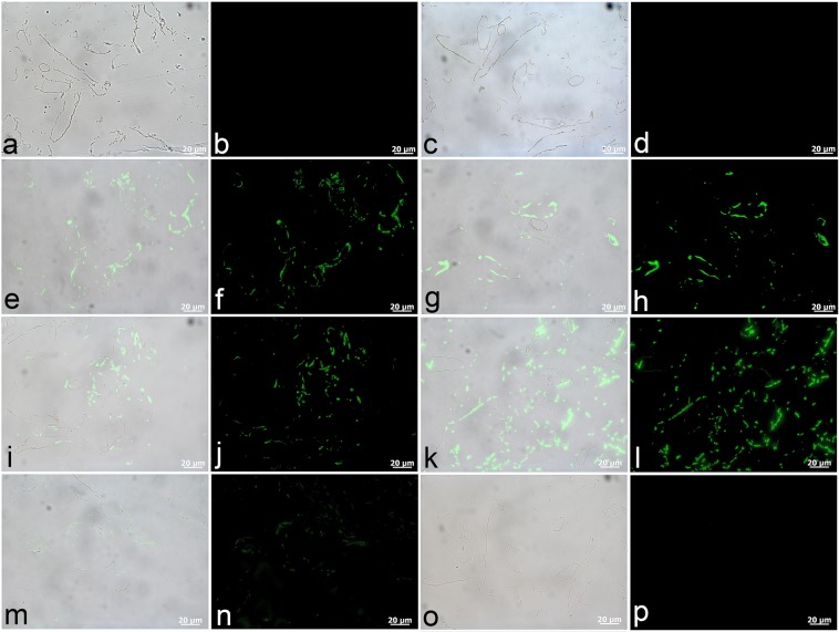
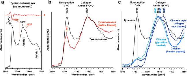
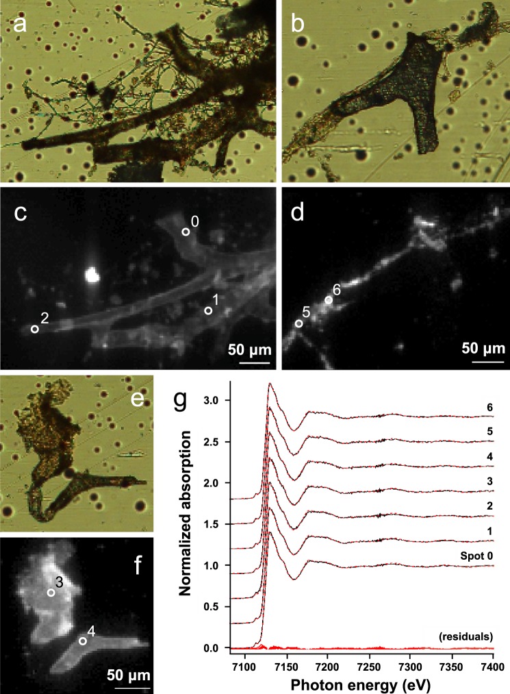
The idea that original soft tissue structures and the native structural proteins comprising them can persist across geological time is controversial, in part because rigorous and testable mechanisms that can occur under natural conditions, resulting in such preservation, have not been well defined. Here, we evaluate two non-enzymatic structural protein crosslinking mechanisms, Fenton chemistry and glycation, for their possible contribution to the preservation of blood vessel structures recovered from the cortical bone of a Tyrannosaurus rex (USNM 555000 [formerly, MOR 555]). We demonstrate the endogeneity of the fossil vessel tissues, as well as the presence of type I collagen in the outermost vessel layers, using imaging, diffraction, spectroscopy, and immunohistochemistry. Then, we use data derived from synchrotron FTIR studies of the T. rex vessels to analyse their crosslink character, with comparison against two non-enzymatic Fenton chemistry- and glycation-treated extant chicken samples. We also provide supporting X-ray microprobe analyses of the chemical state of these fossil tissues to support our conclusion that non-enzymatic crosslinking pathways likely contributed to stabilizing, and thus preserving, these T. rex vessels. Finally, we propose that these stabilizing crosslinks could play a crucial role in the preservation of other microvascular tissues in skeletal elements from the Mesozoic.


Conflict of interest statement

The authors declare no competing interests.

Figures

Figure 1
Figure 1
Amide I sub-band localisation of untreated and treated chicken type I collagen in SR-FTIR spectra. Sub-bands (β-sheet, ~1633 cm−1; triple-helix, ~1658–1660 cm−1; intermolecular, ~1683–1690 cm−1) are indicated in the figures. Red traces denote second derivatives of experimental curves. Although the intermolecular sub-band typically presents at lower wavenumber, the identified value was the nearest local minimum in each of the second derivative traces and consistently appears across all samples; therefore, in this sample, the intermolecular sub-band was indexed at 1697–1699 cm−1.
Figure 2
Figure 2
Microscopy images of T. rex vascular tissue and associated analysis of fibrillar collagen banding. (a) Transmitted VLM of T. rex soft tissue shows an extensive network of hollow, pliable, vascular structure and typical brown hue. (b) SEM image of the surface of a vessel. (c) Magnified image of (b) detailing features consistent with collagen fibre bundles (collagen fibril, “f”; collagen fibre, “CF”). Average fibril width was measured as 110 nm, and average fibre width, 1.0 μm. (d) TEM image of fibrous features observed in a longitudinal vessel cross-section. Intensity profiles of banded texture in (e) boxes 1 and 2 in c and (f) boxes 3, 4, 5 in (d) with example peak-to-peak distances (SEM average, ~74 nm; TEM, ~56 nm) called out in red. See Fig. S6 for precise d-spacing values determined using SAXS. For comparison to a modern blood vessel network in bone, see Fig. 5b of ref..
Figure 3
Figure 3
SR-FTIR full spectra of isolated T. rex vascular tissue and chicken type I collagen (no treatment). All key bands for the identification of protein (Amide I, Amide II, Amide III) are present in the dinosaur tissue spectrum. The T. rex spectrum also presents a strong non-peptide carbonyl (C=O) band at 1739 cm−1 and a carbohydrate band at ~1010 cm−1.
Figure 4
Figure 4
T. rex tissues exhibit positive antibody binding to protein components of extant vascular tissue. (a,c,e,g,i,k,m,o) Are composite images in which fluorescence corresponding to antibody-antigen complexes is overlain upon VLM images of vessel sections, with adjacent images (b,d,f,h,j,l,n,p) captured using a fluorescent filter. (a–d) No spurious binding was observed for negative controls in which vessels were exposed to secondary antibodies raised against the host species of all other antibodies used, i.e., mouse (a,b) and rabbit (c,d). (e,f) Positive binding of dinosaur vessels to actin antibodies can be seen in thin, evenly distributed layers, and (g,h) more broadly distributed binding is apparent for muscle tropomyosin antibodies. Antibodies to both (i,j) type I collagen and (k,l) elastin bind positively to these T. rex vessels. (m,n) Antibodies raised against ostrich haemoglobin exhibit comparatively lower binding intensity. (o,p) No reactivity of dinosaur vessels to antibodies against bacterial peptidoglycan was observed.
Figure 5
Figure 5
SR-FTIR analysis of T. rex vascular tissue, NaBH4 reduced T. rex vascular tissue, chicken type I collagen without treatment, and chicken type I collagen treated with Fenton reagent and iron-catalysed glycation. (a,b) Average FTIR spectra in the non-peptide carbonyl and protein amide I regions for all five samples. (a) Significant reduction in the non-peptide carbonyl band follows treatment of T. rex vascular tissue with NaBH4, which reduces (immature) peptide crosslinks. The blue-shifted Amide I band of the dinosaur tissue, Fenton reagent-treated chicken type I collagen, and Fe-catalysed glycation-treated chicken type I collagen indicate increasing α-helix structure (~1660 cm−1) as the higher-energy triple-helix and intermolecular sub-bands (see Fig. 1 for method of identification) increasingly predominate the spectra. The development of aldehydic carbonyl, ketoaldehyde, and/or immature ketoimine bands in both treated chicken tissues is consistent with the strong carbonyl band in the dinosaur tissue.
Figure 6
Figure 6
X-ray microprobe analysis of iron in the T. rex vascular tissues. (a,b,e) Optical microscope images of vessel tissues and (c,d,f) corresponding iron μ-XRF distribution maps recorded at 10 keV. Brighter pixels correspond to higher Fe content. All scale bars are 50 μm. Additional elemental maps of regions (a) and (b) can be found in Fig. S5. In (b,d) the vessel structure is not an organic tissue but a mineralised cast rich in Ba and S (see Fig. S5). Such fine-scale variation in preservation underscores the notion that preservation depends on the microenvironment. Numbered white circles indicate locations of Fe μ-XANES analysis. (g) Stacked normalised Fe K-edge extended XANES spectra of spots 0–6. Fits are shown in red dashed lines, with corresponding residuals plotted at the bottom. All spectra match to goethite (α-FeO(OH)) with normalised sum-square values ranging from 0.59 to 1.93·10−4. For comparison, an example set of the iron bearing reference spectra used are displayed in Fig. S7.

References

    1. Schweitzer MH, Wittmeyer JL, Horner JR, Toporski JK. Soft-Tissue Vessels and Cellular Preservation in Tyrannosaurus rex. Science. 2005;307:1952–1955. doi: 10.1126/science.1108397. - DOI - PubMed
    1. Schweitzer MH, Wittmeyer JL, Horner JR. Soft tissue and cellular preservation in vertebrate skeletal elements from the Cretaceous to the present. P. Roy. Soc. B-Biol. Sci. 2007;274:183–197. doi: 10.1098/rspb.2006.3705. - DOI - PMC - PubMed
    1. Schweitzer MH, et al. Biomolecular Characterization and Protein Sequences of the Campanian Hadrosaur B. canadensis. Science. 2009;324:626–631. doi: 10.1126/science.1165069. - DOI - PubMed
    1. Lindgren J, et al. Microspectroscopic Evidence of Cretaceous Bone Proteins. PLoS ONE. 2011;6:e19445. doi: 10.1371/journal.pone.0019445. - DOI - PMC - PubMed
    1. Zhuoxin Y, An B, Ramshaw JAM, Brodsky B. Bacterial collagen-like proteins that form triple-helical structures. J. Struct. Biol. 2014;3:451–461. - PMC - PubMed

Publication types