. 2019 Oct 30;9(1):15700.
doi: 10.1038/s41598-019-52189-3.

Postoperative Hypoparathyroidism in Thyroid Surgery: Anatomic-Surgical Mapping of the Parathyroids and Implications for Thyroid Surgery

Affiliations

Postoperative Hypoparathyroidism in Thyroid Surgery: Anatomic-Surgical Mapping of the Parathyroids and Implications for Thyroid Surgery

Florian Burger et al. Sci Rep. .

Abstract

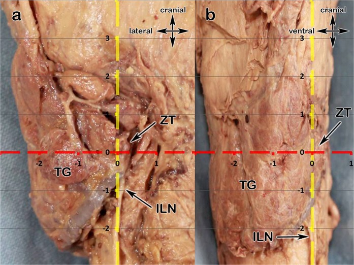
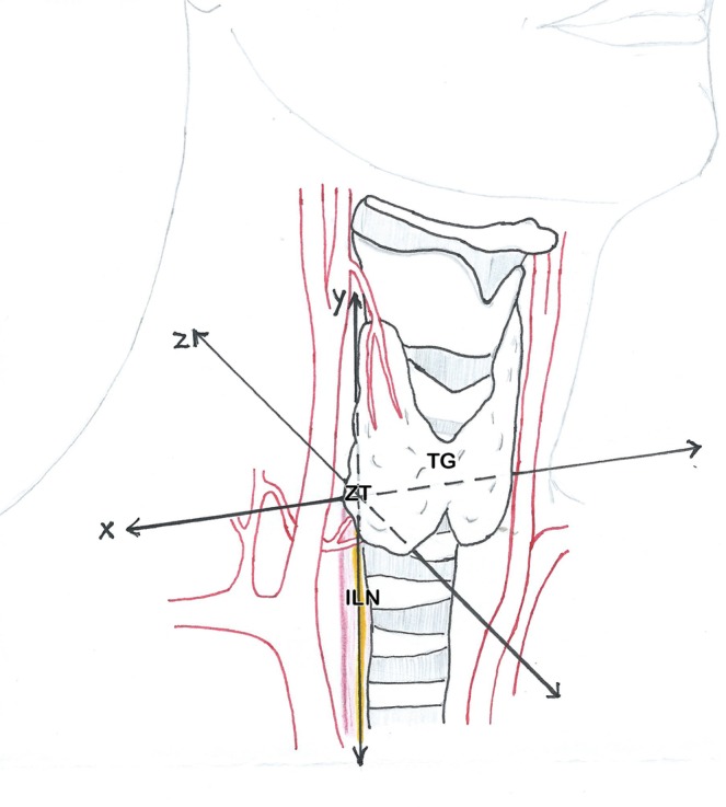
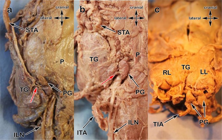
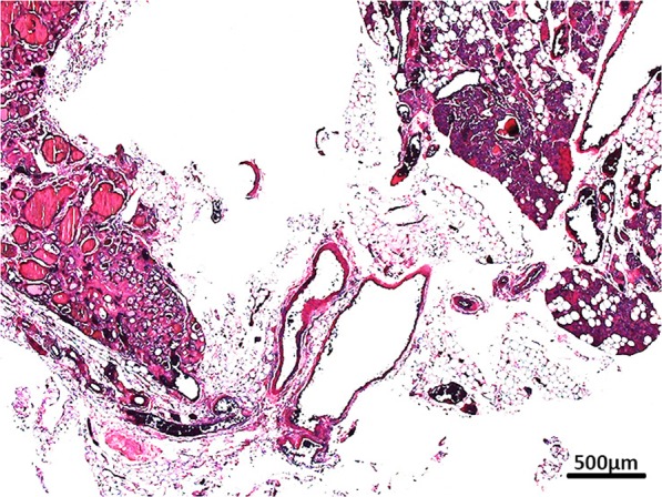
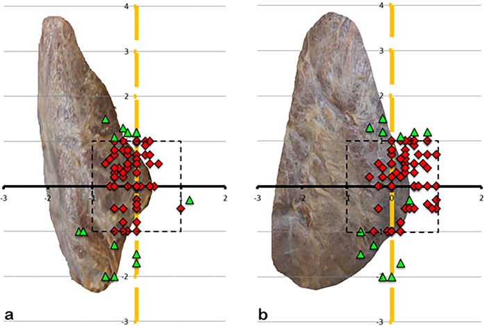
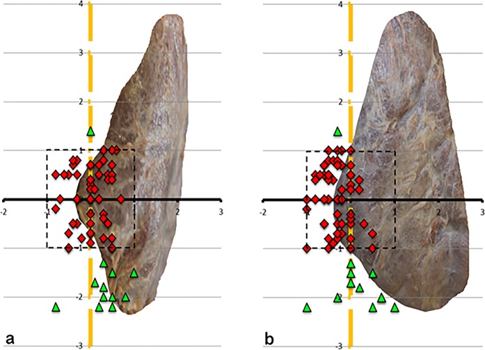
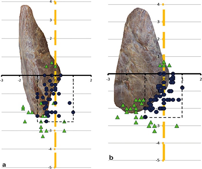
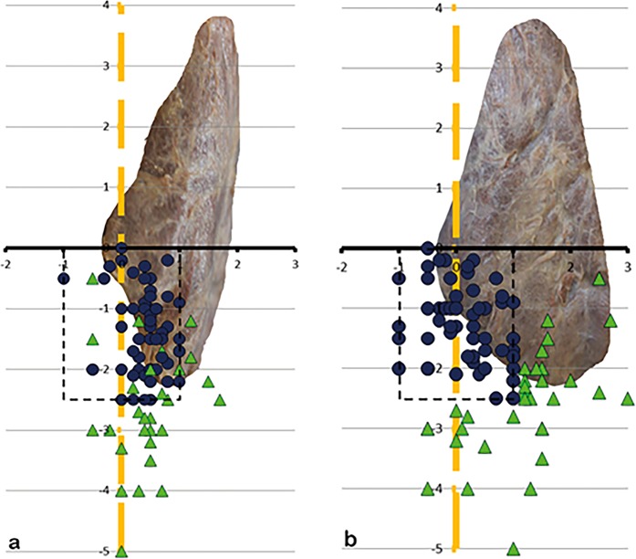
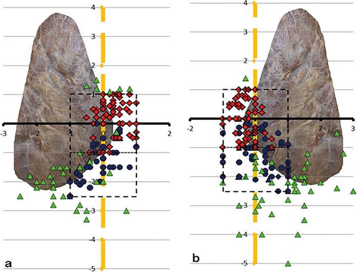
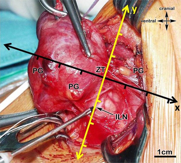
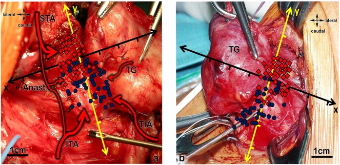
Hypoparathyroidism remains one of the most common complications in thyroid surgery. This study aims for an improved understanding of the complexity of the blood supply and the localisation of the parathyroids compared to the two most important intraoperative landmarks: the inferior laryngeal nerve (ILN) and Zuckerkandl's tubercle (ZT). We examined 103 laryngeal compounds to classify the blood supply and the localisation of the parathyroids. For intraoperative localisation we defined a Cartesian coordinate system with the ZT plane as x-axis and the course of the inferior laryngeal nerve as y-axis. The inferior thyroid artery (ITA) mainly supplies the parathyroids, whereas the superior thyroid artery provides a backup supply. It must be pointed out that 8.2% of parathyroids receive their blood directly from the thyroid gland. 73.5% of all parathyroids lie within 1 cm of the ILN and 1 cm cranial and 2.5 cm caudal to the ZT plane. Our described perimeters mark the most crucial areas during surgery and provide the surgeon with an anatomic mapping showing areas of special carefulness needed. One should keep bearing in mind all possible blood supply types of the parathyroids and therefore all branches should be handled with care.


Conflict of interest statement

The authors declare no competing interests.

Figures

Figure 1
Figure 1
(a,b) Showing the anatomical mapping (Cartesian coordinate system); 1a View from dorsal onto the left TG, 2b view from lateral onto the left TG; thyroid gland (TG), level of the Zuckerkandl tubercle (ZT) – x-axis, inferior laryngeal nerve (ILN) – y-axis.
Figure 2
Figure 2
Scetch of the surgical situs; x - medial-lateral axis; y - cranial-caudal axis; z - anterior-posterior axis, ZT - Zuckerkandl tubercle, ILN - inferior laryngeal nerve (yellow).
Figure 3
Figure 3
(a,b) 3a Type a, 3b Type b; pharynx (P), thyroid gland (TG), inferior laryngeal nerve (ILN), inferior thyroid artery (ITA), superior thyroid artery (STA), anastomose (AN); parathyroids (PG), single arrows show supplying arteries.
Figure 4
Figure 4
(ac) 4a Type x: blood supply by the STA, 4b blood supply directly out of the TG, 4c blood supply by the TIA; right thyroid lobe (RL), left thyroid lobe (LL), thyroid ima artery (TIA), inferior laryngeal nerve (ILN), parathyroids (PG), single arrows show supplying arteries.
Figure 5
Figure 5
Histological prove of parathyroid tissue and supplying vessels.
Figure 6
Figure 6
(a,b) Showing the position of the left SPG, (a) view from dorsal, (b) view from lateral, x-axis ZT-level, y-axis ILN-level, 1-cm scale. The red squares are showing the positioning of the superior parathyroids within 1 cm perimeter (dotted rectangle), the green triangles outside 1 cm perimeter.
Figure 7
Figure 7
(a,b) Showing the position of the right SPG, (a) view from dorsal, (b) view from lateral.
Figure 8
Figure 8
(a,b) Showing the position of the left IPG, (a) view from dorsal, (b) view from lateral, x-axis ZT-level, y-axis ILN-level, 1-cm scale. The blue dots are showing the positioning of the inferior parathyroids within 1 cm perimeter of the ILN-level and within 2,5 cm perimeter (dotted rectangle) on the ZT-level, the green triangles outside the perimeter.
Figure 9
Figure 9
(a,b) Showing the position of the right IPG, (a) view from dorsal, (b) view from lateral.
Figure 10
Figure 10
(a,b) Picture of the arrangement of the SPG and IPG on 10 a the left and 10 b the right side when seen from lateral (SPG – squares, are within the perimeter (dotted rectangle); IPG – dots, are inside the perimeter, triangles, regardless of being SPG or IPG, are outside).
Figure 11
Figure 11
Intraoperative case, with the coordinate system attached, of a left thyroid lobe showing 3 parathyroids. The lower ones on the left. 1 parathyroid (dark coloured) lies intracapsular (histological proved) and is already changing its colour.
Figure 12
Figure 12
(a,b) Gives an overview and connects our results of the artery pattern and the location of the parathyroids to intraoperative field. (a) Overview of the location on the right side including blood supply: view from lateral on the right thyroid lobe, the red squares are showing the positioning of the superior parathyroids within 1 cm perimeter, the blue dots the positioning of the inferior parathyroids within 1 cm perimeter of the inferior laryngeal nerve-level (yellow-axis) and within 2,5 cm perimeter on the Zuckerkandl tubercle-level (black-axis); possible artery pattern of the parathyroids: blood supply by the inferior thyroid artery (ITA); blood supply by anastomoses between inferior thyroid artery and superior thyroid artery (Anast.); blood supply by the superior thyroid artery (STA); blood supply directly out of the thyroid gland (TG); blood supply by a thyroid ima artery (TIA). (b) Overview of the location on the left side (possible artery pattern same as on the right): view from lateral on the left thyroid lobe, the red squares are showing the positioning of the superior parathyroids within 1 cm perimeter, the blue dots the positioning of the inferior parathyroids within 1 cm perimeter of the inferior laryngeal nerve-level (yellow) and within 2,5 cm perimeter on the Zuckerkandl tubercle-level (black); TG…thyroid gland.

References

    1. Thomusch O, et al. The impact of surgical technique on postoperative hypoparathyroidism in bilateral thyroid surgery: a multivariate analysis of 5846 consecutive patients. Surgery. 2003;133:180–185. doi: 10.1067/msy.2003.61. - DOI - PubMed
    1. Lang BH, Chan DT, Chow FC, Wong KP, Chang RY. The Association of Discolored Parathyroid Glands and Hypoparathyroidism Following Total Thyroidectomy. World J Surg. 2016;40:1611–1617. doi: 10.1007/s00268-016-3462-9. - DOI - PubMed
    1. Promberger R, et al. Intra-and postoperative parathyroid hormone-kinetics do not advocate for autotransplantation of discolored parathyroid glands during thyroidectomy. Thyroid. 2010;20:1371–1375. doi: 10.1089/thy.2010.0157. - DOI - PubMed
    1. Trupka, A. & Wicke, C. Nebenschilddrüsenprotektion zur Vermeidung des postoperativen Hypoparathyreoidismus. Dralle H. Endokrine Chirurgie-Evidenz und Erfahrung. Stuttgart: Schattauer, 117–136 (2014).
    1. Barczynski M, Cichon S, Konturek A, Cichon W. Applicability of intraoperative parathyroid hormone assay during total thyroidectomy as a guide for the surgeon to selective parathyroid tissue autotransplantation. World J Surg. 2008;32:822–828. doi: 10.1007/s00268-007-9405-8. - DOI - PubMed

MeSH terms