. 2019 Oct 24:16:39.
doi: 10.1186/s12983-019-0337-6. eCollection 2019.

The innervation of the male copulatory organ of spiders (Araneae) - a comparative analysis

Affiliations

The innervation of the male copulatory organ of spiders (Araneae) - a comparative analysis

Tim M Dederichs et al. Front Zool. .

Abstract

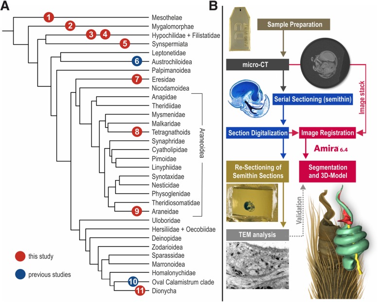
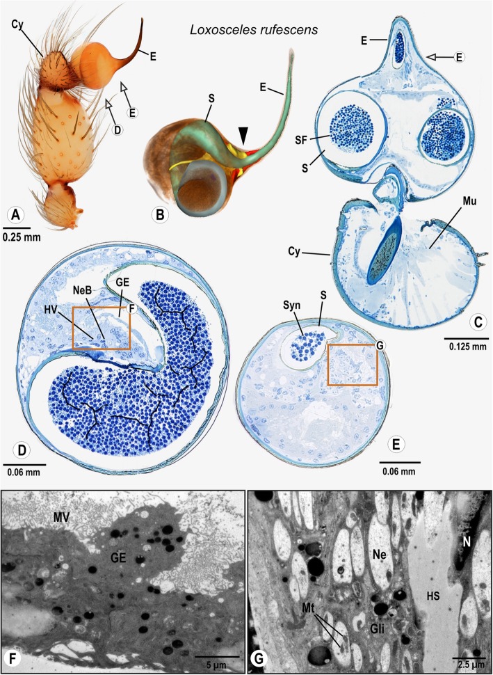
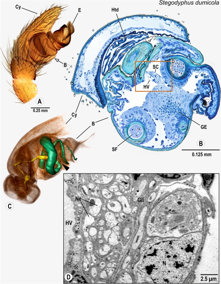
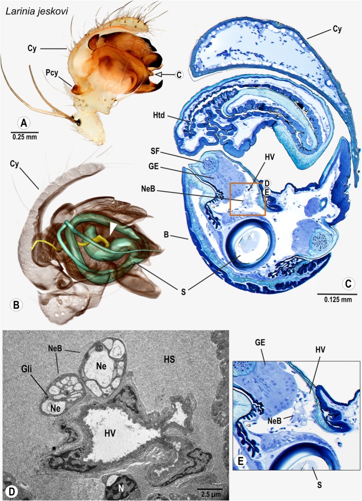
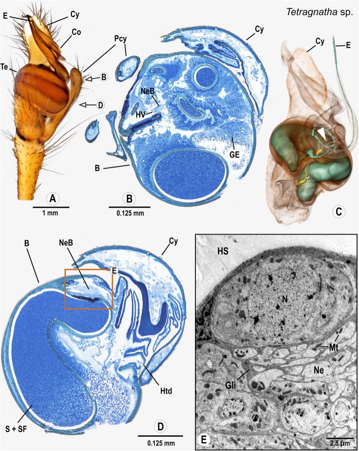
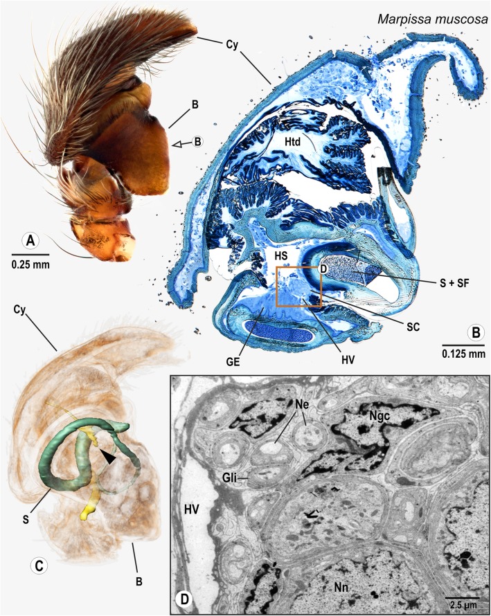
Background: Nervous tissue is an inherent component of the many specialized genital structures for transferring sperm directly into the female's body. However, the male copulatory organ of spiders was considered a puzzling exception. Based on the recent discovery of nervous tissue in the pedipalps of two distantly related spider species, we investigated representatives of all major groups across the spider tree of life for the presence of palpal nerves. We used a correlative approach that combined histology, micro-computed tomography and electron microscopy.

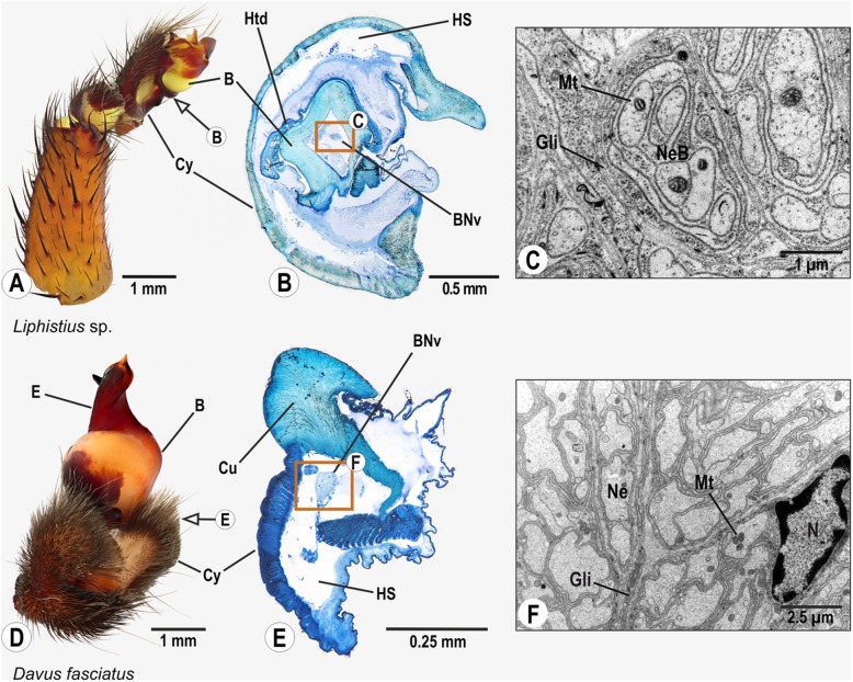
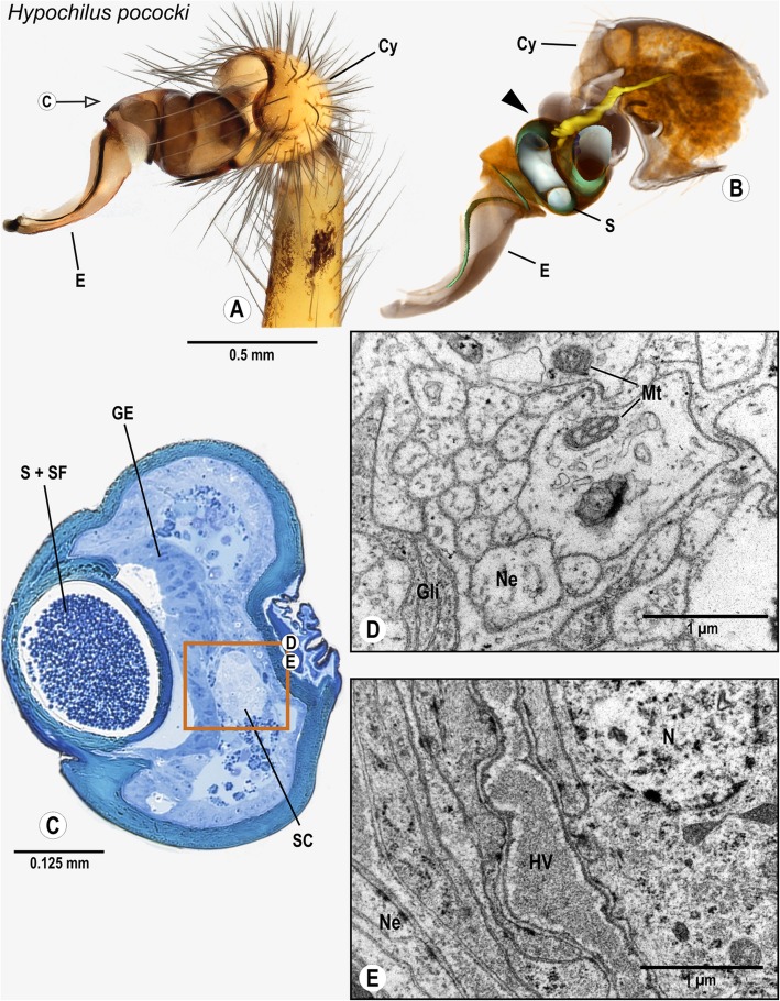
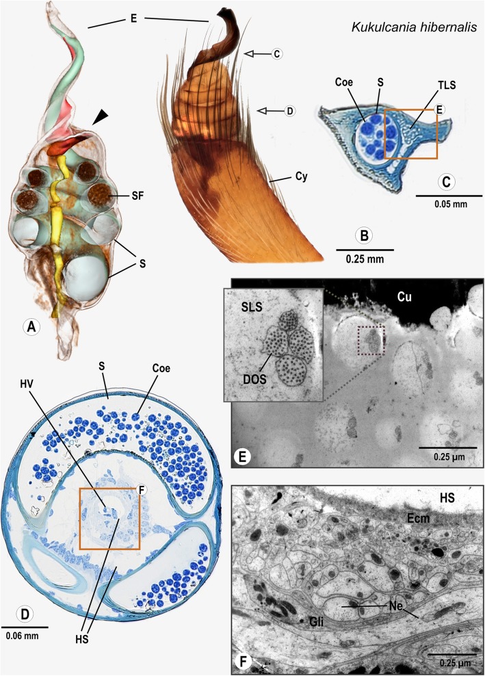
Results: We show that the copulatory organ is innervated in all species investigated. There is a sensory organ at the base of the sperm transferring sclerite in several taxa and nervous tissue occurs close to the glandular tissue of the spermophor, where sperm are stored before transfer.

Conclusions: The innervation of the copulatory organ by the bulb nerve and associated efferent fibers is part of the ground pattern of spiders. Our findings pave the way for unraveling the sensory interaction of genitalia during mating and for the still enigmatic mode of uptake and release of sperm from the male copulatory organ.

Keywords: Bulb nerve; Copulation; Copulatory mechanism; Intromittent organ; Palpal organ; Pedipalp; Sensory organ; Sexual selection; Spiders.


Conflict of interest statement

Competing interestsThe authors declare that they have no competing interests.

Figures

Fig. 1
Fig. 1
(a) Selected taxa for the present study (red) and previous studies (blue) on the innervation of the male copulatory organ of spiders. . 1: Liphistius sp.; 2: Davus fasciatus; 3: Hypochilus pococki; 4: Kukulcania hibernalis; 5: Loxosceles rufescens; 6: Hickmania troglodytes [26]; 7: Stegodyphus dumicola; 8: Tetragnatha extensa; 9: Larinia jeskovi; 10: Philodromus cespitum [27]; 11: Marpissa muscosa. (b) Flowchart of the correlative imaging approach used in this study
Fig. 2
Fig. 2
Palpal organs of Liphistius sp., and Davus fasciatus; external morphology (a,d), histology (b,e), and ultrastructure as documented by TEM (c,f). Arrows indicate planes chosen for semithin sections (b,e). Boxes in B and E show areas in the palpal organ where profiles of the bulb nerve were found. TEM micrographs in C and F show ultrastructural details of the bulb nerve. Abbreviations: B Bulb, BNv Bulb nerve, Cu Cuticle, Cy Cymbium, E Embolus, Gli Glial cell processes, HS Haemolymph space, Htd Haematodocha, Mt Mitochondrium, Ne Neurite, NeB Neurite bundle, N Nucleus of glial cell
Fig. 3
Fig. 3
Palpal organ of Hypochilus pococki; external morphology (a), histology (c), ultrastructure documented by TEM (d, e), and 3D reconstruction of the spermophor (green) and nervous tissue (yellow) as based on segmentation of histological image stacks (b). The arrow indicates the plane chosen for semithin section (a). The arrowhead marks the terminals of the bulb nerve and associated neurons (b). The box marks the cluster of neuronal somata, and ultrastructural details of neurites bundles (d) associated with cluster of neuronal somata (e). Abbreviations: HV Haemolymph Vessel, Cy Cymbium, E Embolus, GE Glandular epithelium, Gli Glial cell processes, Mt Mitochondria, Ne Neurite, N Nucleus of a neuron, S Spermophor, SC Cluster of neuronal somata, SF Seminal fluid
Fig. 4
Fig. 4
Palpal organ of Kukulcania hibernalis; external morphology (b), histology (c, d) ultrastructure as documented by TEM (e, f) and 3D reconstruction of the spermophor (green), nervous tissue (yellow) and internalized sensilla/“tube-like-structures” (red) as based on segmentation of histological image stacks (a). Arrows indicate the planes chosen for semithin sectioning (b) and the arrowhead marks the terminals of the bulb nerve (a). Box in C marks the sector with internalized aggregated sensilla magnified in (e), whereas box in (d) frames the region around the bulb nerve magnified partly in (f). Inset in (e) highlights four dendritic outer segments assembled in an internalized sensillum. Abbreviations: Coe Coenospermium, Cy Cymbium, DOS Dendritic outer segments, Ecm Extracellular Matrix, E Embolus, Gli Glial cell processes, HS Haemolymph space, HV Haemolymph vessel, Ne Neurite, S Spermophor, SF Seminal fluid, SLS Sensillum lymph space, TLS aggregated tube-like structures
Fig. 5
Fig. 5
Palpal organ of Loxosceles rufescens; external morphology (a), histology (ce) ultrastructure as documented by TEM (f, g), and 3D reconstruction of the spermophor (green), nervous tissue (yellow) and distinct cell clusters/“sensor epidermal tissue” (red) as based on segmentation of histological image stacks (b). Arrows indicate planes chosen for semithin sections (a) and the arrowhead marks the terminals of the bulb nerve (b). Box in (d) indicates the location of glandular tissue in the bulb, ultrastructural details are given in (F). Box in (e) marks the branches of the bulb nerve, highly magnified in G. Abbreviations: Cy Cymbium, E Embolus, GE Glandular epithelium, Gli Glial cell processes, HS Haemolymph space HV Haemolymph Vessel, Mu Muscle, Mt Mitochondria, MV Brush of microvilli, Ne Neurite, NeB Neurite Bundle, N Nucleus, S Spermophor, SF Seminal fluid, Syn Synspermium
Fig. 6
Fig. 6
Palpal organ of Stegodyphus dumicola; external morphology (a), histology (b), ultrastructue as documented by TEM (d), and 3D reconstruction of the spermophor (green) and nervous tissue (yellow) as based on segmentation of histological image stacks (c). Arrow in (a) indicates the plane chosen for semi-thin cross section and arrowhead in (c) marks terminals of the bulb nerve. Box in (b) shows central position of bulb nerve in the palpal organ, surrounded by neuronal somata, ultrastructural details of boxed area are illustrated in (d). Abbreviations: B Bulbus, Cy Cymbium, E Embolus, GE Glandular epithelium, Gli Glial cell processes, HS Haemolymph space, Htd Haematodocha, HV Haemolymph Vessel, Mt Mitochondria, Ne Neurites, NeB Neurite Bundle, N Nucleus of a neuron, S Spermophor, SC Cluster of neuronal somata
Fig. 7
Fig. 7
Palpal organ of Larinia jeskovi; external morphology (a), histology (c, e), ultrastructure as documented by TEM (d), and 3D reconstruction of the spermophor (green) and nervous tissue (yellow) as based on segmentation of histological image stacks (b). Arrow in (a) indicates plane chosen for semi-thin cross section (c). Arrowhead in (b) marks terminals of the bulb nerve. Box in (c) shows position and arrangement of neurite bundles branching off the bulb nerve, boxed sector is magnified in (e). Ultrastructural details of the same sector are given in (d), note the presence of two distinct neurite bundles adhering a haemolymph vessel. Abbreviations: B Bulbus, Cy Cymbium, GE Glandular epithelium, Gli Glial cell processes, HS Haemolymph space, Htd Haematodocha, HV Haemolymph Vessel, Ne Neurite, NeB Neurite Bundles, Pcy Paracymbium, SF Seminal fluid
Fig. 8
Fig. 8
Palpal organ of Tetragnatha montana.; external morphology showing the general organization (a) as well as histology (b, d), ultrastructure as documented by TEM (e), and 3D reconstruction of the spermophor (green) and nervous tissue (yellow) of Tetragnatha extensa as based on segmentation of histological image stacks (c). Arrows in (a) indicate planes for semi-thin cross sections taken at distal end (b) and midlevel (e) of the bulb. Arrowhead marks terminals of bulb nerve (c). Note that the embolus in (a) rests in a ridge of the conductor and therefore differs from the one depicted in (c). Box shows a sector where neuronal somata and a neurite bundle branched off the bulb nerve are present, part of this sector is shown in (e) magnified to ultrastructural level. Abbreviations: B Bulbus, Co Conductor, Cy Cymbium, E Embolus, GE Glandular epithelium, Gli Glial cell processes, HS Haemolymph space, Htd Haematodocha, HV Haemolymph Vessel, Mt Mitochondria, N Nucleus, Ne Neurite, NeB Neurite Bundle, Pcy Paracymbium, S Spermophor, SF Seminal fluid, Te Tegulum
Fig. 9
Fig. 9
Palpal organ of Marpissa muscosa; external morphology (a), histology (b), ultrastructure as documented by TEM (d), and 3D reconstruction of the spermophor (green) and nervous tissue (yellow) as based on segmentation of histological image stacks (c). Arrow in (a) indicates plane for semi-thin cross section at midlevel of the bulb (a), arrowhead in (c) points at terminals of the bulb nerve. Box in (b) marks position of neuronal somata and closely adjoined neurite bundles branched off bulb nerve. (d) provides insights into ultrastructure of neuronal somata and neurite bundles. Abbreviations: B Bulbus, Cy Cymbium, GE Glandular epithelium, Gli Glial cell processes, HS Haemolymph space, Htd Haematodocha, HV Haemolymph Vessel, Mt Mitochondria, Ngc Nucleus of glial cell, Nn Nucleus of neuron, Ne Neurite, S Spermophor, SC Cluster of neuronal somata, SF Seminal fluid

References

    1. Eberhard WG. Sexual selection and animal genitalia. Cambridge: Harvard University Press; 1985.
    1. Kelly DA, Moore BC. The morphological diversity of intromittent organs. Integr Comp Biol. 2016;56:630–634. doi: 10.1093/icb/icw103. - DOI - PubMed
    1. Robson G. On the hectocotylus of the Cephalopoda—a reconsideration. J Molluscan Stud. 1926;17:117–122.
    1. Weygoldt P. Arthropoda—Chelicerata: sperm transfer. Reproductive Biology of the Invertebrates. 1990;4:77–119.
    1. Ewers-Saucedo C, Hayer S, Brandis D. Functional morphology of the copulatory system of box crabs with long second gonopods (Calappidae, Eubrachyura, Decapoda, Crustacea) J Morphol. 2015;276:77–89. doi: 10.1002/jmor.20322. - DOI - PubMed

LinkOut - more resources