. 2019 Oct 8;5(10):e02532.
doi: 10.1016/j.heliyon.2019.e02532. eCollection 2019 Oct.

Clinical parameters affecting multipotent adult progenitor cells in vitro

Affiliations

Clinical parameters affecting multipotent adult progenitor cells in vitro

Margaret L Jackson et al. Heliyon. .

Abstract

Background: Human multipotent adult progenitor cells (MAPC®) are an emerging therapy for traumatic brain injury (TBI); however, clinically translating a therapy involves overcoming many factors in vivo which are not present in pre-clinical testing. In this study we examined clinical parameters in vitro that may impact cell therapy efficacy.

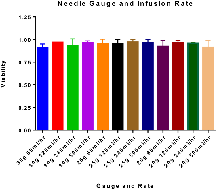
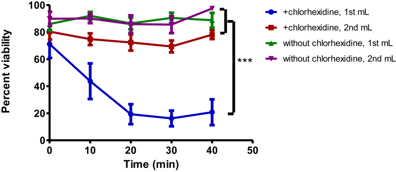
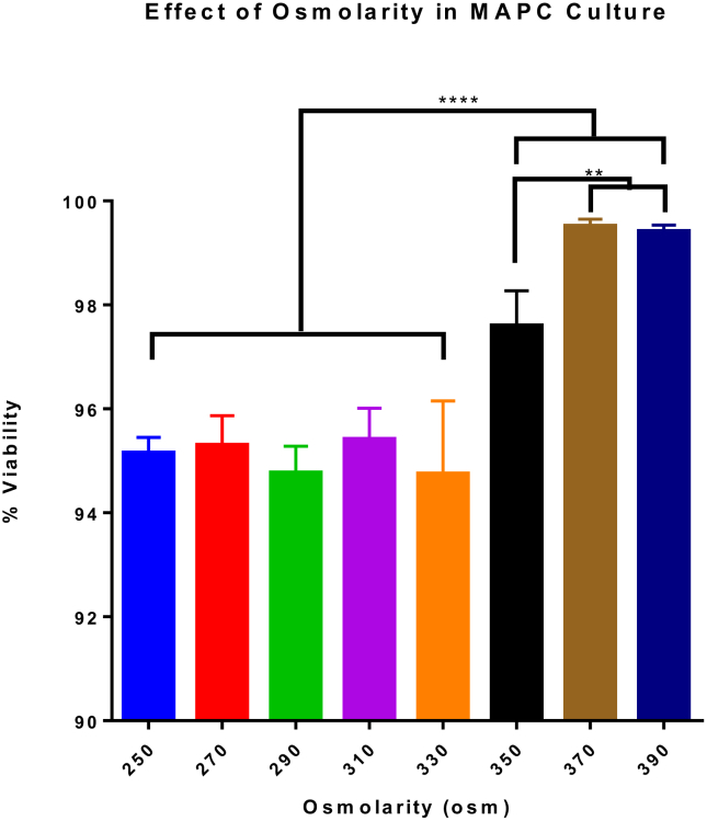
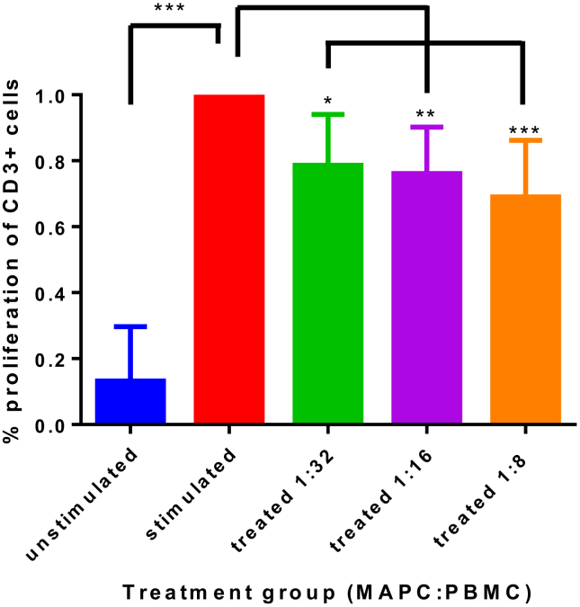
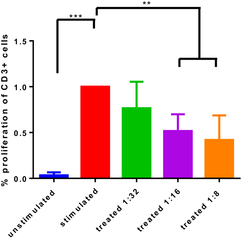
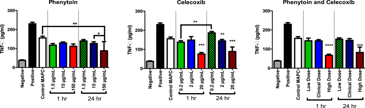
Methods: MAPC were infused through varying gauged needles and catheters with and without chlorhexidine, and their viability tested with trypan blue exclusion. MAPC were co-cultured with phenytoin and celecoxib at relevant clinical concentrations for 1 h and 24 h. Anti-inflammatory potency was tested using a stimulated rat splenocyte co-culture and ELISA for TNF-α production. MAPC were cultured under different osmolar concentrations and stained with propidium iodide for viability. Anti-inflammatory potency was tested by co-culture of MAPC with naïve lymphocytes activated by CD3/CD28 beads, and Click-iT® Plus EdU was used to quantify proliferation by flow cytometry.

Results: The mean viability of the MAPC infused via needles was 95 ± 1%; no difference was seen with varying flow rate, but viability was notably reduced by chlorhexidine. MAPC function was not impaired by co-culture with phenytoin, celecoxib, or combination with both. Co-culture with phenytoin showed a decrease in TNF-α production as compared to the MAPC control. MAPC cultured at varying osmolar concentrations all had viabilities greater than 90% with no statistical difference between them. Co-culture of MAPC with CD3/CD28 activated PBMCs showed a significant reduction in proliferation as measured by EdU uptake.

Discussion: Needle diameter, phenytoin, celecoxib, and a relevant range of osmolarities do not impair MAPC viability or anti-inflammatory potency in vitro.

Keywords: Cell biology; Cell therapy; Immunology; Inflammation; Medicine; Regenerative medicine; Stem cell research; Stem cells research; Translational medicine; Traumatic brain injury.


Figures

Fig. 1
Fig. 1
MAPC viability after infusion through needles. This data complements Table 1. MAPC were infused via needles of different size and at different rates. Neither needle gauge nor infusion rate significantly altered viability of MAPC. The data presented in this table represents the mean percent viability ±SD. Statistical analysis was performed by Two-Way ANOVA. N = 2.
Fig. 2
Fig. 2
Chlorhexidine effect on MAPC viability. Percent viability versus time of the first and second mL of a 1 × 10⁶ MAPC/mL solution when infused through a chlorhexidine-impregnated central venous catheter versus the same type of catheter which had not been treated with chlorhexidine. Data show a significant decrease in cell viability in the first mL infused by a chlorhexidine-impregnated catheter as compared to the second mL infused through a chlorhexidine-impregnated catheter, and a catheter that had not been treated with chlorhexidine (p < 0.001). There was no statistical difference between the viability of the first and second mL of a catheter that had not been treated with chlorhexidine and the second mL of solution infused through a chlorhexidine-impregnated catheter. Values represent mean ± SD. Statistical analysis performed by Two-Way ANOVA. N = 4 per group.
Fig. 3
Fig. 3
Effect of osmolarity on MAPC infusion. MAPC were cultured at osmolar concentrations ranging from 250 osm to 390 osm and measured for percent viability. Specifically, MAPC cultured at higher osmolar concentrations (350, 370, 390 osm) yielded the highest viability and were significantly higher (p value <0.0001) than those cultured at lower concentrations. These results indicate that increasing osmolar concentrations in culture can enhance viability in MAPC. Values represent mean ± SD. Statistical analysis performed by One-Way ANOVA. N = 6.
Fig. 4
Fig. 4
MAPC effect on T cell proliferation. The percent of T-cells proliferating as normalized to the CD3/CD28 stimulated control are presented. The basal proliferation is indicated by the unstimulated control. CD3/CD28 stimulated PBMC proliferate less robustly at all MAPC activation groups (1:32, p < 0.05; 1:16, p < 0.01; 1:8, p < 0.001), with a dose-dependent trend ranging from 21% to 31% reduction in proliferation as MAPC concentration increases. Application of MAPC to activated PBMC results in a suppression of PBMC proliferation ability. Values represent mean ± SD. Statistical analysis performed by One-Way ANOVA. N = 5 per group.
Fig. 5
Fig. 5
MAPC effect on T cell proliferation, under the influence of clinical parameters. Results of co-culture proliferation assay as normalized to positive control. MAPC were first infused via a 30 gauge needle, then co-cultured with phenytoin and celecoxib for 24 h, after which the media was changed and MAPC were plated with activated healthy donor PBMC. There was a statistically significant decrease in proliferation in the higher doses (1:16 and 1:8 treatment groups, p < 0.001). Anti-inflammatory potency of the needle-infused and drug-treated MAPC to reduce proliferation was not statistically different from the MAPC which were not treated with needle-infusion or drug co-culture. Values represent mean ± SD. Statistical analysis performed by One-Way ANOVA. N = 5 per group.
Fig. 6
Fig. 6
TNF-α suppression by MAPC co-cultured with phenytoin, celecoxib or combination, at low, clinical, or high doses. MAPC Splenocyte TNF-α Inhibition Assay Data showing TNF- α production as measured by ELISA. Rat splenocytes were activated with lipopolysaccharide and then treated with MAPCs in culture. They were also treated with either celecoxib, phenytoin, or both as shown. There was a statistically significant difference between the activated control and all groups treated with MAPCs. In addition, there was a synergistic decrease in TNF- α production in the groups that were treated with phenytoin as compared to MAPCs alone. Values represent mean ± SD. Statistical analysis performed by One-Way ANOVA. N = 3 per group.

References

    1. Prevention, C. F. D. C. a. Surveillance Report of Traumatic Brain Injury-Related Emergency Department Visits, Hospitalizations, and Deaths—United States, 2014. 2019. https://stacks.cdc.gov/view/cdc/78062 - PMC - PubMed
    1. Prevention, C. F. D. C. a. Traumatic Brain Injury & Concussion: Get the Facts. 2019.
    1. Cox C.J., Juranek J., Bedi S. Clinical trials in traumatic brain injury: cellular therapy and outcome measures. Transfusion. 2019;59:858–868. - PubMed
    1. Walker P.A. Intravenous multipotent adult progenitor cell therapy for traumatic brain injury: preserving the blood brain barrier via an interaction with splenocytes. Exp. Neurol. 2010;225:341–352. - PMC - PubMed
    1. Walker P.A. Bone marrow–derived stromal cell therapy for traumatic brain injury is neuroprotective via stimulation of non-neurologic organ systems. Surgery. 2012;152:790–793. - PubMed

LinkOut - more resources