Review
. 2019 Feb 13:4:120-131.
doi: 10.1016/j.bioactmat.2019.01.002. eCollection 2019 Dec.

Self-assemble peptide biomaterials and their biomedical applications

Affiliations
Review

Self-assemble peptide biomaterials and their biomedical applications

Jun Chen et al. Bioact Mater. .

Erratum in

Abstract

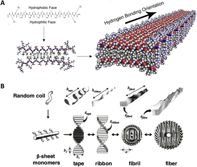
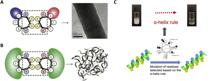
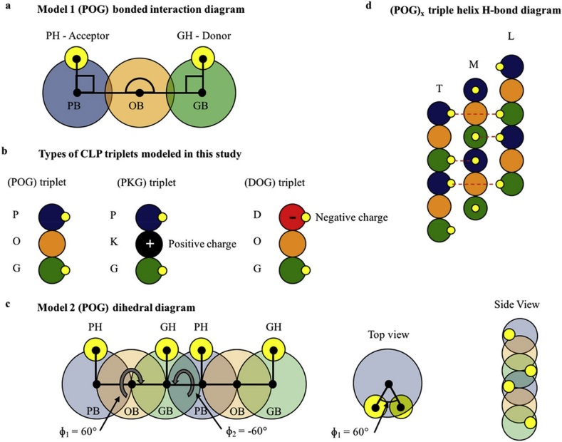
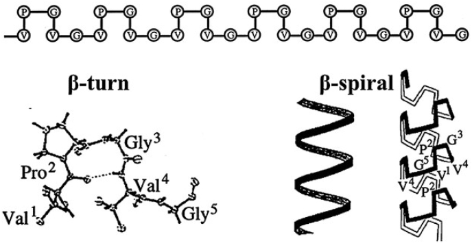
Inspired by self-assembling peptides found in native proteins, deliberately designed engineered peptides have shown outstanding biocompatibility, biodegradability, and extracellular matrix-mimicking microenvironments. Assembly of the peptides can be triggered by external stimuli, such as electrolytes, temperature, and pH. The formation of nanostructures and subsequent nanocomposite materials often occur under physiological conditions. The respective properties of side chains in each amino acids provide numerous sites for chemical modification and conjugation choices of the peptides, enabling various resulting supramolecular nanostructures and hydrogels with adjustable mechanical and physicochemical properties. Moreover, additional functionalities can be easily induced into the hydrogels, including shear-thinning, bioactivity, self-healing, and shape memory. It further broaden the scope of application of self-assemble peptide materials. This review outlines designs of self-assembly peptide (β-sheet, α-helix, collagen-like peptides, elastin-like polypeptides, and peptide amphiphiles) with potential additional functionalities and their biomedical applications in bioprinting, tissue engineering, and drug delivery.


Figures

Fig. 1
Fig. 1
(A) β-Sheet forming short peptides with alternating ionic complementary properties [17]. (Copyright © 2017 American Chemical Society) (B) Short amphiphilic β-sheet peptides that self-assemble into anti-parallel nanotapes and further aggregate into ribbons and higher order structures. In a recent paper, shorter sequences (P9-6 and P7-6) with aliphatic hydrophobic resides (in green) were demonstrated to form fibrillar structures [18]. (Copyright (2001) National Academy of Sciences.)
Fig. 2
Fig. 2
Hydrogelating SAF design principles. A, Thick SAF designs, specific charged interactions between certain b and c positions lead to peptide alignment and fibre thickening. B, Thin hSAFs, specific interactions at all b, c and f sites were replaced with weaker, more-general interactions, to result in smaller, more flexible, bundles of thinner fibres [31]. (Reproduced and adapted with permission from Nature) C, Hypothesis of self-assembly from peptide monomers to supramolecular networks of Schematic model based on the α-helix rule [32]. (Reproduced and adapted with permission from Nature).
Fig. 3
Fig. 3
(a) Diagram of model 1 coarse-grained (POG) triplet with bonded interactions shown by lines and angles connecting specific bead types. (b) Types of CLP triplets used in the simulations studied in this work. (c) Diagram of model 2 coarse-grained CLP strand with the dihedral angles listed. (d) (POG)x triple helical H-bond diagram highlighting the donor–acceptor interaction and the offset of the individual strands where T represents the trailing strand, M represents the middle strand, and L represents the leading strand [33]. (Copyright © 2018 American Chemical Society.).
Fig. 4
Fig. 4
Poly(VPGVG) adopts a β‐spiral structure at temperatures above Tt. The dedeVPGVG repeats form β‐turns, stabilized by intramolecular hydrogen bonds between the backbones of the first and fourth residues of the pentapeptide. These β‐turns arrange into helical β‐spirals, which are represented with and without displaying the structure of the β‐turns in the turns of the helix. Reprinted with permission from Urry D. W., Physical chemistry of biological free energy transduction as demonstrated by elastic protein‐based polymers [56]. (Copyright © 1997 American Chemical Society.).
Fig. 5
Fig. 5
Design of peptide amphiphile into biomimetic structures: Molecular model of a peptide amphiphile (A), nanofiber: a 12-mer peptide identified through phage display library screening is coupled to a hydrophobic fatty acid chain (C16), which undergoes self-assembly into nanofibers (B), and Resulting materials can serve as soft- and hard-tissue guiding scaffolds (C) [45]. (Copyright © 2015 American Chemical Society.).
Fig. 6
Fig. 6
(a) Chemical structures of various amphiphiles (P1—P4). (b) Pictures of glass vials containing metallo-hydrogels obtained from different proportions of the P3 and nickel salt (NiCl2). (c) Multi-stimuli responsiveness shown by the hydrogel obtained from P3 [88]. (Copyright ©The Royal Society of Chemistry 2014.).
Fig. 7
Fig. 7
Injectable ELP–HA hydrogels. a) ELP–HA is composed of hydrazine-modified elastin-like protein (ELP-HYD) and aldehyde-modified hyaluronic acid (HA-ALD). b) Schematic of ELP–HA hydrogel formation. c) Photographs demonstrating the injectability and rapid self-healing of ELP–HA hydrogels [48]. (Copyright © 2017, John Wiley and Sons).
Fig. 8
Fig. 8
Chemical structures of amoc-capped peptides [99]. (Copyright © 2018 american chemical society).
Fig. 9
Fig. 9
3D neurosphere formation by NSCs in hSAF gels. 3D reconstruction of z-stack fluorescent images on (A) laminin, (B) undecorated hSAF, and (C) RGDS-decorated hSAF gels. (D) 3D reconstruction of z-stack fluorescent images of cells on RGDS-decorated gels showing neurosphere connections. DAPI-stained cell nuclei (blue) and nestin expression (green). (A–D) Grid scales: 24.75 μm [26]. (Copyright © 2015 American Chemical Society).
Fig. 10
Fig. 10
Cell viability and proliferation analysis of A) hBMSCs and B) hACs in their respective medium and biomaterials by Alamar Blue assay. The results are represented as the mean ± standard deviation for n = 3. Statistically significant differences are denoted by symbols; *p < 0.05, **p < 0.01 and ***p < 0.001 (Silk‐CC and PA‐RGDS‐CC indicate the scaffolds which were transferred to the CC medium) [112]. (Copyright © 2018, John Wiley and Sons).
Fig. 11
Fig. 11
(a) pDal molecular structure. (b) cylindrical and twisted nanofibers as seen using TEM (red arrows indicate twisted nanofibers). (c) snapshots from molecular simulations of the self-assembly of pDal nanofibers starting from a random configuration. (d) 2 nm length cross section of the nanofiber formed during simulation, revealing the wrapping around of the peptide moiety (shown in purple) around the fiber axis; the hydrophobic core shown in cyan. (e) pairwise radial distribution function for the TYR backbone bead with respect to all other amino acid backbone beads in a nanofiber. (f) Idealized nanofiber structure with the pDal molecules arranged. (g) Pairwise radial distribution function g(r) between the ARG backbone bead and the TYR backbone bead, calculated over five different time periods of the simulation [115]. (Copyright © 2013 American Chemical Society).

References

    1. Gutteridge A., Thornton J.M. Understanding nature's catalytic toolkit. Trends Biochem. Sci. 2005;30(11):622–629. - PubMed
    1. Koutsopoulos S. Self-assembling peptide nanofiber hydrogels in tissue engineering and regenerative medicine: progress, design guidelines, and applications. J. Biomed. Mater. Res. 2016;104(4):1002–1016. - PubMed
    1. Mandal D., Nasrolahi Shirazi A., Parang K. Self-assembly of peptides to nanostructures. Org. Biomol. Chem. 2014;12(22):3544–3561. - PMC - PubMed
    1. Wang H. Cellular membrane enrichment of self-assembling d-peptides for cell surface engineering. ACS Appl. Mater. Interfaces. 2014;6(12):9815–9821. - PubMed
    1. Wen Y. Retaining antibodies in tumors with a self-assembling injectable system. Mol. Pharm. 2013;10(3):1035–1044. - PubMed

LinkOut - more resources