Tracking muscle wasting and disease activity in facioscapulohumeral muscular dystrophy by qualitative longitudinal imaging
- PMID: 31668022
- PMCID: PMC6903444
- DOI: 10.1002/jcsm.12473
Tracking muscle wasting and disease activity in facioscapulohumeral muscular dystrophy by qualitative longitudinal imaging
Abstract
Background: Facioscapulohumeral muscular dystrophy (FSHD) is one of the most frequent late-onset muscular dystrophies, characterized by progressive fatty replacement and degeneration involving single muscles in an asynchronous manner. With clinical trials at the horizon in this disease, the knowledge of its natural history is of paramount importance to understand the impact of new therapies. The aim of this study was to assess disease progression in FSHD using qualitative muscle magnetic resonance imaging, with a focus on the evolution of hyperintense lesions identified on short-tau inversion recovery (STIR+) sequences, hypothesized to be markers of active muscle injury.
Methods: One hundred genetically confirmed consecutive FSHD patients underwent lower limb muscle magnetic resonance imaging at baseline and after 365 ± 60 days in this prospective longitudinal study. T1 weighted (T1w) and STIR sequences were used to assess fatty replacement using a semiquantitative visual score and muscle oedema. The baseline and follow-up scans of each patient were also evaluated by unblinded direct comparison to detect the changes not captured by the scoring system.
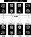
Results: Forty-nine patients showed progression on T1w sequences after 1 year, and 30 patients showed at least one new STIR+ lesion. Increased fat deposition at follow-up was observed in 13.9% STIR+ and in only 0.21% STIR- muscles at baseline (P < 0.001). Overall, 89.9% of the muscles that showed increased fatty replacement were STIR+ at baseline and 7.8% were STIR+ at 12 months. A higher number of STIR+ muscles at baseline was associated with radiological worsening (odds ratio 1.17, 95% confidence interval 1.06-1.30, P = 0.003).
Conclusions: Our study confirms that STIR+ lesions represent prognostic biomarkers in FSHD and contributes to delineate its radiological natural history, providing useful information for clinical trial design. Given the peculiar muscle-by-muscle involvement in FSHD, MRI represents an invaluable tool to explore the modalities and rate of disease progression.
Keywords: Biomarkers; FSHD; Facioscapulohumeral muscular dystrophy; Muscle MRI; Muscle wasting; STIR hyperintensity.
© 2019 The Authors Journal of Cachexia, Sarcopenia and Muscle published by John Wiley & Sons Ltd on behalf of Society on Sarcopenia, Cachexia and Wasting Disorders.
Conflict of interest statement
Pursuant to the terms of a Master Academic Services Agreement with the Catholic University of the Sacred Heart, M.M. and E.R. provided central reading services for MRI scans generated in aTyr's clinical trials of Resolaris (ATYR1940). E.R. was the site principal investigator for some of these trials. F.L., P.O., M.R.B., A.P., and G.T. have nothing to report.
Figures


References
MeSH terms
LinkOut - more resources
Full Text Sources

