. 2019 Nov:29:99-113.
doi: 10.1016/j.molmet.2019.08.019. Epub 2019 Aug 28.

PAR2 controls cholesterol homeostasis and lipid metabolism in nonalcoholic fatty liver disease

Affiliations

PAR2 controls cholesterol homeostasis and lipid metabolism in nonalcoholic fatty liver disease

Rajashree Rana et al. Mol Metab. 2019 Nov.

Abstract

Objective: Increases in hepatic and plasma cholesterol occur in patients with nonalcoholic fatty liver disease (NAFLD), although the reason for this is not well understood. We investigated whether Protease-Activated Receptor 2 (PAR2) plays a role in cholesterol and lipid homeostasis in NAFLD.

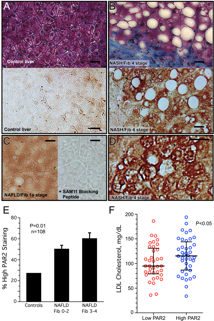
Methods: Human liver biopsies (n = 108) were quantified for PAR2 expression from NAFLD cases randomly selected and stratified by liver fibrosis stage, the primary predictor for clinical outcomes, while controlling for age, gender, and BMI between fibrosis groups. Demographic data and laboratory studies on plasma samples were obtained within 6 months of liver biopsy. Wild-type and PAR2-KO (C57BL/6 F2rl1-/-) mice were fed either normal or high fat diet for 16 weeks and plasma and liver assayed for lipids and soluble metabolites.

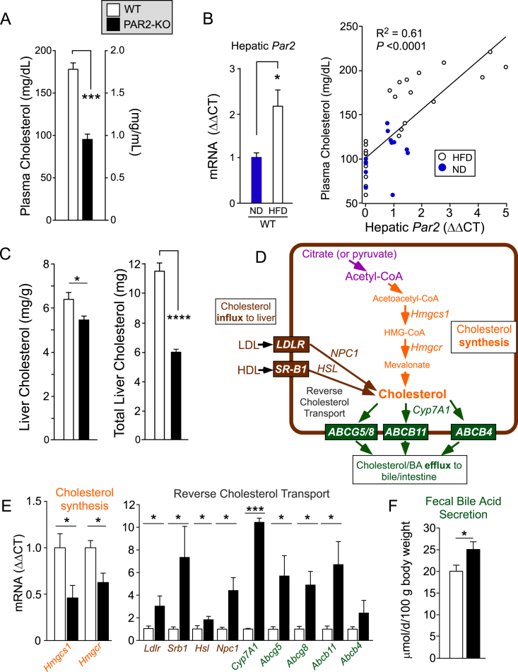
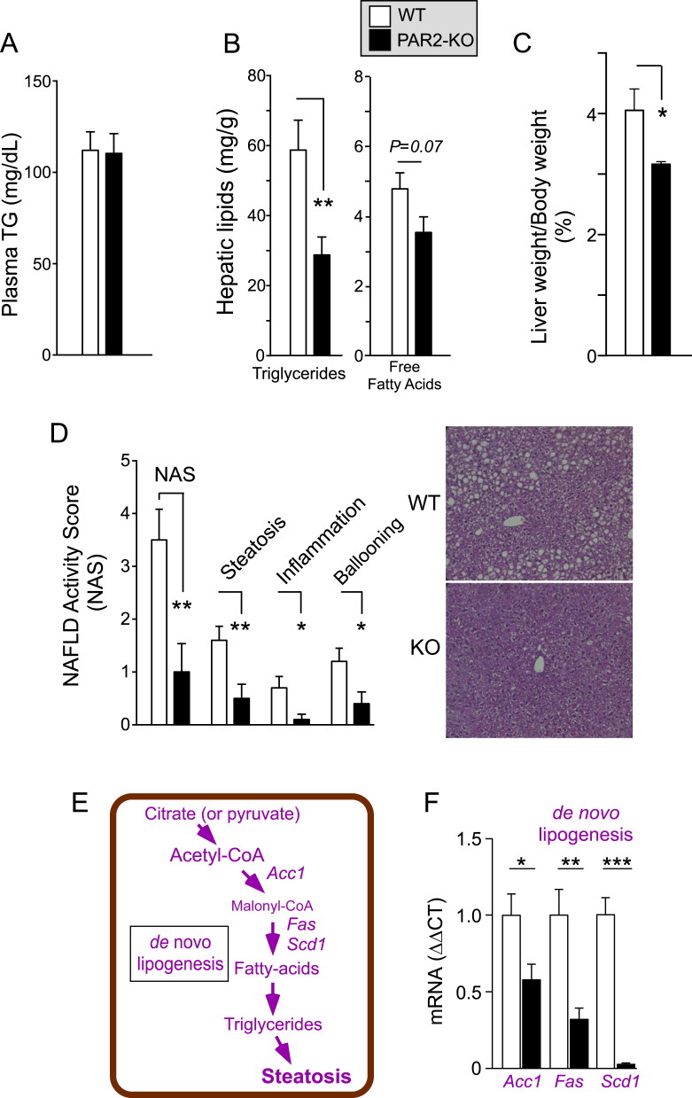
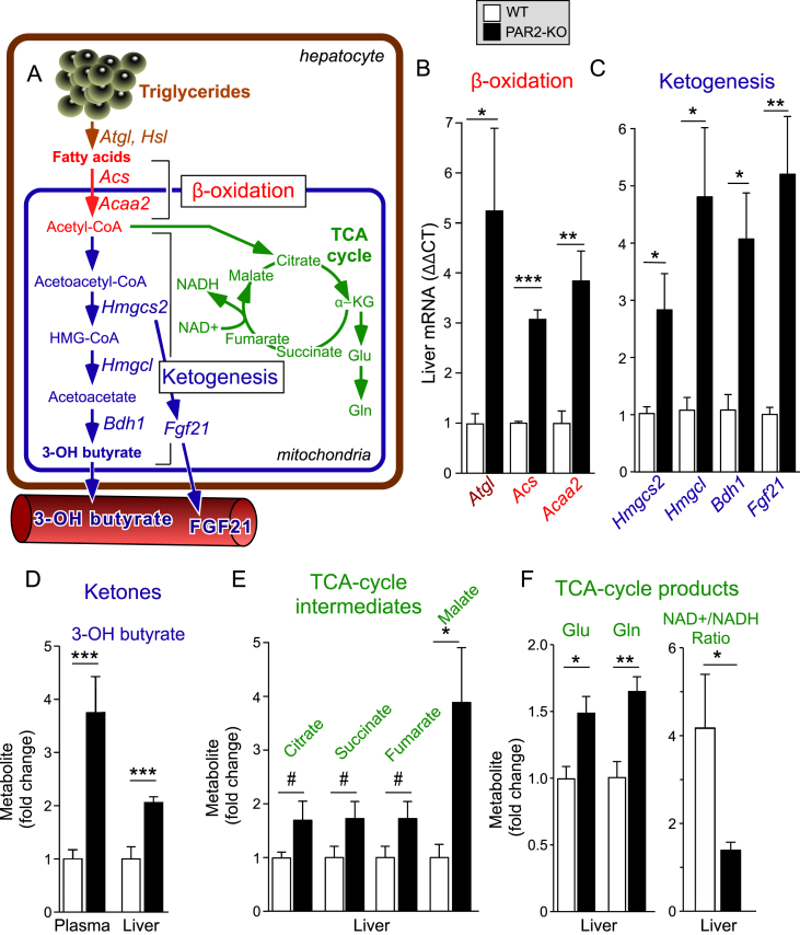
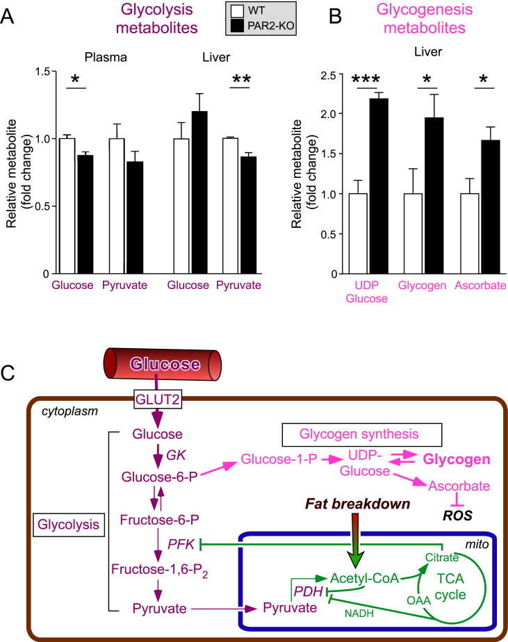
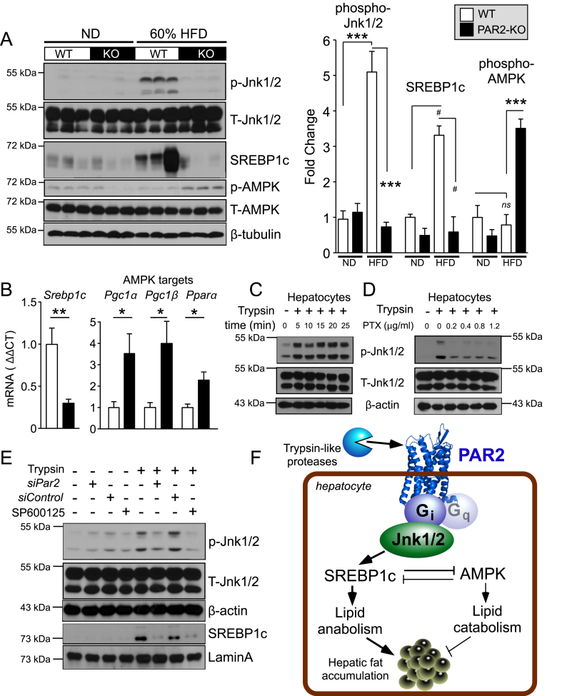
Results: Severity of NAFLD and plasma cholesterol levels significantly correlated with hepatocyte PAR2 expression in NAFLD patients. Conversely, PAR2 deficiency in mice resulted in reduced expression of key hepatic genes involved in cholesterol synthesis, a 50% drop in plasma and total liver cholesterol, and induced a reverse cholesterol transport system that culminated in 25% higher fecal bile acid output. PAR2-deficient mice exhibited enhanced fatty acid β-oxidation with a ketogenic shift and an unexpected increase in liver glycogenesis. Mechanistic studies identified Gi-Jnk1/2 as key downstream effectors of protease-activated PAR2 in the regulation of lipid and cholesterol homeostasis in liver.

Conclusions: These data indicate that PAR2 may be a new target for the suppression of plasma cholesterol and hepatic fat accumulation in NAFLD and related metabolic conditions.

Keywords: Cholesterol; Energy metabolism; Fatty liver; NASH; Protease-activated receptor 2.


Figures

Figure 1
Figure 1
PAR2 expression is enhanced in the livers of patients with NAFLD and NASH and is associated with high LDL cholesterol. Liver biopsies were obtained from 93 subjects with the diagnosis of NAFLD/NASH with fibrosis scores ranging from 0 to 4, along with 15 control subjects with normal liver biopsies. (A) Photomicrographs (400x) of control liver biopsy and (B) fibrosis stage 4 NASH subject biopsy stained with Masson's trichrome to show blue collagen matrix deposition and macrovesicular steatosis (white empty spaces) in top panels. PAR2 staining (brown-SAM11 Ab) of control liver and fibrosis stage 4 NASH patient shown below. (C) PAR2 staining of NAFLD patient with mild stage 1a fibrosis (left panel), also in the presence of the SAM11 blocking peptide, SLIGKVDGTSVTG (right panel) (400x). (D) PAR2 staining of fibrosis stage 4 NASH patient at 800× magnification depicting strong PAR2 staining at periphery/plasma membrane of individual hepatocytes. (E) PAR2 staining of liver biopsies was scored as high or low in a blinded manner in control subjects (n = 15), NAFLD/NASH subjects with fibrosis score of 0–2 (n = 50), and NASH subjects with severe fibrosis scores of 3–4 (n = 43). A logistic regression model of PAR2 staining as the dependent variable and NAFLD fibrosis stage (adjusted for sex and diabetes) as the independent variable was used to analyze the data. (F) LDL cholesterol levels of NAFLD/NASH subjects with low PAR2 versus high PAR2 staining of liver biopsies. Data was analyzed by Pearson's chi-squared test (Mood's median test) for statistical significance with medians and interquartile ranges shown. Black bar = 25 μm.
Figure 2
Figure 2
PAR2 deficiency suppresses hypercholesterolemia and enhances reverse cholesterol transport in liver in HFD-fed mice. Male C57BL/6 wild type (WT-open bars) and PAR2-KO (KO-black bars) mice were fed 60% high fat diet (HFD, n = 10–14) for 16 weeks. (A) At the 16 week endpoint, plasma was obtained from non-fasted mice at Z+4 time point and assayed for total cholesterol. (B) Quantitative PCR (ΔΔCT) of hepatic Par2 mRNA from mice fed normal diet (ND) or HFD for 16 weeks, normalized to β-actin. Plasma cholesterol significantly correlated with hepatic Par2 expression from mice fed both ND and HFD. Linear regression analysis was performed and least-squares lines, R2 values and P values for the slope are shown. (C) Livers from mice in A were harvested and weighed at the 16 week endpoint and assayed for mg cholesterol per g of liver tissue and total liver cholesterol content. (D) Hepatic transporters and enzymes controlling cholesterol flux and synthesis pathways affected by PAR2-deficiency in livers of mice fed a HFD diet for 16 weeks. (E) Quantitative PCR (ΔΔCT) of mRNA from mouse liver (normalized to β-actin mRNA) for Hmgcs1 (3-hydroxy-3-methylglutaryl-Coenzyme A synthase 1), Hmgcr (3-hydroxy-3-methylglutaryl-Coenzyme A reductase), Hsl (Hormone Sensitive Lipase), Npc1 (NPC intracellular cholesterol transporter 1), Abcg5 (ATP-binding cassette, sub-family G member 5), Abcg8 (ATP-binding cassette, sub-family G member 8), Abcb11 (ATP-binding cassette, sub-family B, member 11), Abcb4 (ATP-binding cassette, sub-family B, member 4), Ldlr (low-density lipoprotein receptor), Srb1 (scavenger receptor class B, member 1) in WT and PAR2-KO mice fed HFD for 16 weeks. (F) Mean fecal bile acid content from HFD-fed mice in A during a two-week collection period. All data are represented as mean ± SEM; Unpaired 2-tailed T-tests were performed,*P < 0.05, ***P < 0.001, ****P < 0.0001.
Figure 3
Figure 3
PAR2 deficiency significantly reduces NAS score in NAFLD and expression of genes involved in de novo lipogenesis in mice. Male C57BL/6 wild type and PAR2-KO mice were fed 60% high fat diet (HFD, n = 10–14) for 16 weeks. (A) At the 16 week endpoint, plasma was obtained from non-fasted mice (Z+4) and assayed for triglycerides. (B) Livers from mice in A were harvested at the 16 week endpoint and assayed for triglycerides and free fatty acids. (C) Mean %Liver/Body weight at 16 weeks. (D) NAFLD-Activity Score (NAS) comprising steatosis, inflammation, and ballooning was determined in a blinded manner from hematoxylin and eosin (H&E) stained sections of mouse liver from WT and PAR2-KO mice fed HFD for 16 weeks. (E) Hepatic enzymes controlling de novo lipogenesis. (F) Quantitative PCR (ΔΔCT) mRNA from mouse liver (normalized to β-actin mRNA) for Acc1 (acetyl-CoA carboxylase 1), Fas (fatty-acid synthase) and Scd1 (stearoyl-Coenzyme A desaturase 1). Data represent mean ± SEM from 4 to 5 mice per gene analyzed. Unpaired 2-tailed T-tests were performed; *P < 0.05, **P < 0.01, ***P < 0.001.
Figure 4
Figure 4
PAR2 deficiency significantly upregulates hepatic genes involved in fatty acid β-oxidation and induces a ketogenic shift in HFD-fed mice. (A) Metabolic pathways controlling fatty acid breakdown and ketone synthesis affected by PAR2-deficiency in livers of mice fed a HFD diet for 16 weeks. (B–C) Quantitative PCR (ΔΔCT) of mRNA from mouse liver (normalized to β-actin mRNA): Atgl (Adipose triglyceride lipase), Acs (long chain fatty acyl-CoA ligase or synthetase), Acaa2 (acetyl-Coenzyme A acyltransferase 2), Hmgcs2 (3-hydroxy-3-methylglutaryl-Coenzyme A synthase 2), Hmgcl (3-hydroxy-3-methylglutaryl-Coenzyme A lyase) and Bdh1 (3-hydroxybutyrate dehydrogenase, type 1), Fgf21 (fibroblast growth factor 21) in WT and PAR2-KO mice fed HFD for 16 weeks. Data for Hsl shown in Figure 2E. (D–F) Plasma and/or liver metabolites in WT vs PAR2-KO mice fed HFD for 16 weeks was quantified by NMR-metabolomic methods, Glu = glutamate, Gln = glutamine. Data represent mean ± SEM from 3 to 10 mice per gene and 6–9 mice per metabolite analyzed; unpaired 2-tailed T-tests were performed, *P < 0.05, **P < 0.01, ***P < 0.001, #P = 0.1.
Figure 5
Figure 5
PAR2 deficiency induces glycogenesis and ascorbate production in livers of HFD-fed mice. (A–B) Plasma and liver glycolysis and glycogenesis metabolites in WT vs PAR2-KO mice fed HFD for 16 weeks were quantified by NMR-metabolomic methods. (C) Glycolysis and glycogenesis pathways and regulation in the livers of PAR2-deficient mice. Data represent mean ± SEM from 6 to 9 mice per metabolite analyzed; unpaired 2-tailed T-tests were performed, *P < 0.05, **P < 0.01, ***P < 0.001.
Figure 6
Figure 6
PAR2-Gi activates JNK-SREBP-1c pathways that induce hepatic lipid accumulation in HFD-fed mice. (A) Western blots of liver lysates from WT and PAR2-KO mice fed either normal chow diet (ND) or high fat diet (HFD) for 16 weeks. Densitometric quantification of western blots on left for Phospho (P)-Jnk1/2 normalized to total (T)-Jnk1/2, active 65 kDa proteolytic fragment of SREBP-1c normalized to β-tubulin, and Phospho (P)-AMPK normalized to total-AMPK. (B) Quantitative PCR (ΔΔCT) of mRNA from mouse liver (n = 5) normalized to β-actin mRNA of Srebp1c and AMPK targets: Pgc1α (peroxisome proliferative activated receptor, gamma, coactivator 1 alpha), Pgc1β (peroxisome proliferative activated receptor, gamma, coactivator 1 beta) and Pparα (peroxisome proliferator activated receptor alpha). (C–D) Trypsin (10 nM) induces phospho-Jnk1/2 signaling in HuH7 hepatoma cells, which is suppressed by the Gi inhibitor, pertussis toxin (PTX); 15 min time-point used in D. (E) Par2 siRNA (500 nM) knockdown inhibits trypsin-induced phosphorylation of Jnk1/2 (15 min) in HuH7 cells. Trypsin induces nuclear translocation of SREBP-1c, which is inhibited by siPAR2 and by the Jnk1/2 inhibitor SP600125. β-actin and Lamin A were used as loading controls for whole cell and nuclear lysate western blots, respectively. (F) Mechanism of PAR2-Gi regulation of lipid metabolism in hepatocytes by activating Jnk1/2 and SREBP-1c pathways. One-way ANOVA followed by Tukey's post-hoc test (A) or unpaired 2-tailed T-test (B) was performed, *P < 0.05, **P < 0.01, ***P < 0.001, #P = 0.06.

References

    1. Samuel V.T., Shulman G.I. Nonalcoholic fatty liver disease as a nexus of metabolic and hepatic diseases. Cell Metabolism. 2018;27(1):22–41. - PMC - PubMed
    1. Younossi Z.M. Non-alcoholic fatty liver disease - a global public health perspective. Journal of Hepatology. 2018;70(3):531–544. - PubMed
    1. Min H.K., Kapoor A., Fuchs M., Mirshahi F., Zhou H., Maher J. Increased hepatic synthesis and dysregulation of cholesterol metabolism is associated with the severity of nonalcoholic fatty liver disease. Cell Metabolism. 2012;15(5):665–674. - PMC - PubMed
    1. Shearer A.M., Rana R., Austin K., Baleja J.D., Nguyen N., Bohm A. Targeting liver fibrosis with a cell-penetrating protease-activated receptor-2 (PAR2) pepducin. Journal of Biological Chemistry. 2016;291(44):23188–23198. - PMC - PubMed
    1. Michael E.S., Kuliopulos A., Covic L., Steer M.L., Perides G. Pharmacological inhibition of PAR2 with the pepducin P2pal-18S protects mice against acute experimental biliary pancreatitis. American Journal of Physiology Gastrointestinal and Liver Physiology. 2013;304(5):G516–G526. - PMC - PubMed

Publication types

MeSH terms

Substances