. 2019 Oct 31;179(4):909-922.e12.
doi: 10.1016/j.cell.2019.09.030. Epub 2019 Oct 24.

Structure of the Decorated Ciliary Doublet Microtubule

Affiliations

Structure of the Decorated Ciliary Doublet Microtubule

Meisheng Ma et al. Cell. .

Abstract

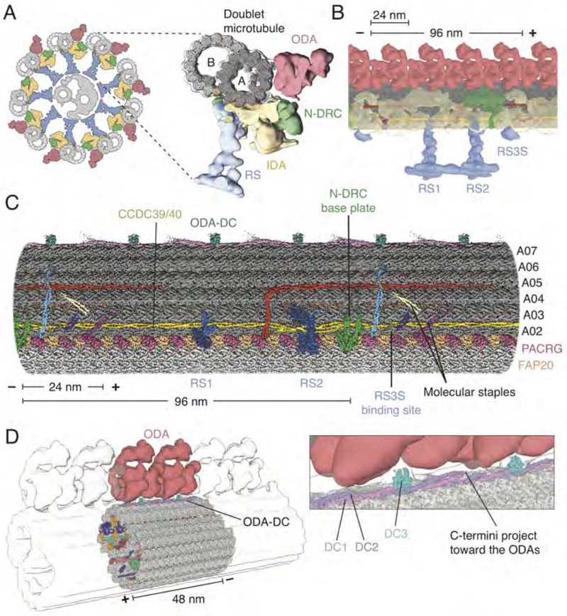
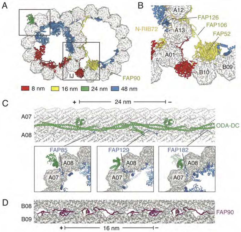
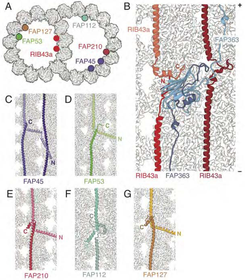
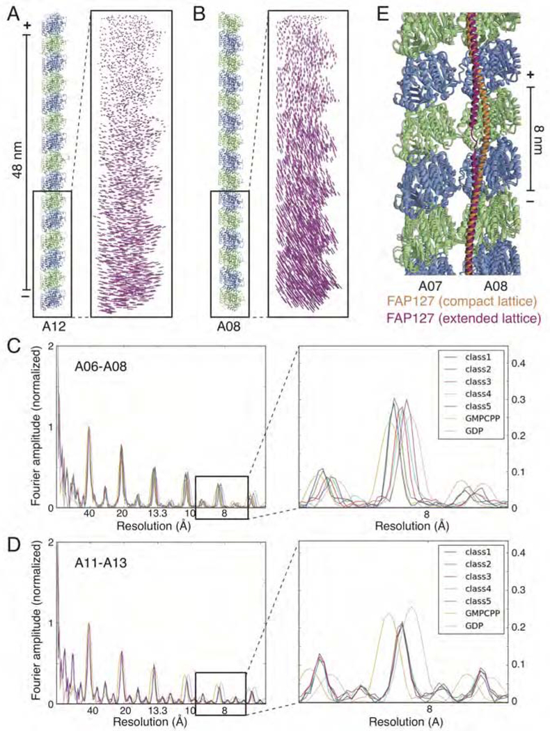
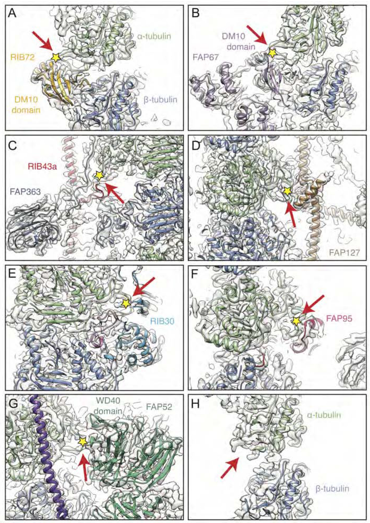
The axoneme of motile cilia is the largest macromolecular machine of eukaryotic cells. In humans, impaired axoneme function causes a range of ciliopathies. Axoneme assembly, structure, and motility require a radially arranged set of doublet microtubules, each decorated in repeating patterns with non-tubulin components. We use single-particle cryo-electron microscopy to visualize and build an atomic model of the repeating structure of a native axonemal doublet microtubule, which reveals the identities, positions, repeat lengths, and interactions of 38 associated proteins, including 33 microtubule inner proteins (MIPs). The structure demonstrates how these proteins establish the unique architecture of doublet microtubules, maintain coherent periodicities along the axoneme, and stabilize the microtubules against the repeated mechanical stress induced by ciliary motility. Our work elucidates the architectural principles that underpin the assembly of this large, repetitive eukaryotic structure and provides a molecular basis for understanding the etiology of human ciliopathies.

Keywords: axoneme; cilia; cryo-EM; doublet microtubule; tubulin.


Conflict of interest statement

Declaration of interests

The authors declare no competing interests.

Figures

Figure 1.
Figure 1.. Structure of the 96-nm Repeat of the Doublet Microtubule and Relationship to the Axoneme
(A) Left, schematic representation of the cross-section of the axoneme from C. reinhardtii showing nine doublet microtubules surrounding a central pair of singlet microtubules (grey). Attached to the doublet microtubules are the radial spokes (RS; blue), inner dynein arm (IDA; yellow), nexin-dynein regulatory complex (N-DRC; green) and outer dynein arm (ODA; red). Right, subtomogram average (EMD-6872) (Kubo et al., 2018) of the axoneme with our map of the 96-nm doublet microtubule repeat (grey) docked inside. (B) Longitudinal view of the doublet microtubule docked into the subtomogram average of the axoneme (EMD-6872). (C) Density map of the 96-nm repeat showing axonemal proteins decorating the external surface of the A tubule. (D) The ODA-DC repeats every 24 nm and, based on fitting the structure into the subtomogram average (EMD-6872), is the main attachment point for the ODA. Inset, detail of the interaction between the ODA and ODA-DC. In all panels, the minus (−) and plus (+) ends of the doublet microtubule are indicated at the ends of the scale bar. See also Figures S1, S2, S3, S4 and Tables S1, S2, S3 and S4.
Figure 2.
Figure 2.. The 48-nm Internal Repeat Structure
(A). Two slices through the doublet microtubule showing the microtubule inner proteins (MIPs), each uniquely colored. Protein labeling continues in panel B. (B) Three longitudinal sections through the A tubule of the doublet microtubule. From left-to-right: the first section shows protofilaments A10-A01, the second section shows protofilaments A03-A08, and the third section shows protofilaments A06-B01. In all panels, the minus (−) and plus (+) ends of the doublet microtubule are indicated and the seam of the A tubule is marked with an asterisk. See also Figures S1, S2, S4 and Tables S1, S2, S3 and S4.
Figure 3.
Figure 3.. MTPs form Networks of Different Periodicities
(A) Slice through the doublet microtubule showing non-tubulin components colored by periodicity. Boxes represent regions highlighted in panels B and C. (B) The cluster of proteins around the inner junction (IJ) with 16-nm repeat length. The RIB72 N-terminal domain (N-RIB72) is shown in orange as although it follows 16-nm repeat, the rest of the protein has 8-nm periodicity. (C) Top, longitudinal view of the outer dynein arm docking complex (ODA-DC) bound in the external cleft between protofilaments A07 and A08. MIPs FAP85, FAP129 and FAP182 protrude through the cleft of the microtubule and make contact with the ODA-DC. Bottom, cross-sections showing the interactions between 48-nm repeat MIPs and the 24-nm repeat ODA-DC. (D) Longitudinal view showing the N-terminal half of FAP90 occupying the external cleft between protofilaments B08 and B09. See also Figures S4 and S7.
Figure 4.
Figure 4.. Maintenance of Longitudinal Periodicity through End-to-end Self-association
(A) The positions of the fMIPs bound to the 48-nm doublet microtubule. There are two copies of RIB43a and FAP45. (B) Overview of RIB43 and FAP363 bound to the ribbon showing end-to-end association. (C-G) Details of the end-to-end self-association between molecules of (C) FAP45, (D) FAP53, (E) FAP210, (F) FAP112, and (G) FAP127. In all longitudinal sections the plus (+) end of the microtubule is facing upwards.
Figure 5.
Figure 5.. Tubulin Lattice Variation for Different Regions of the Doublet Microtubule
(A) Left, protofilament A12 of the ribbon, with the right side facing the central axis of the A tubule. Right, vector display (Cα displacement for each residue of α- and β-tubulin) of protofilament A12 between the two most distinct classes (out of five) following 3D classification of protofilaments A11–13 shows a lateral movement in the cross-section plane. (B) Left, protofilament A08, in the same orientation as (A). Right, vector display of protofilament A08 between the two most distinct classes from 3D classification of protofilaments A06-A08 shows a longitudinal extension/compaction in addition to the lateral movement as seen in (A). (C) Layer line profile for the 5 different classes from 3D classification of protofilaments A06-A08. The magnified section shows variable longitudinal spacings of tubulin heterodimers among the 5 different classes, from 82.1 to 83.5 Å, which is comparable to the difference between cytosolic microtubules bound with GMPCPP (84 Å, gold, mimicking the GTP-bound state) and GDP (81.6 Å, pink). (D) Layer line profile for the 5 different classes from 3D classification of protofilaments A11-A13 (the ribbon region). The magnified section shows that the longitudinal spacings of tubulin heterodimers among the 5 different classes have a consistent value around 82.6 Å. (E) FAP127 adopts different conformations when bound to different lattice dimensions of protofilaments A07-A08. To accommodate the longer lattice dimensions, a helical region of FAP127 straightens and unwinds.
Figure 6.
Figure 6.. MIPs at the Junctions between Tubules
(A) Three slabs (labeled 1–3) through the doublet microtubule showing the locations of MIPs at the seam and outer junction (OJ). The OJ is penetrated by three MIPs; FAP127 (shown in slab 1), FAP53 (slab 2) and FAP141 (slab 3). The seam is recognized by a number of MIPs including the kinase FAP67. See also Figures S5 and S6. (B) Alternating copies of FAP20 and PACRG connect the A and B tubules at the inner junction (IJ). One copy of PACRG adjacent to the nexin-dynein regulatory complex (N-DRC) is absent every 96 nm. (C) Details of the interactions among FAP20, PACRG and the tubulin heterodimer viewed from the microtubule lumen. Ten residues of the typically disordered C-terminal tail of β-tubulin are resolved. See also Figures S5 and S6.
Figure 7.
Figure 7.. Interactions between the K40 Loop of α-tubulin and MIPs
(A-G) Interaction between the αK40 loop on (A) protofilament A01 and the DM10 domain of RIB72, (B) protofilament A09 and the DM10 domain of FAP67, (C) protofilament A13 and RIB43a and an extended region of FAP363, (D) protofilament A07 and FAP127, (E) protofilament A12 and RIB30, (F) protofilament A11 and FAP95, (G) protofilament B09 and FAP52. (H) The αK40 loop is unresolved in undecorated regions, such as on protofilament A02. In all panels, the αK40 loop is indicated with a red arrow and the K40 residue with a yellow star.

Comment in

References

    1. Afonine PV, Poon BK, Read RJ, Sobolev OV, Terwilliger TC, Urzhumtsev A, and Adams PD (2018). Real-space refinement in PHENIX for cryo-EM and crystallography. Acta Crystallogr D Struct Biol 74, 531–544. - PMC - PubMed
    1. Albee AJ, Kwan AL, Lin H, Granas D, Stormo GD, and Dutcher SK (2013). Identification of cilia genes that affect cell-cycle progression using whole-genome transcriptome analysis in Chlamydomonas reinhardtti. G3 (Bethesda) 3, 979–991. - PMC - PubMed
    1. Alushin GM, Lander GC, Kellogg EH, Zhang R, Baker D, and Nogales E (2014). High-resolution microtubule structures reveal the structural transitions in αβ-tubulin upon GTP hydrolysis. Cell 157, 1117–1129. - PMC - PubMed
    1. Antony D, Becker-Heck A, Zariwala MA, Schmidts M, Onoufriadis A, Forouhan M, Wilson R, Taylor-Cox T, Dewar A, Jackson C, et al. (2013). Mutations in CCDC39 and CCDC40 are the major cause of primary ciliary dyskinesia with axonemal disorganization and absent inner dynein arms. Hum. Mutat 34, 462–472. - PMC - PubMed
    1. Austin-Tse C, Halbritter J, Zariwala MA, Gilberti RM, Gee HY, Hellman N, Pathak N, Liu Y, Panizzi JR, Patel-King RS, et al. (2013). Zebrafish Ciliopathy Screen Plus Human Mutational Analysis Identifies C21orf59 and CCDC65 Defects as Causing Primary Ciliary Dyskinesia. Am. J. Hum. Genet 93, 672–686. - PMC - PubMed

Publication types

Substances