Review
. 2020 Mar:33:83-101.
doi: 10.1016/j.molmet.2019.08.021. Epub 2019 Oct 10.

Adapt and conquer: Metabolic flexibility in cancer growth, invasion and evasion

Affiliations
Review

Adapt and conquer: Metabolic flexibility in cancer growth, invasion and evasion

Peter Kreuzaler et al. Mol Metab. 2020 Mar.

Abstract

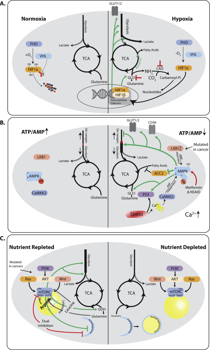
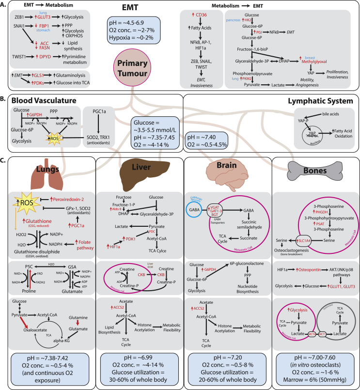
Background: It has been known for close to a century that, on average, tumors have a metabolism that is different from those found in healthy tissues. Typically, tumors show a biosynthetic metabolism that distinguishes itself by engaging in large scale aerobic glycolysis, heightened flux through the pentose phosphate pathway, and increased glutaminolysis among other means. However, it is becoming equally clear that non tumorous tissues at times can engage in similar metabolism, while tumors show a high degree of metabolic flexibility reacting to cues, and stresses in their local environment.

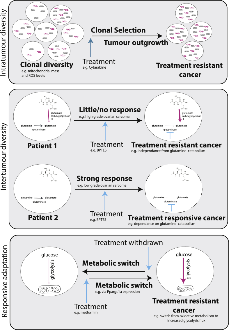
Scope of the review: In this review, we want to scrutinize historic and recent research on metabolism, comparing and contrasting oncogenic and physiological metabolic states. This will allow us to better define states of bona fide tumor metabolism. We will further contextualize the stress response and the metabolic evolutionary trajectory seen in tumors, and how these contribute to tumor progression. Lastly, we will analyze the implications of these characteristics with respect to therapy response.

Major conclusions: In our review, we argue that there is not one single oncogenic state, but rather a diverse set of oncogenic states. These are grounded on a physiological proliferative/wound healing program but distinguish themselves due to their large scale of proliferation, mutations, and transcriptional changes in key metabolic pathways, and the adaptations to widespread stress signals within tumors. We find evidence for the necessity of metabolic flexibility and stress responses in tumor progression and how these responses in turn shape oncogenic progression. Lastly, we find evidence for the notion that the metabolic adaptability of tumors frequently frustrates therapeutic interventions.

Keywords: Central carbon metabolism; Metabolic flexibility; Tumour metabolism; Tumourigenesis.


Figures

Figure 1
Figure 1
Metabolic adaptations to acute metabolic stresses in the tumor microenvironment changes central carbon metabolism flow and exposes vulnerabilities. A) Under normoxic conditions proline residues on HIF1ɑ are hydroxylated by PHDs, thus creating recognition sites for VHL, which ubiquitinates HIF1ɑ marking it for proteasomal degradation. Lack of oxygen prevents hydroxylation and degradation. HIF1ɑ stabilization leads to activation of a transcriptional program that increased glycolytic flux and shunts pyruvate towards glucose. Glutamine is funneled into the Krebs cycle and frequently used for fatty acid synthesis through reductive flux. Due to the higher turnover of glutamine, targeting GLS1 or CAD are trialed as therapeutic approaches. The former precludes a source of nutrients, the latter leads to an accumulation of ammonia. B) A high ATP/AMP ratio leads to binding of ATP to AMPK thus rendering it inactive. Growth stimuli and oncogenes can engage growth programs unabatedly. A drop of this ratio, leads to AMP binding to AMPK, which allows LKB1 to phosphorylate and activate it. AMPK can quench biosynthetic pathways and engage energy production, by boosting nutrient utilization in the Krebs cycle, including glucose, glutamine, and fatty acids, as well as nutrient uptake via transporters. LKB1 is frequently mutated in lung and cervical cancers, leading to failed engagement of LKB1. Persistent stress can, however, re-engage AMPK via a Calcium dependent AMP independent pathway carried out by CaMKK2. Thus, tumors frequently retain parts of this stress response pathway. Drugs including metformin and non-steroidal anti-inflammatory drugs (NSAID) have been shown to re-engage AMPK as well and are in clinical trial as therapies. C) The mTOR complex needs signals both from afferent proliferative pathways as well as nutrient sensors to be fully active. Once fully active, it engages a biosynthetic Warburg like metabolism with enhanced Glycolysis, pyruvate shunting into lactate, and increased glutaminolysis. mTor strongly inhibits autophagy, and inhibition of mTor induces autophagy leading to cell survival. Dual inhibition of mTor and autophagy is a promising drug combination. Note that some upstream activators can have mTor independent effects on metabolism that are not depicted. mTor is rarely mutated itself, allowing tumors to sense and react to nutrient depletion in tumors. When mTor is inhibited due to lack of nutrients, metabolism becomes more oxidative and autophagy is engaged, until amino acid levels have been replenished.
Figure 2
Figure 2
Metabolic adaptations contribute to every step of metastasis progression. The physiological pH, oxygen concentration, and notable nutrient availability in each organ are summarized in blue boxes. A. Metabolic adaptations in the primary tumor during EMT and metastasis instigation. EMT transcription factors have been shown to directly signal to metabolic enzymes (left), and in turn, metabolic transformations can also directly upregulate pro-EMT signaling (right). B. Metabolic adaptations in blood vasculature and the lymphatic system. Circulating tumor cells are faced with increased oxygen availability and therefore upregulate anti-ROS pathways. In the lymph, a novel role for bile acid metabolism has been shown to promote fatty acid oxidation via YAP activation. C. Differential metabolic adaptations observed in metastases to the four most common sites of dissemination: lungs, liver, brain and bones. The metabolic adaptations shown here have been observed in models of different cancer types however the metabolic environment of the organ being colonized may influence metabolic adaptations of metastases through similar mechanisms. Due to continuous oxygen exposure, metastases to the lung upregulate several different mechanisms to combat oxidative stress and promote oxidative phosphorylation for energy and biomass production. Liver metastases make use of the availability of alternative carbon sources by upregulating fructose and acetate metabolism, and exploit the host organ's extracellular matrix for synthesizing creatine phosphate, which then contributes to ATP production in the metastasis cells. Metastases to the brain also adapt to use alternative available fuel sources including acetate and GABA and also promote PPP for nucleotide biosynthesis. Finally, metastases to the bone adapt their metabolism to help them exploit the natural bone-breaking osteoclast cells in order to make more room for metastasis expansion. It has been shown that bone metastasis cells excrete serine and lactate to promote osteoclastogenesis. Furthermore, bone metastases excrete the bone-specific signaling protein osteopontin which upregulates glucose intake for glycolysis. Figure 2 references: Oxygen concentrations in healthy tissues [263], in tumors [264]. Glucose concentrations [265,266], in blood [267], utilization by liver [268], utilization by brain [269,270]. Intratissue pH [[271], [272], [273]], intratumor pH [274], bone pH [273], bone marrow pH [275].
Figure 3
Figure 3
Metabolic diversity and adaptability enable tumors to resist treatment. Diversity may occur within a tumor, where treatment selects for clones most able to overcome treatment, or between tumors, resulting in differential responses between patients due to inherent metabolic differences. Tumor cells may also rewire their metabolism as a direct response to treatment to minimize the effect on the cell while retaining their ability to revert to their nascent metabolism when the treatment program ends.

References

    1. Warburg O., Posener K., Negelein E. The metabolism of cancer cells. Biochemische Zeitschrift. 1924;152:319–344.
    1. Gullino P.M., Clark S.H., Grantham F.H. The interstitial fluid of solid tumors. Cancer Research. 1964;24:780–794. - PubMed
    1. Spick C., Herrmann K., Czernin J. 18F-FDG PET/CT and PET/MRI perform equally well in cancer: evidence from studies on more than 2,300 patients. Journal of Nuclear Medicine: Official Publication, Society of Nuclear Medicine. 2016;57(3):420–430. - PMC - PubMed
    1. Mithieux G. New data and concepts on glutamine and glucose metabolism in the gut. Current Opinion in Clinical Nutrition and Metabolic Care. 2001;4(4):267–271. - PubMed
    1. van Hall G. Lactate kinetics in human tissues at rest and during exercise. Acta Physiologica. 2010;199(4):499–508. - PubMed

Publication types