Review
. 2020 Jan:323:113090.
doi: 10.1016/j.expneurol.2019.113090. Epub 2019 Oct 25.

Analyzing chemotherapy-induced peripheral neuropathy in vivo using non-mammalian animal models

Affiliations
Review

Analyzing chemotherapy-induced peripheral neuropathy in vivo using non-mammalian animal models

Anthony M Cirrincione et al. Exp Neurol. 2020 Jan.

Abstract

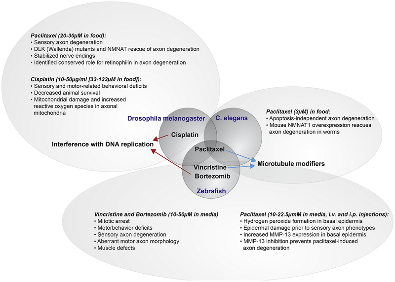
Non-mammalian models of CIPN remain relatively sparse, but the knowledge gained from the few published studies suggest that these species have great potential to serve as a discovery platform for new pathways and underlying genetic mechanisms of CIPN. These models permit large-scale genetic and pharmacological screening, and they are highly suitable for in vivo imaging. CIPN phenotypes described in rodents have been confirmed in those models, and conversely, genetic players leading to axon de- and regeneration under conditions of chemotherapy treatment identified in these non-mammalian species have been validated in rodents. Given the need for non-traditional approaches with which to identify new CIPN mechanisms, these models bear a strong potential due to the conservation of basic mechanisms by which chemotherapeutic agents induce neurotoxicity.

Keywords: Axon degeneration; CIPN; Chemotherapy-induced peripheral neuropathy; Drosophila; Non-mammalian; Review; Zebrafish C. elegans; in vivo imaging.


Conflict of interest statement

Conflict of interest

The authors declare no conflicts of interest.

Figures

Figure 1.
Figure 1.. Overview of identified mechanisms in non-mammalian models leading to chemotherapy-induced neurotoxicity.
Three model species shown in the middle bubbles (Drosophila, C. elegans, and zebrafish) have been assessed for neurotoxic effects derived from various chemotherapeutic agents known to cause CIPN in humans. All species were studied for their response to paclitaxel treatment (microtubule stabilizer), whereas cisplatin (DNA-replication interfering) was uniquely studied in Drosophila and vincristine (microtubule destabilizer) and bortezomib (proteasome inhibitor) were uniquely studied in zebrafish. The outcomes of each of these studies are shown in the outer bubbles.
Figure 2.
Figure 2.. In vivo analysis of paclitaxel-induced axon damage in Drosophila.
This figure was reprinted with permission from Figures 1 and 2 (Brazill et al., 2018b). (A) A: Thermal nociceptive profile of third instar larvae (120 hours after egg laying) Larvae were stimulated with indicated temperatures following vehicle-containing food exposure for 24 hours. Points indicate individual larvae. No response was counted if larvae did not withdraw from heat stimulus after 20 seconds. D: Profile in larvae treated with paclitaxel under same conditions. 42C showed a robust withdrawal response in all larvae tested. (B) A/B: Confocal Projections of fixed, filet-dissected vehicle or paclitaxel-treated larvae showing class IV dendritic arborization (C4da) neuronal compartments labeled by CD4:tdGFP. A2/B2: C4da axon terminals project in a ladder-like pattern in the ventral nerve cord. scale bar: 10μm A3/B3: Dorsal dendrite projections of C4da ddaC neurons shown with cell body near the center at the bottom of the frame. Scale bar: 50μm. C: Quantification of area of dendritic field shows a reduced field upon paclitaxel treatment. D: Quantification of number of terminal branches shows an increase upon paclitaxel treatment suggestive of dendrite-stabilizing effects of paclitaxel.
Figure 3.
Figure 3.
This figure was with permission reprinted from Figure 5 (Mao et al., 2016). In (b) worm mechanosensory neurons and axons are labeled by zdIs5[mec-4::GFP] (scale bar: 100 μm; scale bar in inset: 10 μm). Representative images from the tail region comparing DMSO buffer versus taxol. Note fragmented (arrow) and beaded (arrowhead) axons in the taxol condition. (c) Taxol-induced axon degeneration was partially rescued by Neuro-Nmnat1[gcIs35(neuro-m-nonN-Nmnat1]).
Figure 4.
Figure 4.. In vivo analysis of paclitaxel-induced neurotoxicity in zebrafish.
This figure is in part re-used from (Lisse et al., 2016). (A) Tg(isl2b:GFP) zebrafish larva with fluorescent sensory neurons in the head and body, which innervate the epidermis. (B) Larval zebrafish treated with either DMSO control vehicle (top) or 22μM paclitaxel (bottom) after 1, 3, and 4 days following treatment begin. Sensory axons selectively degenerate in the distal tail fin of zebrafish around 3–4 days when treated with paclitaxel but not DMSO.

References

    1. Addington J, Freimer M, 2016. Chemotherapy-induced peripheral neuropathy: an update on the current understanding. F1000Res 5. - PMC - PubMed
    1. Amos LA, Löwe J, 1999. How Taxol stabilises microtubule structure. Chem Biol 6, R65–69. - PubMed
    1. Araki T, Sasaki Y, Milbrandt J, 2004. Increased nuclear NAD biosynthesis and SIRT1 activation prevent axonal degeneration. Science 305, 1010–1013. - PubMed
    1. Areti A, Yerra VG, Naidu V, Kumar A, 2014. Oxidative stress and nerve damage: role in chemotherapy induced peripheral neuropathy. Redox Biol 2, 289–295. - PMC - PubMed
    1. Argyriou AA, Iconomou G, Kalofonos HP, 2008. Bortezomib-induced peripheral neuropathy in multiple myeloma: a comprehensive review of the literature. Blood 112, 1593–1599. - PubMed

Publication types

Substances