. 2020 Jan:28:101345.
doi: 10.1016/j.redox.2019.101345. Epub 2019 Oct 19.

Metformin protects against PM2.5-induced lung injury and cardiac dysfunction independent of AMP-activated protein kinase α2

Affiliations

Metformin protects against PM2.5-induced lung injury and cardiac dysfunction independent of AMP-activated protein kinase α2

Junling Gao et al. Redox Biol. 2020 Jan.

Abstract

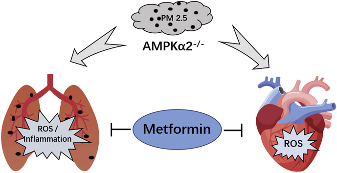
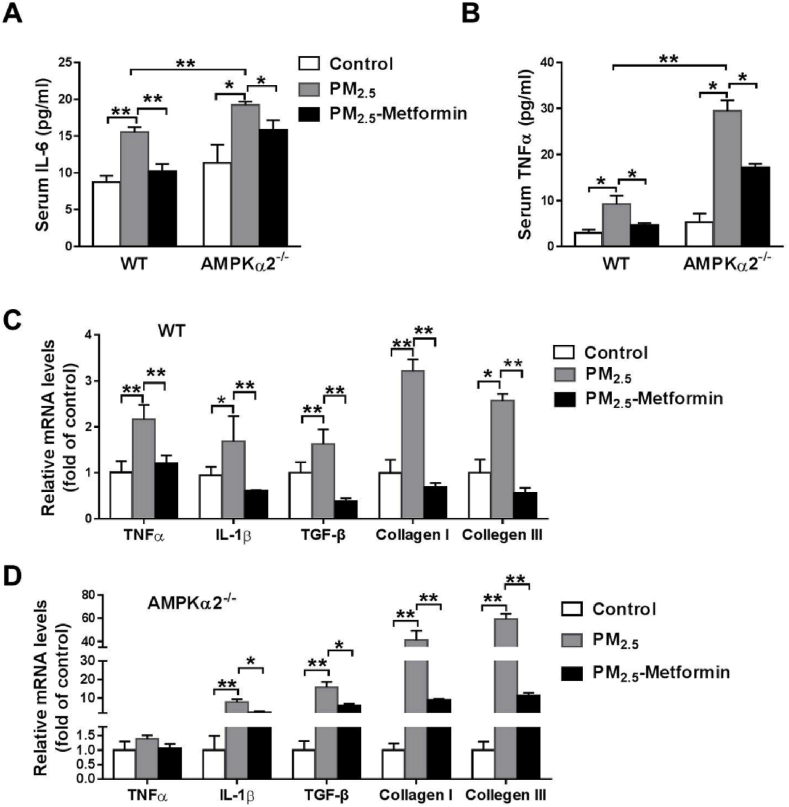
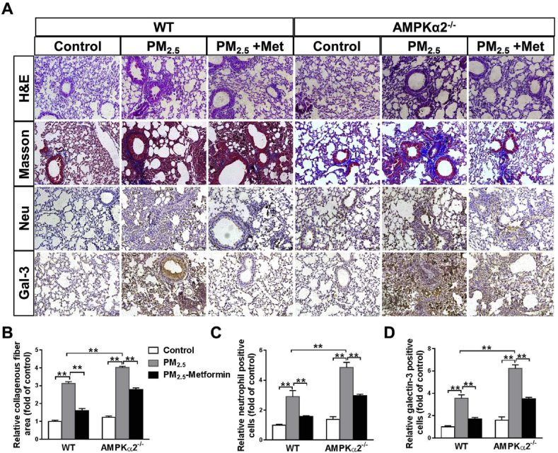
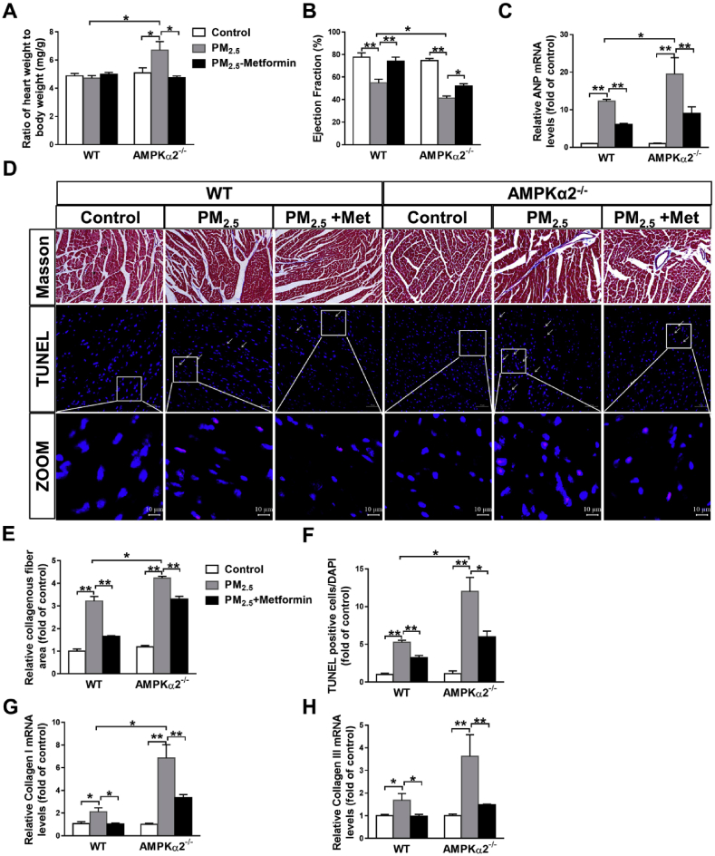
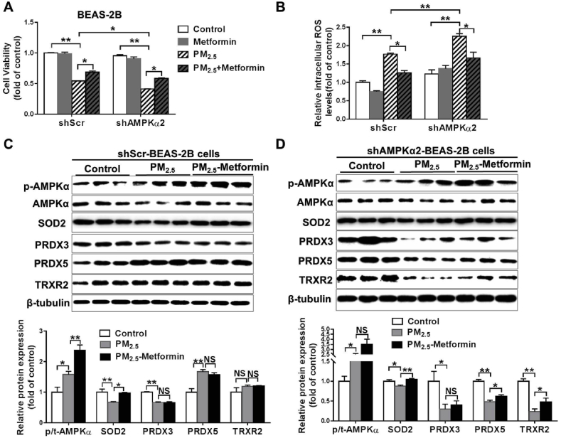
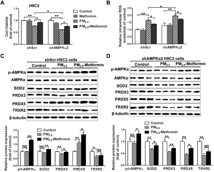
Fine particulate matter (PM2.5) airborne pollution increases the risk of respiratory and cardiovascular diseases. Although metformin is a well-known antidiabetic drug, it also confers protection against a series of diseases through the activation of AMP-activated protein kinase (AMPK). However, whether metformin affects PM2.5-induced adverse health effects has not been investigated. In this study, we exposed wild-type (WT) and AMPKα2-/- mice to PM2.5 every other day via intratracheal instillation for 4 weeks. After PM2.5 exposure, the AMPKα2-/- mice developed more severe lung injury and cardiac dysfunction than were developed in the WT mice; however the administration of metformin was effective in attenuating PM2.5-induced lung injury and cardiac dysfunction in both the WT and AMPKα2-/- mice. In the PM2.5-exposed mice, metformin treatment resulted in reduced systemic and pulmonary inflammation, preserved left ventricular ejection fraction, suppressed induction of pulmonary and myocardial fibrosis and oxidative stress, and increased levels of mitochondrial antioxidant enzymes. Moreover, pretreatment with metformin significantly attenuated PM2.5-induced cell death and oxidative stress in control and AMPKα2-depleted BEAS-2B and H9C2 cells, and was associated with preserved expression of mitochondrial antioxidant enzymes. These data support the notion that metformin protects against PM2.5-induced adverse health effects through a pathway that appears independent of AMPKα2. Our findings suggest that metformin may also be a novel drug for therapies that treat air pollution associated disease.

Keywords: AMPKα2; Cardiac dysfunction; Lung injury; Metformin; PM(2.5).


Conflict of interest statement

The authors declare that they have no known competing financial interests or personal relationships that could have appeared to influence the work reported in this paper.

Figures

Image 1
Graphical abstract
Fig. 1
Fig. 1
Metformin alleviates PM2.5-induced systemic and pulmonary inflammation. WT and AMPKα2−/− mice were administered PBS or 10 mg/kg PM2.5 every other day via intratracheal instillation for 4 weeks. During the entire experimental period, some of the PM2.5-exposed mice were treated with metformin in the drinking water (300 mg/kg/day). After PM2.5 exposure, serum IL-6 and TNFα levels (A–B), and mRNA levels of inflammatory and fibrotic genes in the lungs of WT and AMPKα2−/− mice were measured (C–D). N = 5; data are presented as the mean ± SEM; * indicates p < 0.05, ** indicates p < 0.01.
Fig. 2
Fig. 2
Metformin ameliorates PM2.5-induced pulmonary fibrosis and inflammation. (A) Representative lung sections from the PBS- and PM2.5-exposed WT and AMPKα2−/− mice with or without metformin (Met) treatment were stained with hematoxylin and eosin (H&E), Masson's trichrome, and antibodies specific for neutrophils and macrophages (galectin-3, Gal-3) (brown staining). Scale bar = 100 μm. The relative collagenous fiber area (B), and the number of Gal-3 positive cells (C) and neutrophils (D) were quantified. N = 4; ** indicates p < 0.01. (For interpretation of the references to colour in this figure legend, the reader is referred to the Web version of this article.)
Fig. 3
Fig. 3
Metformin attenuates PM2.5-induced pulmonary oxidative stress and cell death. After PM2.5 exposure, the levels of 3′-nitrotyrosine (3′-NT) (A) and 4-hydroxynonenal (4-HNE) (B) in lung tissue were measured. Lung sections from the control and PM2.5-exposed mice were stained with DAPI (blue) and TUNEL assay kit dye (red) (scale bar = 20 μm, arrows point to TUNEL-positive cells), and the TUNEL-positive cells were quantified (C, D). Lysates of lung tissue were examined by western blotting to determine the expression levels of total and phosphorylated AMPKα, superoxide dismutase 2 (SOD2), peroxiredoxin 3(PRDX3), PRDX5, thioredoxin reductase 2 (TRXR2) and Bcl-2. β-Tubulin was used as a loading control (E, F). N = 3–5; data are presented as the mean ± SEM; * indicates p < 0.05, ** indicates p < 0.01. (For interpretation of the references to colour in this figure legend, the reader is referred to the Web version of this article.)
Fig. 4
Fig. 4
Metformin ameliorates PM2.5-induced cardiac dysfunction, myocardial fibrosis and cardiomyocyte apoptosis. After PM2.5 exposure, the heart weight to body weight ratio (A), LV ejection fraction (B) and mRNA levels of atrial natriuretic peptide (ANP) (C) were measured. Representative heart sections from the control and PM2.5- or PM2.5 with metformin-treated WT and AMPKα2−/− mice were stained with Masson's trichrome (scale bar = 100 μm) and TUNEL assay kit dye (red) plus DAPI (blue) (scale bar = 50 μm) (D). The fibrotic area (E) and the TUNEL-positive cells were quantified (F) were quantified. The mRNA levels of myocardial collagen I and III were measured (G, H). N = 4–7; data are presented as the mean ± SEM; * indicates p < 0.05, ** indicates p < 0.01. (For interpretation of the references to colour in this figure legend, the reader is referred to the Web version of this article.)
Fig. 5
Fig. 5
Metformin attenuates myocardial oxidative stress in PM2.5-exposed mice. After PM2.5 exposure, heart tissue was collected, and the myocardial 3′-nitrotyrosine (3′-NT) (A) and 4-HNE levels were measured. The heart sections were stained with Mitosox (scale bar = 20 μm), and the relative Mitosox fluorescence intensity was quantified (C, D). Lysates of the heart tissue were examined by western blotting for the expression of p-AMPKα, SOD2, PRDX5, PRDX3, TRXR2 and TRX2. β-Tubulin was used as a loading control (E, F). N = 4; data are presented as the mean ± SEM; * indicates p < 0.05, ** indicates p < 0.01.
Fig. 6
Fig. 6
Metformin attenuates PM2.5-induced cell death and oxidative stress in BEAS-2B cells. Scramble shRNA (shScr)- and AMPKα2-specific shRNA (shAMPKα2)-stably transfected BEAS-2B cells were pretreated with PBS or 1 mM metformin for 2 h and then exposed to 50 μg/ml PM2.5 for 24 h. Cell viability (A) and intracellular ROS levels (B) were then determined. Lysates of the control and PM2.5-exposed shScr (C) or shAMPKα2 cells (D) with or without metformin pretreatment were examined by western blotting to determine the expression of phosphorylated AMPKα, SOD2, PRDX3, PRDX5 and TRXR2. β-Tubulin was used as a loading control. N = 3–4; data are presented as the mean ± SEM; * indicates p < 0.05, ** indicates p < 0.01, NS, not significant.
Fig. 7
Fig. 7
Metformin ameliorates PM2.5-induced cell death and oxidative stress in H9C2 cells. H9C2 cells stably transfected with shScr or shAMPKα2 were pretreated with PBS or 1 mM metformin for 2 h and then exposed to 50 μg/ml PM2.5 for 24 h. Cell viability (A) and intracellular ROS levels (B) were then determined. Lysates of the control and the PM2.5-exposed shScr- or shAMPKα2-H9C2 cells with or without of metformin pretreatment were examined by western blotting to determine the expression levels of phosphorylated AMPKα, SOD2, PRDX3, PRDX5 and TRXR2. β-Tubulin was used as a loading control (C-D). N = 3–4, data are mean ± SEM; * indicates p < 0.05, ** indicates p < 0.01, NS, not significant.

References

    1. Hopke P.K., Cohen D.D., Begum B.A., Biswas S.K., Ni B., Pandit G.G., Santoso M., Chung Y.S., Rahman S.A., Hamzah M.S., Davy P., Markwitz A., Waheed S., Siddique N., Santos F.L., Pabroa P.C., Seneviratne M.C., Wimolwattanapun W., Bunprapob S., Vuong T.B., Duy Hien P., Markowicz A. Urban air quality in the Asian region. Sci. Total Environ. 2008;404(1):103–112. - PubMed
    1. Deng X., Zhang F., Rui W., Long F., Wang L., Feng Z., Chen D., Ding W. PM2.5-induced oxidative stress triggers autophagy in human lung epithelial A549 cells. Toxicol. In Vitro : An International Journal Published in Association with BIBRA. 2013;27(6):1762–1770. - PubMed
    1. Tian L., Zhang W., Lin Z.Q., Zhang H.S., Xi Z.G., Chen J.H., Wang W. Impact of traffic emissions on local air quality and the potential toxicity of traffic-related particulates in Beijing, China. Biomed. Environ. Sci. : BES (Biomed. Environ. Sci.) 2012;25(6):663–671. - PubMed
    1. Li R., Kou X., Geng H., Xie J., Yang Z., Zhang Y., Cai Z., Dong C. Effect of ambient PM(2.5) on lung mitochondrial damage and fusion/fission gene expression in rats. Chem. Res. Toxicol. 2015;28(3):408–418. - PubMed
    1. Kirrane E.F., Luben T.J., Benson A., Owens E.O., Sacks J.D., Dutton S.J., Madden M., Nichols J.L. A systematic review of cardiovascular responses associated with ambient black carbon and fine particulate matter. Environ. Int. 2019;127:305–316. - PMC - PubMed

Publication types

MeSH terms