Randomized Controlled Trial
. 2019:24:102042.
doi: 10.1016/j.nicl.2019.102042. Epub 2019 Oct 18.

Evidence for decreased Neurologic Pain Signature activation following thoracic spinal manipulation in healthy volunteers and participants with neck pain

Affiliations
Randomized Controlled Trial

Evidence for decreased Neurologic Pain Signature activation following thoracic spinal manipulation in healthy volunteers and participants with neck pain

Kenneth A Weber Ii et al. Neuroimage Clin. 2019.

Abstract

Background context: Spinal manipulation (SM) is a common treatment for neck and back pain, theorized to mechanically affect the spine leading to therapeutic mechanical changes. The link between specific mechanical effects and clinical improvement is not well supported. SM's therapeutic action may instead be partially mediated within the central nervous system.

Purpose: To introduce brain-based models of pain for spinal pain and manual therapy research, characterize the distributed central mechanisms of SM, and advance the preliminary validation of brain-based models as potential clinical biomarkers of pain.

Study design: Secondary analysis of two functional magnetic resonance imaging studies investigating the effect of thoracic SM on pain-related brain activity: A non-controlled, non-blinded study in healthy volunteers (Study 1, n = 10, 5 females, and mean age = 31.2 ± 10.0 years) and a randomized controlled study in participants with acute to subacute neck pain (Study 2, n = 24, 16 females, mean age = 38.0 ± 15.1 years).

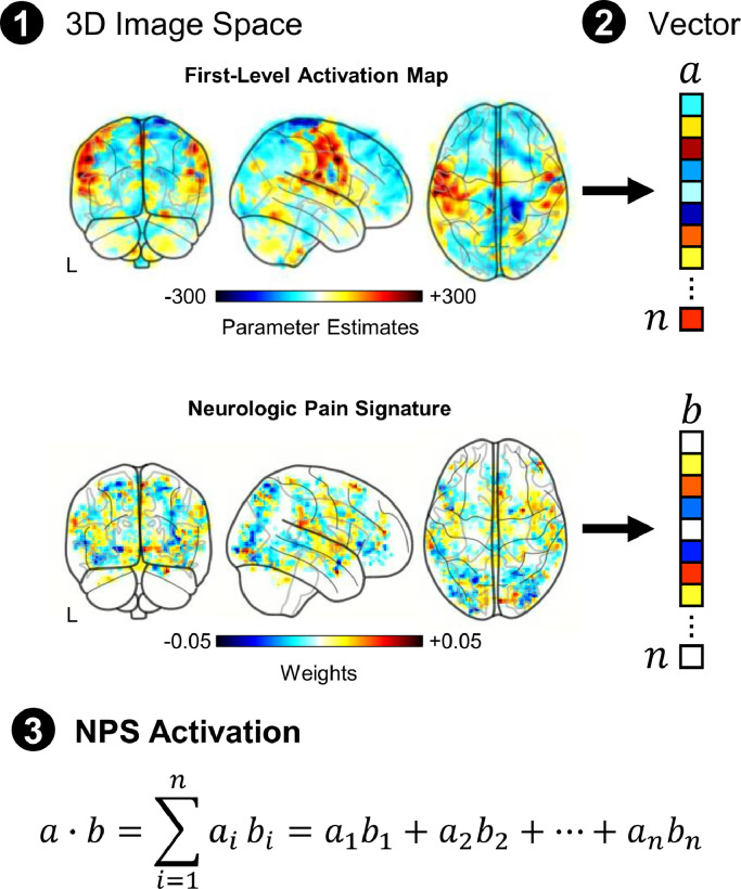
Methods: Functional magnetic resonance imaging was performed during noxious mechanical stimulation of the right index finger cuticle pre- and post-intervention. The effect of SM on pain-related activity was studied within brain regions defined by the Neurologic Pain Signature (NPS) that are predictive of physical pain.

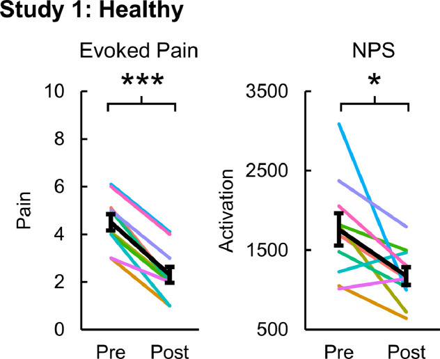
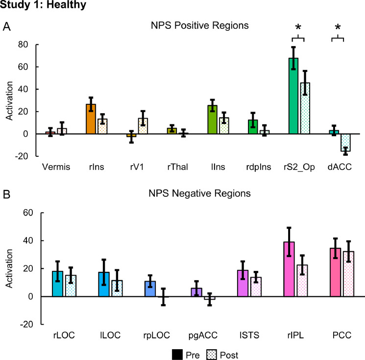
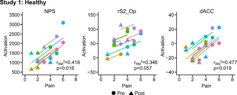
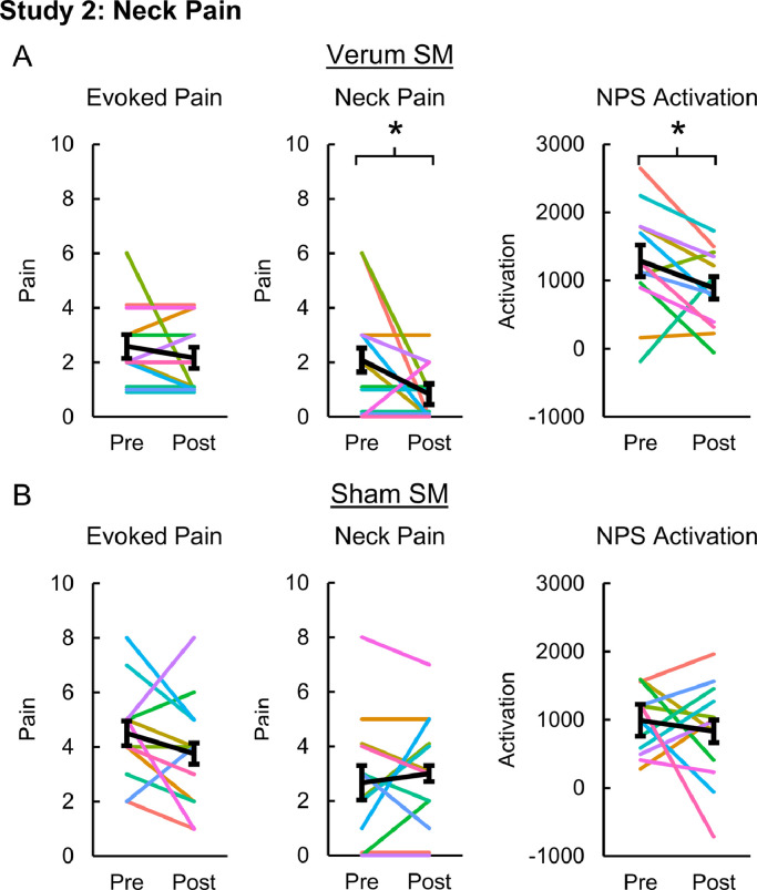
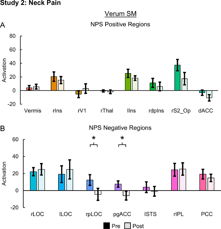
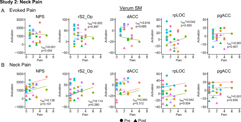
Results: In Study 1, evoked mechanical pain (p < 0.001) and NPS activation (p = 0.010) decreased following SM, and the changes in evoked pain and NPS activation were correlated (rRM2 = 0.418, p = 0.016). Activation within the NPS subregions of the dorsal anterior cingulate cortex (dACC, p = 0.012) and right secondary somatosensory cortex/operculum (rS2_Op, p = 0.045) also decreased following SM, and evoked pain was correlated with dACC activity (rRM2 = 0.477, p = 0.019). In Study 2, neck pain (p = 0.046) and NPS (p = 0.033) activation decreased following verum but not sham SM. Associations between evoked pain, neck pain, and NPS activation, were not significant and less clear, possibly due to inadequate power, methodological limitations, or other confounding factors.

Conclusions: The findings provide preliminary evidence that SM may alter the processing of pain-related brain activity within specific pain-related brain regions and support the use of brain-based models as clinical biomarkers of pain.

Keywords: Functional magnetic resonance imaging; Humans; Neck pain; Neuroimaging; Pain; Pain measurement; Randomized controlled trial; Spinal manipulation.


Conflict of interest statement

Drs. Weber, Wager, Mackey and Elliott report grants from the National Institutes of Health during the conduct of this study. Dr. Wager reports a Small Business Innovation Research grant with WaviMed. Dr. Wager is also on the scientific advisory board of Curable Health, Inc., has consulted for GSK and Cognifisense, has performed contract work for PainQX, and has been issued two patents: US 2016/0,054,409 fMRI-based Neurologic Signature of Physical Pain (PCT/US14/3353) and US 2018/0,055,407 Neurophysiological Signatures for Fibromyalgia (CU4199B-PPA1). Dr. Elliott is an advisory member for the board of directors of the Journal of Orthopaedic and Sports Physical Therapy, an editorial board member for the Journal of Orthopaedic and Sports Physical Therapy, an editorial board member for Musculoskeletal Science and Practice, and an advisory board member for Spine. Drs. Liu and Sparks declare no competing interests.

Figures

Fig 1
Fig. 1
To investigate the effect of SM on evoked mechanical pain-related brain activity, we focused on activation within brain regions predictive of physical pain using the Neurologic Pain Signature (NPS). The NPS is a multivariate brain activation pattern that has been shown to be sensitive and specific to experimentally-evoked pain at the individual level (Wager et al., 2013). The NPS activation was calculated by taking the dot product of the NPS pattern weights and the stimulus parameter estimate images from each participant's first-level analysis. The dot product provides a single scalar value to quantify the NPS activation (arbitrary units).
Fig 2
Fig. 2
Evoked pain and Neurologic Pain Signature (NPS) activation pre- and post-intervention for the healthy volunteers receiving verum spinal manipulation (SM). Both evoked pain and NPS activation decreased following SM. NPS activation is in arbitrary units. One-tailed paired samples t-tests, *p<0.05, and ***p<0.001. Error bars = ± standard error.
Fig 3
Fig. 3
Neurologic Pain Signature (NPS) subregion activation pre- and post-intervention for the healthy volunteers receiving verum spinal manipulation (SM). A) Activation within the NPS positive regions of the right secondary somatosensory cortex/operculum (rS2_Op) and the dorsal anterior cingulate cortex (dACC) decreased following SM. B) No significant changes were identified within any of the NPS negative regions. NPS activation is in arbitrary units. Two-tailed paired samples t-tests, *p < 0.05. Error bars = ± standard error. rIns = right mid-insula, rV1 = right primary visual cortex, rThal = right thalamaus, lIns = left mid-insula, rdplns = right dorsal posterior insula, rLOC = right lateral occipital complex, lLOC = left lateral occipital complex, rpLOC = right posterior lateral occipital complex, pgACC = pregenual anterior cingulate cortex, lSTS = left superior temporal sulcus, rIPL = right inferior parietal lobule, and PCC = posterior cingulate cortex.
Fig 4
Fig. 4
Scatter plots for the repeated measures correlations (RMCORR) between evoked pain and Neurologic Pain Signature (NPS) activation and NPS subregion activation for the healthy volunteers receiving verum spinal manipulation (SM). The observations from the same participant are shown in the same color, and the colored lines show the RMCORR fit for each participant (i.e., fixed slopes and varying intercepts). Evoked pain was positively correlated to NPS activation. Only correlations within subregions showing significant differences in Study 1 were explored. Within the subregions, evoked pain was positively correlated to the dorsal anterior cingulate cortex (dACC) activation and tended to be positively correlated to right somatosensory cortex (rS2_Op) activation. NPS activation was hypothesized to be positively correlated with evoked pain, so one-tailed tests were performed. Two-tailed tests were performed for the exploratory analysis within the NPS subregions. NPS activation is in arbitrary units.
Fig 5
Fig. 5
Evoked pain, neck pain, and Neurologic Pain Signature (NPS) activation pre- and post-intervention for the neck pain participants receiving verum (A) and sham (B) spinal manipulation (SM). Neck pain and NPS activation decreased following verum SM but not evoked pain. No significant changes in evoked pain, neck pain, or NPS activation were seen following sham SM. NPS activation is in arbitrary units. One-tailed paired samples t-tests, *p < 0.05. Error bars = ± standard error.
Fig 6
Fig. 6
Neurologic Pain Signature (NPS) subregion activation pre- and post-intervention for the neck pain participants receiving verum spinal manipulation (SM). A) No significant changes were identified within any of the NPS positive regions. B) Activation within the NPS negative regions of the right posterior lateral occipital complex (rpLOC) and pregenual anterior cingulate cortex (pgACC) decreased following SM. NPS activation is in arbitrary units. Two-tailed paired samples t-tests, *p < 0.05. Error bars = ± standard error. rIns = right mid-insula, rV1 = right primary visual cortex, rThal = right thalamaus, lIns = left mid-insula, rdplns = right dorsal posterior insula, rS2_Op = right secondary somatosensory cortex/operculum, dACC = dorsal anterior cingulate cortex, rLOC = right lateral occipital complex, lLOC = left lateral occipital complex, lSTS = left superior temporal sulcus, rIPL = right inferior parietal lobule, and PCC = posterior cingulate cortex.
Fig 7
Fig. 7
Scatter plots for the repeated measures correlations (RMCORR) between evoked pain (A) and neck pain (B) and Neurologic Pain Signature (NPS) activation and NPS subregion activation for the neck pain participants receiving verum spinal manipulation (SM). The observations from the same participant are shown in the same color, and the colored lines show the RMCORR fit for each participant (i.e., fixed slopes and varying intercepts). Only correlations within subregions showing significant differences in Studies 1 and 2 were explored. While no significant correlations were identified for evoked pain or neck pain in the neck pain participants receiving verum SM, a non-significant positive weak correlation between neck pain and NPS activation (p = 0.107) was present. NPS activation is in arbitrary units. NPS activation was hypothesized to be positively correlated with evoked pain and neck pain, so one-tailed tests were performed. Two-tailed tests were performed for the exploratory analysis within the NPS subregions. rS2_Op=right secondary somatosensory cortex/operculum, dACC=dorsal anterior cingulate cortex, rpLOC=right posterior lateral occipital complex, and pgACC=pregenual anterior cingulate cortex.

References

    1. Andersson J.L.R., Jenkinson M., Smith S. 2010. Non-linear registration, aka spatial normalisation. FMRIB technical report TR07JA2.
    1. Apkarian A.V., Krauss B.R., Fredrickson B.E., Szeverenyi N.M. Imaging the pain of low back pain: functional magnetic resonance imaging in combination with monitoring subjective pain perception allows the study of clinical pain states. Neurosci. Lett. 2001;299(1–2):57–60. - PubMed
    1. Ashburner J. SPM: a history. Neuroimage. 2012;62(2):791–800. - PMC - PubMed
    1. Bagarinao E., Johnson K.A., Martucci K.T. Preliminary structural mri based brain classification of chronic pelvic pain: A MAPP network study. Pain. 2014;155(12):2502–2509. - PMC - PubMed
    1. Bakdash J.Z., Marusich L.R. Repeated measures correlation. Front Psychol. 2017;8:456. - PMC - PubMed

Publication types