

Micro-CT and histological investigation of the spatial pattern of feto-placental vascular density
- PMID: 31670095
- PMCID: PMC6892277
- DOI: 10.1016/j.placenta.2019.09.014
Micro-CT and histological investigation of the spatial pattern of feto-placental vascular density
Abstract
Introduction: There are considerable variations in villous morphology within a normal placenta. However, whether there is a reproducible spatial pattern of variation in villous vascular density is not known. Micro-CT provides three-dimensional volume imaging with spatial resolution down to the micrometre scale. In this study, we applied Micro-CT and histological analysis to investigate the degree of heterogeneity of vascularisation within the placenta.
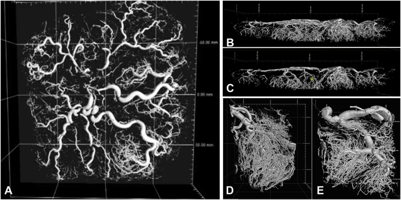
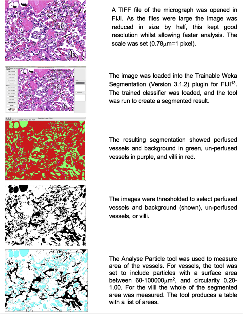
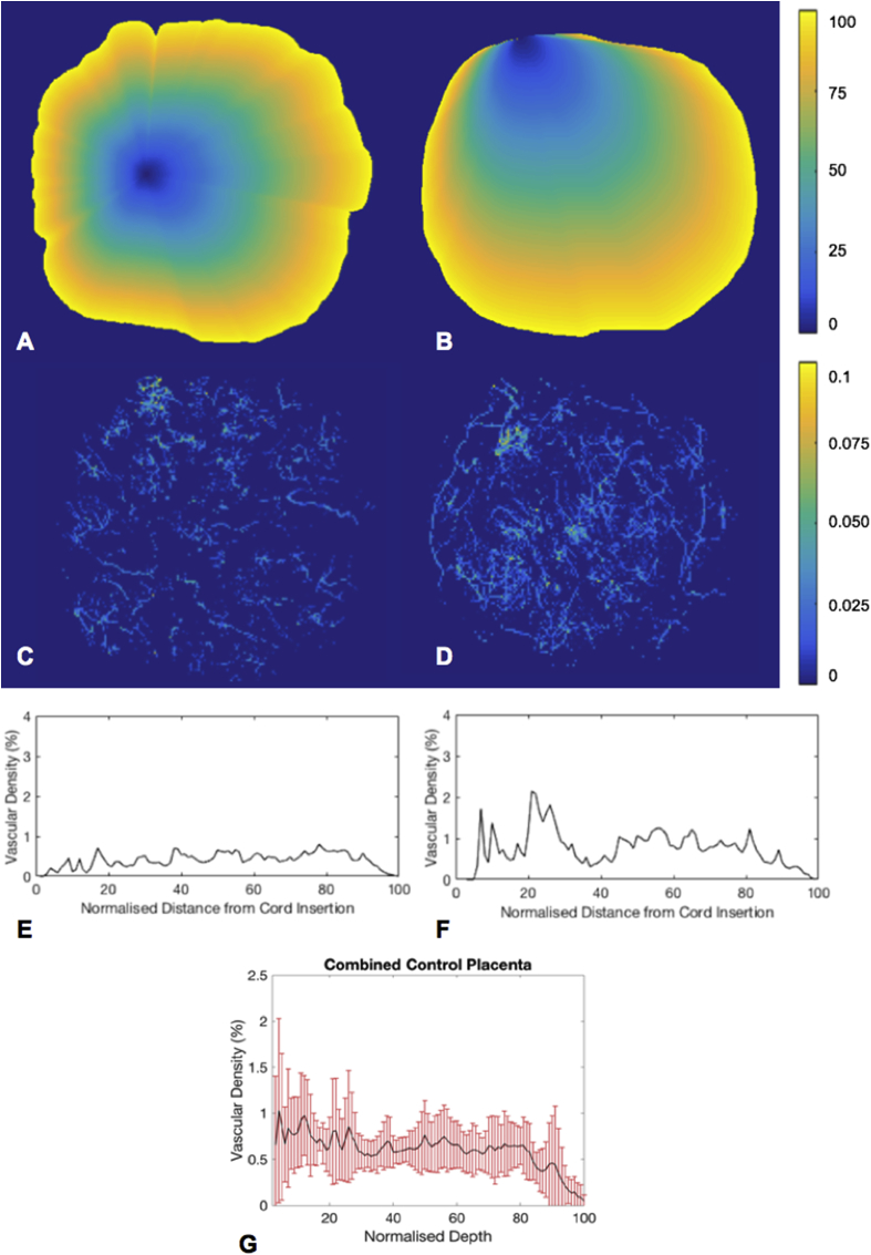
Method: Ten term placentas were collected at elective caesarean section, perfused with contrast agent and imaged whole with Micro-CT. Eight full depth tissue blocks were then taken from each placenta and imaged. Sections were taken for histological analysis. Data was analysed to investigate vascular fill, and vascular density in relation to location from cord insertion to placental edge at each scale.
Results: Whole placental imaging revealed no spatially consistent difference in villous vessel density within the main placental tissue, although there was a great degree of heterogeneity. Both block imaging and histological analysis found a large degree of heterogeneity of vascular density within placentas, but no strong correlation between villous vascular density and block location (rs = 0.066, p = 0.7 block imaging, rs = 0.06, p = 0.6 histological analysis).
Discussion: This work presents a novel method for imaging the human placenta vascular tree using multiscale Micro-CT imaging. It demonstrates that there is a large degree of variation in vascular density throughout normal term human placentas. The three-dimensional data created by this technique could be used, with more advanced computer analysis, to further investigate the structure of the vascular tree.
Copyright © 2019. Published by Elsevier Ltd.
Conflict of interest statement
None.
Figures

References
-
- Carter A.M. Evolution of placental function in mammals: the molecular basis of gas and nutrient transfer, hormone secretion, and immune responses. Physiol. Rev. 2012;92 - PubMed
-
- Kingdom J., Huppertz B., Seaward G., Kaufmann P. vol 92. 2000. pp. 35–43. (Development of the Placental Villous Tree and its Consequences for Fetal Growth). - PubMed
-
- Jackson M.R. Reduced placental villous tree elaboration in small-for-gestational-age pregnancies: relationship with umbilical artery Doppler waveforms. Am. J. Obstet. Gynecol. 1995;172:518–525. - PubMed
-
- SALAFIA C.M., Pezzullo J.C., Minior V.K., Divon M.Y. Placental pathology of absent and reversed end-diastolic flow in growth-restricted fetuses. Obstet. Gynecol. 1997;90:830–836. - PubMed
Publication types
MeSH terms
Grants and funding
LinkOut - more resources
Full Text Sources
