. 2019:24:102025.
doi: 10.1016/j.nicl.2019.102025. Epub 2019 Oct 17.

Tau PET and multimodal brain imaging in patients at risk for chronic traumatic encephalopathy

Affiliations

Tau PET and multimodal brain imaging in patients at risk for chronic traumatic encephalopathy

Orit H Lesman-Segev et al. Neuroimage Clin. 2019.

Abstract

Objective: To characterize individual and group-level neuroimaging findings in patients at risk for Chronic Traumatic Encephalopathy (CTE).

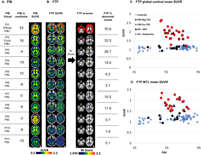
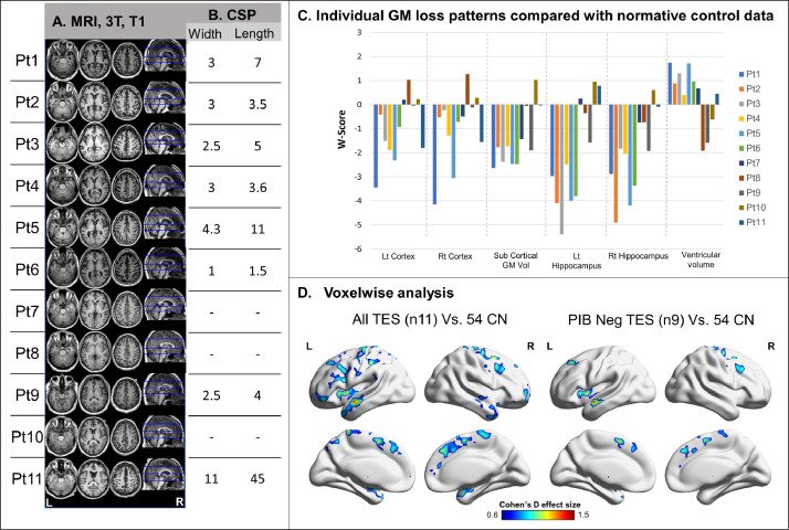
Methods: Eleven male patients meeting criteria for Traumatic Encephalopathy Syndrome (TES, median age: 64) underwent neurologic evaluation, 3-Tesla MRI, and PET with [18F]-Flortaucipir (FTP, tau-PET) and [11C]-Pittsburgh compound B (PIB, amyloid-PET). Six patients underwent [18F]-Fluorodeoxyglucose-PET (FDG, glucose metabolism). We assessed imaging findings at the individual patient level, and in group-level comparisons with modality-specific groups of cognitively normal older adults (CN). Tau-PET findings in patients with TES were also compared to a matched group of patients with mild cognitive impairment or dementia due to Alzheimer's disease (AD).

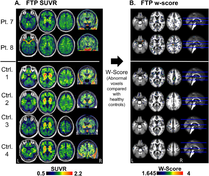
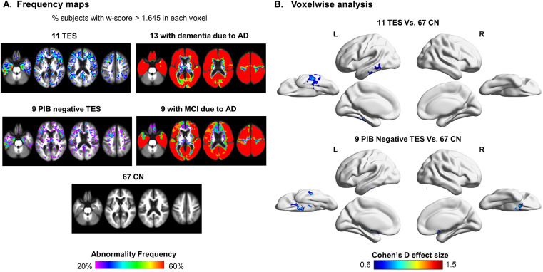
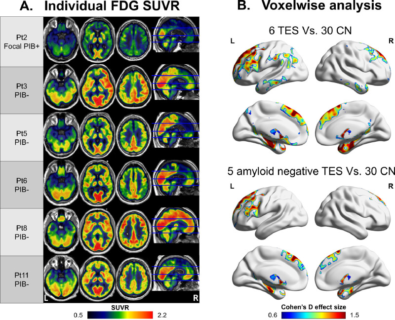
Results: All patients with TES sustained repetitive head injury participating in impact sports, ten in American football. Three patients met criteria for dementia and eight had mild cognitive impairment. Two patients were amyloid-PET positive and harbored the most severe MRI atrophy, FDG hypometabolism, and FTP-tau PET binding. Among the nine amyloid-negative patients, tau-PET showed either mildly elevated frontotemporal binding, a "dot-like" pattern, or no elevated binding. Medial temporal FTP was mildly elevated in a subset of amyloid-negative patients, but values were considerably lower than in AD. Voxelwise analyses revealed a convergence of imaging abnormalities (higher FTP binding, lower FDG, lower gray matter volumes) in frontotemporal areas in TES compared to controls.

Conclusions: Mildly elevated tau-PET binding was observed in a subset of amyloid-negative patients at risk for CTE, in a distribution consistent with CTE pathology stages III-IV. FTP-PET may be useful as a biomarker of tau pathology in CTE but is unlikely to be sensitive to early disease stages.

Keywords: Amyloid; Chronic traumatic encephalopathy (CTE); Imaging; Magnetic resonance imaging (MRI); Positron emission tomography (PET); Tau.


Figures

Fig. 1.
Fig. 1
Amyloid and tau PET patterns in TES, individual patterns. Aβ (PIB)-PET visual read, centiloid value, and SUVR images (A); Tau (FTP)-PET SUVR images, w-score images, and percentage of voxels with w-score>1.645 (B). Mean FTP-SUVR (C) and medial temporal lobe mean SUVR (D) in patients with TES (black triangles – PIB-positive; black circles – PIB-negative), patients with MCI (red circles black outline) or dementia due to AD (red circles no outline) and healthy controls (blue circles). The patient number on the left corresponds to the patient number in Table 1. Images are presented in neurological convention (left of the image is the left side of the patient). Abbreviations- Pt = Patient; TES = traumatic encephalopathy syndrome; PIB = Pittsburgh compound B; PIB+ = positive visual read, PIB- = negative visual read; FTP = flortaucipir; MTL = medial temporal lobe; SUVR = standardized uptake value ratio; W-score = age adjusted z-score.(For interpretation of the references to colour in this figure legend, the reader is referred to the web version of this article.)
Fig. 2.
Fig. 2
Tau (FTP) PET “dot-like” pattern in TES patients and in CN FTP SUVR (A) and w-score (B) images of two patients (patients 7 and 8) and four control subjects that have a non-specific distribution of small clusters with mildly elevated binding. Abbreviations: FTP = flortaucipir; TES = traumatic encephalopathy syndrome; CN – cognitively normal control; Pt. – patient; Ctrl. – control; SUVR - standardized uptake value ratio; W-score = age adjusted z-score.
Fig. 3.
Fig. 3
Tau (FTP) PET in TES compared with CN and AD, group analysis FTP binding frequency maps of the TES cohort (n = 11), the amyloid-negative subgroup of TES patients (n = 9), healthy controls (n = 67), patients with dementia due to AD (n = 13) and patients with MCI due to AD (n = 9). The value in each voxel represents the percentage of subjects that have high binding (defined here as w-score > 1.645) in that voxel (A). Voxelwise two sample t-test analysis reported in Cohen's d effect size of FTP-PET binding in the TES cohort (n = 11) and in the amyloid-negative subgroup of TES patients (n = 9) compared with 67 healthy controls, PUncorrected < 0.005 in the voxel level, Pfwe < 0.05 in the cluster level (B). Images are presented in neurologic convention. PIB = Pittsburg compound B; FTP = flortaucipir; Lat = lateral; Med = medial; Inf = inferior; TES – traumatic encephalopathy syndrome; AD = Alzheimer's disease; MCI = mild cognitive impairment; CN – cognitively normal control; CDR = clinical dementia score; FWE – family wise correction.
Fig. 4.
Fig. 4
Individual and group-level analysis of structural changes in TES patients Axial T1 weighted MRI of all 11 patients with TES (A) including cavum Septum Pellucidum (CSP) width and length in millimeters (B). Individual atrophy patterns as represented by w-scores of bilateral cortical thickness, subcortical gray matter volume, bilateral hippocampal volume, and ventricular volume compared to normalized control data (based on Potvin et-al (Potvin et al., 2017)) (C). Group-level two sample t-test analysis of atrophy patterns (PUncorrected < 0.005 at the voxel level, Pfwe < 0.05 at the cluster level converted to Cohen's d effect size) in the whole TES cohort (n = 11) and the amyloid-negative subgroup only (n = 9) compared with 54 controls. Patient numbers correspond to the same numbers as in Table 1 and in the other figures. TES – traumatic encephalopathy syndrome; CN – cognitively normal control; CSP – cavum septum pellucidum; GM – gray matter; Lt – left; Rt – right; Vol – volume; Pt – patient; FEW – family wise error corrected.
Fig. 5.
Fig. 5
Individual and group-level metabolism pattern FDG-PET SUVR images of six patients with TES that underwent FDG-PET (A) and voxelwise two sample t-test analysis of the all patients and the five amyloid-negative patients compared with 30 controls. Reported in Cohen's d effect size, Puncorrected<0.005; cluster threshold: Pfwe<0.05 (B). Patient numbers correspond to the same numbers as in Table 1 and in the other figures. FDG – fluorodeoxyglucose; SUVR - standardized uptake value ratio; TES – traumatic encephalopathy syndrome; CN – cognitively normal control; FEW – family wise error correction.

Comment in

References

    1. Alosco M.L., Tripodis Y., Fritts N.G. Cerebrospinal fluid tau, αβ, and sTREM2 in former national football league players: modeling the relationship between repetitive head impacts, microglial activation, and neurodegeneration. Alzheimers Dement. J. Alzheimers Assoc. 2018;14(9):1159–1170. - PMC - PubMed
    1. Bang S.A., Song Y.S., Moon B.S. Neuropsychological, metabolic, and GABAA receptor studies in subjects with repetitive traumatic brain injury. J. Neurotrauma. 2015;33(11):1005–1014. - PubMed
    1. Barrio J.R., Small G.W., Wong K.-.P. In vivo characterization of chronic traumatic encephalopathy using [F-18]FDDNP PET brain imaging. Proc. Natl. Acad. Sci. U. S. A. 2015;112(16):E2039–E2047. - PMC - PubMed
    1. Baugh C.M., Robbins C.A., Stern R.A., McKee A.C. Current understanding of chronic traumatic encephalopathy. Curr. Treat. Options Neurol. 2014;16(9):306. - PMC - PubMed
    1. Bevan-Jones W.R., Cope T.E., Jones P.S. [18F]AV-1451 binding in vivo mirrors the expected distribution of TDP-43 pathology in the semantic variant of primary progressive aphasia. J. Neurol. Neurosurg. Psychiatry. September 2017 - PMC - PubMed

Publication types

MeSH terms