Review
. 2020 Jan:65:27-36.
doi: 10.1016/j.mri.2019.09.007. Epub 2019 Oct 25.

Magnetic resonance imaging (MRI) studies of knee joint under mechanical loading: Review

Affiliations
Review

Magnetic resonance imaging (MRI) studies of knee joint under mechanical loading: Review

Saeed Jerban et al. Magn Reson Imaging. 2020 Jan.

Abstract

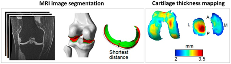
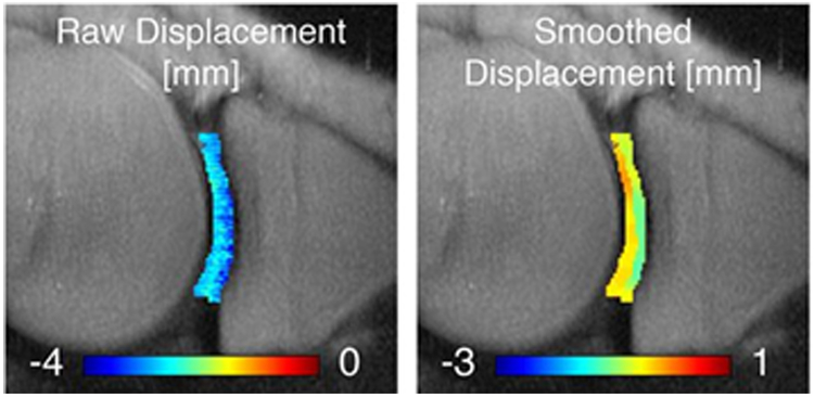
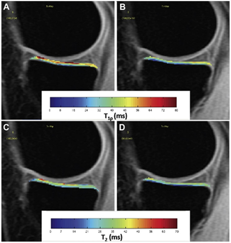
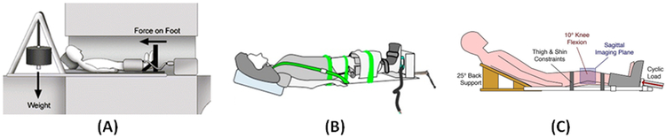
Osteoarthritis (OA) is a very common disease that affects the human knee joint, particularly the articular cartilage and meniscus components which are regularly under compressive mechanical loads. Early-stage OA diagnosis is essential as it allows for timely intervention. The primary non-invasive approaches currently available for OA diagnosis include magnetic resonance imaging (MRI), which provides excellent soft tissue contrast at high spatial resolution. MRI-based knee investigation is usually performed on joints at rest or in a non-weight-bearing condition that does not mimic the actual physiological condition of the joint. This discrepancy may lead to missed detections of early-stage OA or of minor lesions. The mechanical properties of degenerated musculoskeletal (MSK) tissues may vary markedly before any significant morphological or structural changes detectable by MRI. Recognizing distinct deformation characteristics of these tissues under known mechanical loads may reveal crucial joint lesions or mechanical malfunctions which result from early-stage OA. This review article summarizes the large number of MRI-based investigations on knee joints under mechanical loading which have been reported in the literature including the corresponding MRI measures, the MRI-compatible devices employed, and potential challenges due to the limitations of clinical MRI sequences.

Keywords: Cartilage; Knee; Loading; MRI; Meniscus; Osteoarthritis.


Conflict of interest statement

Conflict of interest statement

The authors have no conflicts of interest to declare.

Figures

Figure 1:
Figure 1:
Representative steps for thickness map measurement in tibiofemoral cartilage using MRI images and pixel-wise quantification. Wang et al. used MRI images from 3D-SPGR sequence with approximately 0.3×0.3×1.5 mm3 voxel size to manually segment 3D geometries of bone and articular cartilage. Cartilage thickness was calculated as the shortest distance from the subchondral bone surface to articular surface in segmented cartilages. Contact region was defined as the overlapping areas of the femoral and tibial articular surfaces. A, P, M, and L refer to anterior, posterior, medial and lateral compartments of the cartilages, respectively. This figure is cropped from a figure presented previously by Wang et al. [26]; the reprinting permission is granted through Rightslink system.
Figure 2:
Figure 2:
Axial displacement map in sagittal slice of the tibiofemoral cartilage of medial knee joint under mechanical loading as measured with displacement encoding technique [31,36,37]. The figure is cropped from a previously presented figure by Chan et al. [31] based on Creative Commons permission guidelines (https://creativecommons.org/licenses/by/4.0/).
Figure 3:
Figure 3:
Representative T (top row) and T2 (bottom row) maps of the medial tibial cartilage in the unloaded (A, C) and loaded (B, D) conditions. The figure is a figure previously presented by Souza et al. [47]. The reprinting permission is granted through Rightslink system.
Figure 4:
Figure 4:
(A) Schematic of a cable/pulley setup combined with a back-board and a sliding foot plate on low friction rollers to position the subject [34,44-50]. This schematic was previously presented by Cotofana et al. [44]. Reprinting permission is granted based on the open access license. (B) Schematic of the ratcheting mechanism driving an orthotic boot to apply mechanical load to the subject’s feet, where an MRI-compatible load cell is attached for estimation of the applied load [26,27,83]. This schematic was previously presented by Wang et al. [26] and reprinting permission is granted through the Rightslink system. (C) Schematic of the active loading system utilizing an electro-pneumatic device [31]. The schematic was previously presented by Chan et al. [31] Reprinting is based on Creative Commons use guidelines (https://creativecommons.org/licenses/by/4.0/).

References

    1. Mansour JM. Biomechanics of Cartilage. Kinesiol Mech Pathomechanics Hum Mov 2009:66–79. doi:10.1002/art.23548. - DOI
    1. Sophia Fox a. J, Bedi a., Rodeo S a. The Basic Science of Articular Cartilage: Structure, Composition, and Function. Sport Heal A Multidiscip Approach 2009;1:461–8. doi:10.1177/1941738109350438. - DOI - PMC - PubMed
    1. Chang EY, Du J, Chung CB. UTE imaging in the musculoskeletal system. J Magn Reson Imaging 2015;41:870–83. doi:10.1002/jmri.24713. - DOI - PMC - PubMed
    1. López-Franco M, Gómez-Barrena E. Cellular and molecular meniscal changes in the degenerative knee: a review. J Exp Orthop 2018;5. doi:10.1186/s40634-018-0126-8. - DOI - PMC - PubMed
    1. Chevrier A, Nelea M, Hurtig MB, Hoemann CD, Buschmann MD. Meniscus structure in human, sheep, and rabbit for animal models of meniscus repair. J Orthop Res 2009;27:1197–203. doi:10.1002/jor.20869. - DOI - PubMed

Publication types

MeSH terms