Review
. 2020 Jul;8(4):892-897.
doi: 10.1111/andr.12722. Epub 2019 Nov 20.

Cycles, waves, and pulses: Retinoic acid and the organization of spermatogenesis

Affiliations
Review

Cycles, waves, and pulses: Retinoic acid and the organization of spermatogenesis

Rachel Gewiss et al. Andrology. 2020 Jul.

Abstract

Background: Spermatogenesis in mammals is organized in a manner that maximizes sperm production. The central aspect of this organization is the cycle of the seminiferous epithelium that is characterized by an asynchronous repeating series of germ cell associations. These cell associations are the result of a fixed point of entry into the cycle at regular short time intervals and the longer time required for cells to fully differentiate and exit the cycle.

Objective: This review will examine the current information on the action and metabolism of retinoic acid in the testis, the interaction of retinoic acid (RA) with the cycle and the spermatogenic wave, and the mechanisms that can lead to synchronous spermatogenesis. Finally, the unique applications of synchronous spermatogenesis to the study of the cycle and the mass isolation of specific germ cell populations are described.

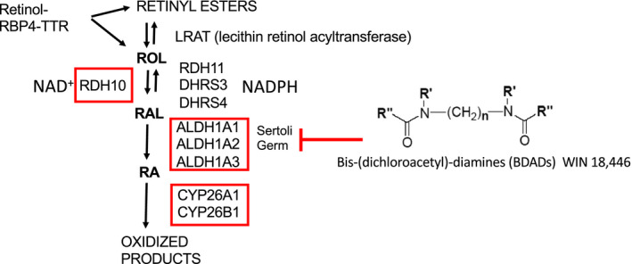
Materials and methods: Retinoic acid metabolism and spermatogonial differentiation have been examined by gene deletions, immunocytochemistry, chemical inhibitors, and mass spectrometry.

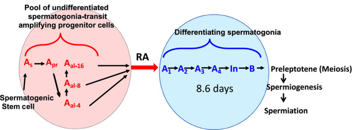
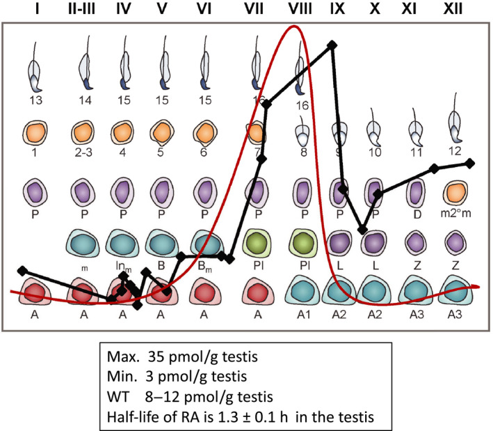
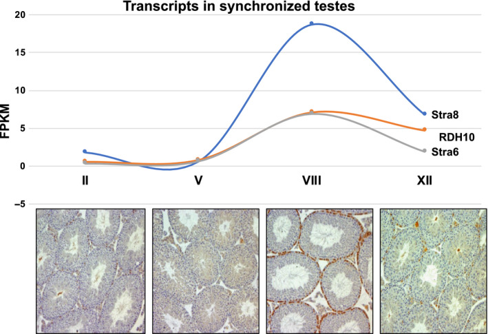
Results, discussion, and conclusion: Both the Sertoli cells and the germ cells have the capacity to synthesize retinoic acid from retinol and in the mouse the entry into the cycle of the seminiferous epithelium, and the subsequent conversion of undifferentiated spermatogonia into differentiating spermatogonia is governed by a peak of RA synthesis occurring at stages VIII-IX of the cycle. Normal asynchronous spermatogenesis can be modified by altering RA levels, and as a result the entire testis will consist of a few closely related stages of the cycle.

Keywords: cycle; retinoic acid; synchronization; wave.


Figures

Figure 1
Figure 1
The effect of retinoic acid on the development of male germ cells. The so‐called ‘undifferentiated’ A spermatogonia originate as A single (As) cells and undergo mitotic division with incomplete cytokinesis to form two A paired (Apr) cells. Further mitotic divisions with incomplete separation form syncytia of A aligned (Aal) cells in chains of 4, 8, and 16 cells (red circle). The undifferentiated spermatogonia that are not self‐renewing spermatogenic stem cells are capable of undergoing the A to A1 transition (spermatogonial differentiation) in response to an RA pulse (red arrow). This differentiation event triggers development through the subtypes of differentiating spermatogonia (A1, A2, A3, A4, Intermediate, and B) until cells develop into pre‐leptotene spermatocytes prepared to enter meiosis (blue circle). At a given point along a seminiferous tubule, a new cohort of undifferentiated spermatogonia will undergo spermatogonial differentiation every 8.6 days in the mouse
Figure 2
Figure 2
Retinoic acid metabolism in the testis. Retinol (Vitamin A) transits to the testis in a complex with retinol binding protein 4 (RBP4) and transthyretin. There lecithin retinol acyltransferase (LRAT) can convert retinol (ROL) into retinyl esters, or ROL can be converted into retinal (RAL). The ROL to RAL conversion is catalyzed by RDH10 in an NAD+‐dependent manner and is the rate‐limiting step in the conversion of ROL to RA. The reverse reaction can be carried out by one of several retinal reductases (RDH11, DHRS3, and DHRS4) and is dependent on NADPH. The second and irreversible step converts RAL to RA and is catalyzed in both Sertoli and germ cells by the aldehyde dehydrogenase 1A (ALDH1A) family of enzymes. This step can be blocked with the BDAD compound WIN 18,446, resulting in an arrest at the A to A1 transition that mimics Rdh10 or Aldh1a1‐3 genetic knockouts in Sertoli and/or germ cells. RA in the testis is degraded into inactive oxidized products by CYP26A1 and CYP26B1. The genes for each of the enzymes enclosed by the red boxes have been deleted in multiple cell types in the testis, and the results are discussed in the text
Figure 3
Figure 3
Retinoic acid pulse across the cycle of the seminiferous epithelium. Red line shows the predicted RA pulse based on previous data, black line shows actual, relative RA values across the cycle in stage‐synchronized testes (data from.21 RA levels peak at late stage VIII/early stage IX and reach a maximum value of 35 picomoles per gram testis (pmoles/g testis). Minimum RA values around 3 pmoles/g testis were observed in stages II through V. In unsynchronized adult testes where all stages of the seminiferous epithelium are present, RA values averaged around 8‐12 pmoles/g testis
Figure 4
Figure 4
Retinoic acid‐responsive gene transcripts across the cycle of the seminiferous epithelium. Stra8 (blue line) showed the greatest transcriptional activity in response to RA signaling, but Rdh10 (orange line) and Stra6 (gray line) transcript levels also directly correlate with the RA pulse. Cross sections for each of the represented stages (II, V, VIII, and XII) of stage‐synchronized testes are shown below the transcript data. Brown‐stained cells at the basement membrane represent STRA8‐positive spermatogonia and pre‐leptotene spermatocytes at the A to A1 transition (Stage VIII)

References

    1. Regaud C. Etudes sur la structure des tubes seminiferes et sur la spermatogenese chez les Mammiferes. Arch Anat Micr. 1901;4(101–156):231‐280.
    1. Leblond CP, Clermont Y. Definition of the stages of the cycle of the seminiferous epithelium in the rat. Ann N Y Acad Sci. 1952;55:548‐573. - PubMed
    1. Griswold MD. Spermatogenesis: the commitment to meiosis. Physiol Rev. 2016;96:1‐17. - PMC - PubMed
    1. Franca LR, Ogawa T, Avarbock MR, Brinster RL, Russell LD. Germ cell genotype controls cell cycle during spermatogenesis in the rat. Biol Reprod. 1998;59:1371‐1377. - PubMed
    1. Perey B, Clermont Y, Leblond CP. The wave of the seminiferous epithelium in the rat. Am J Anat. 1961;108:47‐78.

Publication types