. 2019 Dec 5;4(23):e126917.
doi: 10.1172/jci.insight.126917.

A composite immune signature parallels disease progression across T1D subjects

Affiliations

A composite immune signature parallels disease progression across T1D subjects

Cate Speake et al. JCI Insight. .

Abstract

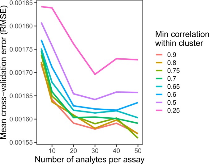
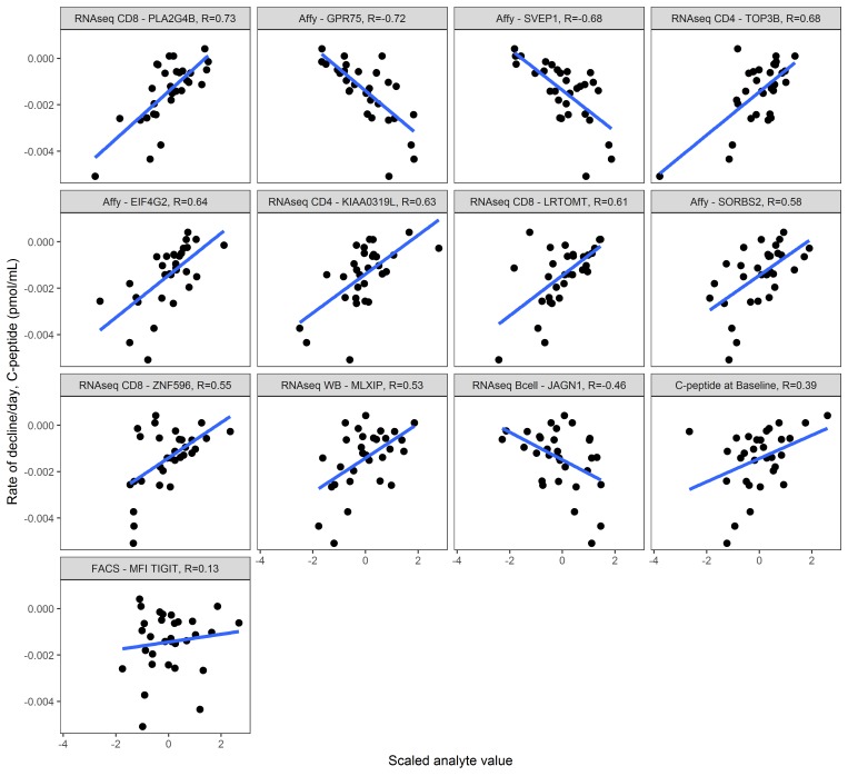
At diagnosis, most people with type 1 diabetes (T1D) produce measurable levels of endogenous insulin, but the rate at which insulin secretion declines is heterogeneous. To explain this heterogeneity, we sought to identify a composite signature predictive of insulin secretion, using a collaborative assay evaluation and analysis pipeline that incorporated multiple cellular and serum measures reflecting β cell health and immune system activity. The ability to predict decline in insulin secretion would be useful for patient stratification for clinical trial enrollment or therapeutic selection. Analytes from 12 qualified assays were measured in shared samples from subjects newly diagnosed with T1D. We developed a computational tool (DIFAcTO, Data Integration Flexible to Account for different Types of data and Outcomes) to identify a composite panel associated with decline in insulin secretion over 2 years following diagnosis. DIFAcTO uses multiple filtering steps to reduce data dimensionality, incorporates error estimation techniques including cross-validation and sensitivity analysis, and is flexible to assay type, clinical outcome, and disease setting. Using this novel analytical tool, we identified a panel of immune markers that, in combination, are highly associated with loss of insulin secretion. The methods used here represent a potentially novel process for identifying combined immune signatures that predict outcomes relevant for complex and heterogeneous diseases like T1D.

Keywords: Autoimmunity; Diabetes; Immunotherapy; Molecular pathology.


Conflict of interest statement

Conflict of interest: EW receives a salary from Celgene, Inc. JMO receives a salary from Gilead Sciences, Inc. MKL has received research support from TxCell, Pfizer, and Bristol Myers Squibb and has patents pending related to alloantigen-specific chimeric antigen receptors (PCT/CA2018/051167 and PCT/CA2018/051174). PSL receives research support from Bristol Myers Squibb and is an inventor on patent US5844095A, “CTLA4Ig fusion proteins.” RG has received consulting income from Juno Therapeutics, Takeda, Infotech Soft, and Celgene, Inc., and has received research support from Janssen Pharmaceuticals and Juno Therapeutics.

Figures

Figure 1
Figure 1. C-peptide decay rate for each subject included in the recent-onset cohort.
C-peptide decay rate for each subject included in the recent-onset cohort (n = 50). Each line is 1 subject. Subjects highlighted in blue and green demonstrate slow and rapid decline, respectively. The subject highlighted in olive showed no detectable insulin secretion at a time point before the end of the study; decay rate calculations used throughout exclude the second time point at the limit of C-peptide detection. Some subjects (example in green/magenta) are similar in age but have disparate C-peptide decay rates.
Figure 2
Figure 2. Schematic of analysis pipeline developed to integrate multiple data types.
Analytes are merged and scaled. Initial univariate filtering leaves only those analytes with at least modest correlations to the outcome for each assay. Subsequent filtering (clustering) identifies best correlated analyte in a given cluster (regardless of assay). From these, a composite model is generated using LASSO with cross-validation. The box plots depict the minimum and maximum values (whiskers), the upper and lower quartiles, and the median. The length of the box represents the interquartile range.
Figure 3
Figure 3. Establishment of optimal parameter settings.
Changing settings within the analytical tool (DIFAcTO) identifies point of lowest cross-validation error associated with higher within-cluster correlation (r = 0.7) and a moderate number of analytes per assay (n = 30). Each line shows the cross-validation error (root mean squared error, RMSE) for a given analyte per assay setting (x axis) at a given minimum correlation within cluster (indicated by color).
Figure 4
Figure 4. Characteristics of analytes selected by tool.
Sensitivity analysis shows that, even with cross-validation implemented in LASSO, 5/17 analytes are not robust to different parameter settings within the tool. These 5, indicated in grayscale, are unlikely to be validated in an independent cohort. Each individual plot’s x and y axes represent settings used to run the analytical tool; the number of analytes per assay setting is on the x axis, and minimum correlation per cluster is on the y axis of each miniplot. Darkest coloring indicates that the analyte was selected by the tool using that combination of x and y settings. Lighter coloring indicates that another analyte in that same cluster was selected by the tool. Light gray indicates that this analyte was not selected using that combination of x and y settings. Each miniplot is labeled by analyte and the assay from which it was originally measured. “Affy” indicates the transcriptional response to T1D serum assay as this is conducted on the Affymetrix platform.
Figure 5
Figure 5. Individual correlations between each selected analyte and insulin secretion.
Immune markers were measured at trial enrollment (within 90 days of diagnosis, n = 30 subjects), and y axis indicates C-peptide decay rate per day over the 2 years after diagnosis. Each miniplot uses the scaled value for the analyte on the x axis. Pearson’s correlation values are listed at the top of each miniplot; miniplots are ordered by absolute correlation value. Regression lines in blue. Note that in this data set, several immune parameters have higher correlation values with rate of C-peptide decay than does C-peptide level at diagnosis (Pearson’s r = 0.39). Assays and analyte names are truncated; full names can be found in Table 3. “Affy” indicates the transcriptional response to T1D serum assay as this is conducted on the Affymetrix platform.

References

    1. Bundy BN, Krischer JP, Type 1 Diabetes TrialNet Study Group A model-based approach to sample size estimation in recent onset type 1 diabetes. Diabetes Metab Res Rev. 2016;32(8):827–834. doi: 10.1002/dmrr.2800. - DOI - PMC - PubMed
    1. Hao W, Gitelman S, DiMeglio LA, Boulware D, Greenbaum CJ, Type 1 Diabetes TrialNet Study Group Fall in C-peptide during first 4 years from diagnosis of type 1 diabetes: variable relation to age, HbA1c, and insulin dose. Diabetes Care. 2016;39(10):1664–1670. doi: 10.2337/dc16-0360. - DOI - PMC - PubMed
    1. Barker A, et al. Age-dependent decline of β-cell function in type 1 diabetes after diagnosis: a multi-centre longitudinal study. Diabetes Obes Metab. 2014;16(3):262–267. doi: 10.1111/dom.12216. - DOI - PubMed
    1. Mallone R, Roep BO. Biomarkers for immune intervention trials in type 1 diabetes. Clin Immunol. 2013;149(3):286–296. doi: 10.1016/j.clim.2013.02.009. - DOI - PubMed
    1. Mathieu C, Lahesmaa R, Bonifacio E, Achenbach P, Tree T. Immunological biomarkers for the development and progression of type 1 diabetes. Diabetologia. 2018;61(11):2252–2258. doi: 10.1007/s00125-018-4726-8. - DOI - PubMed

Publication types

Substances