. 2019 Oct 31;15(10):e1008057.
doi: 10.1371/journal.ppat.1008057. eCollection 2019 Oct.

Astrovirus replication in human intestinal enteroids reveals multi-cellular tropism and an intricate host innate immune landscape

Affiliations

Astrovirus replication in human intestinal enteroids reveals multi-cellular tropism and an intricate host innate immune landscape

Abimbola O Kolawole et al. PLoS Pathog. .

Abstract

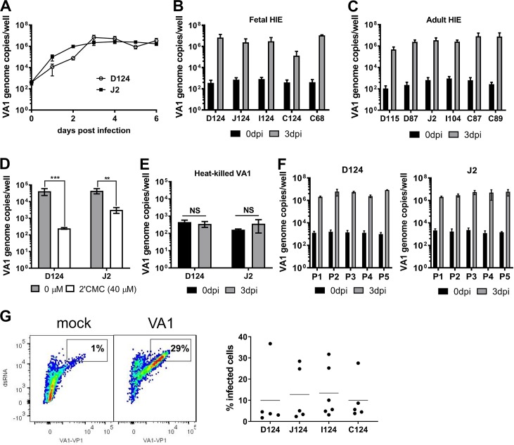
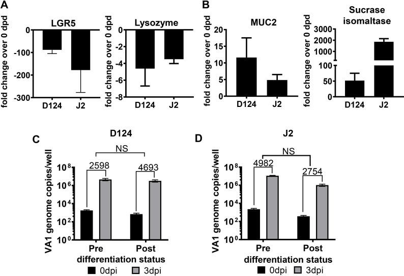
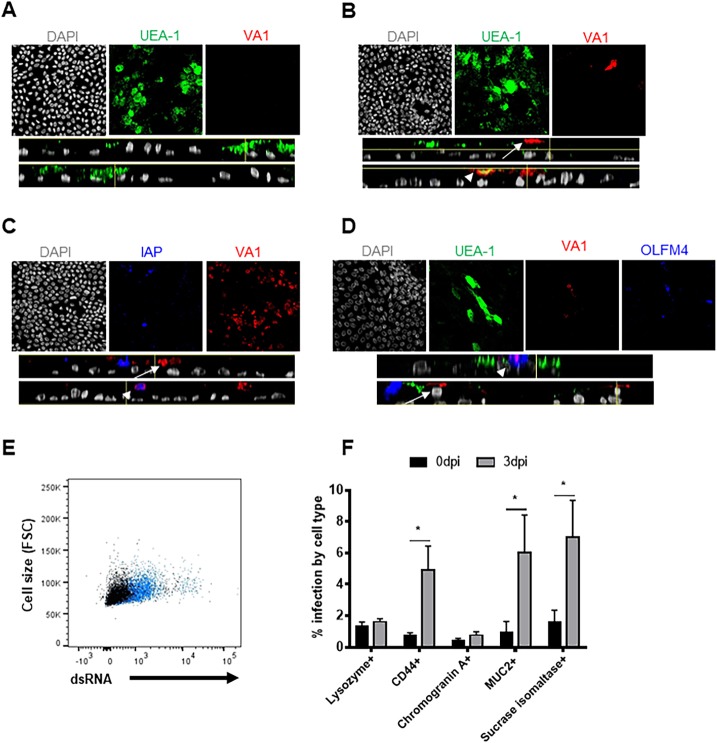
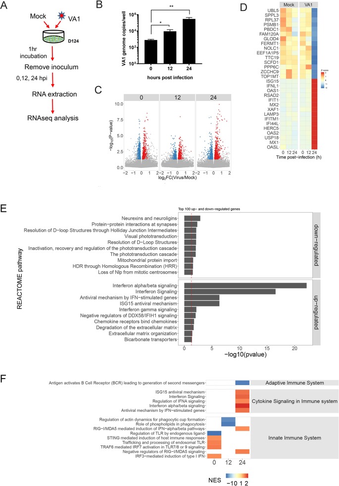
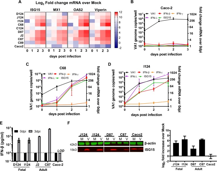
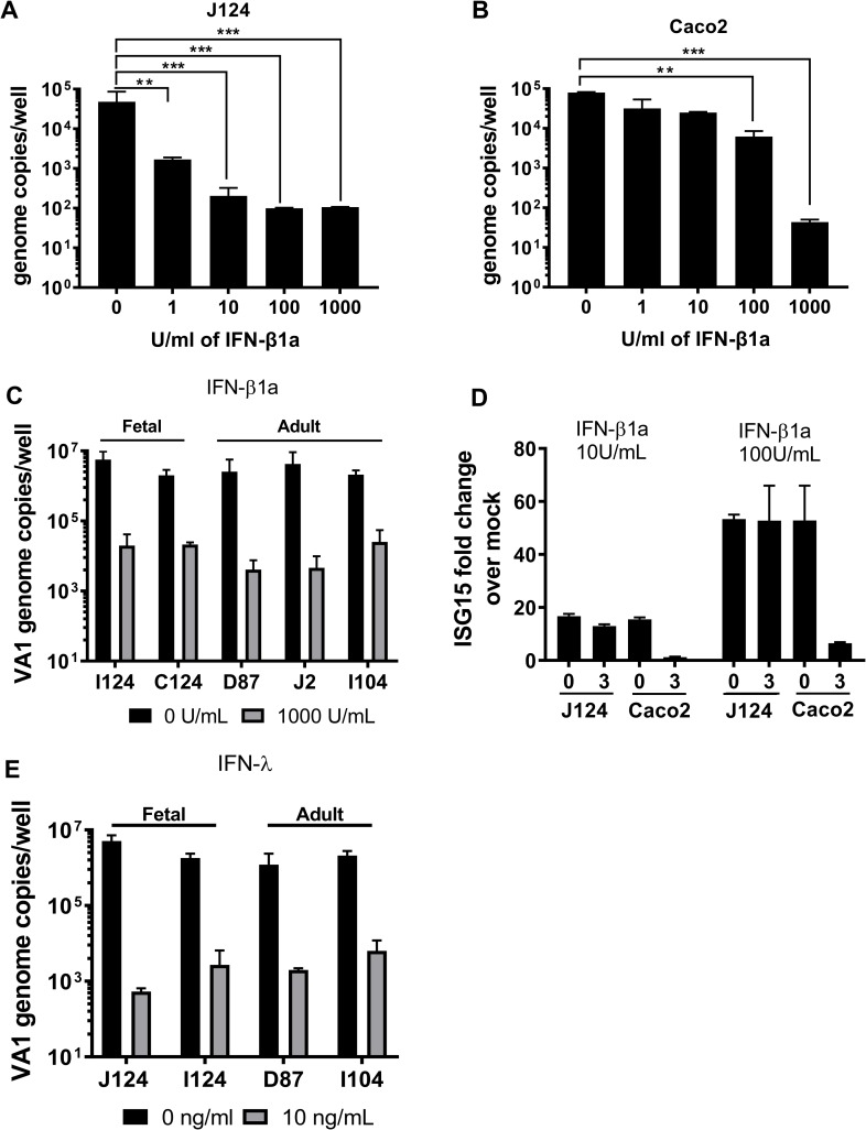
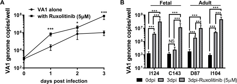
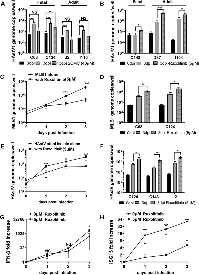
Human astroviruses (HAstV) are understudied positive-strand RNA viruses that cause gastroenteritis mostly in children and the elderly. Three clades of astroviruses, classic, MLB-type and VA-type have been reported in humans. One limitation towards a better understanding of these viruses has been the lack of a physiologically relevant cell culture model that supports growth of all clades of HAstV. Herein, we demonstrate infection of HAstV strains belonging to all three clades in epithelium-only human intestinal enteroids (HIE) isolated from biopsy-derived intestinal crypts. A detailed investigation of infection of VA1, a member of the non-canonical HAstV-VA/HMO clade, showed robust replication in HIE derived from different patients and from different intestinal regions independent of the cellular differentiation status. Flow cytometry and immunofluorescence analysis revealed that VA1 infects several cell types, including intestinal progenitor cells and mature enterocytes, in HIE cultures. RNA profiling of VA1-infected HIE uncovered that the host response to infection is dominated by interferon (IFN)-mediated innate immune responses. A comparison of the antiviral host response in non-transformed HIE and transformed human colon carcinoma Caco-2 cells highlighted significant differences between these cells, including an increased magnitude of the response in HIE. Additional studies confirmed the sensitivity of VA1 to exogenous IFNs, and indicated that the endogenous IFN response of HIE to curtail the growth of strains from all three clades. Genotypic variation in the permissiveness of different HIE lines to HAstV could be overcome by pharmacologic inhibition of JAK/STAT signaling. Collectively, our data identify HIE as a universal infection model for HAstV and an improved model of the intestinal epithelium to investigate enteric virus-host interactions.


Conflict of interest statement

The authors have declared that no competing interests exist.

Figures

Fig 1
Fig 1. VA1 replicates in human intestinal enteroids (HIE).
A) VA1 growth curve. Undifferentiated D124 and J2 HIE were cultured in 2D monolayers and infected with VA1 (MOI of 1). At indicated times, HIE were harvested and viral RNA was extracted and quantified by RT-qPCR. B-C) Undifferentiated HIE derived from indicated segments of fetal and adult intestines were infected with VA1 and analyzed as before. D) VA1 replication is blocked by the nucleoside analogue 2’-C-methylcytidine (2CMC). Undifferentiated D124 and J2 HIE were infected as before and cells treated with 40 μM 2CMC after adsorption. Cells were harvested and viral RNA quantified at 1 dpi. E) Heat-killed VA1 does not replicate in HIE. VA1 was heat-killed for 3 min at 100°C prior to infection of undifferentiated D124 and J2 HIE. Cells were harvested and viral RNA quantified at 3 dpi. F) VA1 remains infectious for 5 passages in HIE. Supernatant from infected D124 or J2 (passage 1, P1) containing ~103 genome copies/ml was used to infect monolayers of D124 and J2, respectively. Cells were harvested and viral RNA quantified at 3 dpi. The procedure was repeated four times (P2-P5). For all experiments, viral genome copies were measured by RT-qPCR. A–F) Data are from ≥ 3 experiments; error = mean ± SD. G) A single cell suspension of VA1-infected (MOI of 1) D124, I124, J124 and C124 HIE was generated at 3 dpi and stained with antibodies against dsRNA and VA1 capsid protein prior to flow cytometry analysis. The left panel is a representative flow plot of mock- and VA1-infected J124 HIE. The right panel represents the percentage of double-stained cells in each segment. Each dot is a biological replicate. Abbreviations: dpi = days postinfection, D = duodenum, I = ileum, J = jejunum, C = colon. The numbers indicate patient identifiers. **P<0.01; ***P<0.001; NS = not significant.
Fig 2
Fig 2. VA1 replication is not affected by the differentiation status of HIE.
A-B) Differentiation status of J2 and D124 HIE monitored at 0 and 6 days post-differentiation (dpd) (after WNT3A removal) by measuring transcripts of (A) down-regulated genes—LGR5 (a stem-cell marker) and Lysozyme (a Paneth cells marker), and (B) up-regulated genes—MUC2 (a goblet cell marker) and sucrase isomaltase (a mature enterocyte marker). Transcript levels were measured by qPCR. Fold change is relative to GAPDH and is statistically significantly different (P<0.05) from the 0 dpd in A) and B). C-D) VA1 replicates in HIE pre- and post- differentiation. C) D124 and D) J2 HIE were differentiated for 6 days, infected with VA1 (MOI of 1), and viral genome titers were measured at the indicated days by RT-qPCR. Fold increase was calculated by dividing 3 dpi virus titer with 0 dpi virus titer. N ≥ 3; error = mean ± SD. Abbreviations: D = duodenum, J = jejunum. The number associated with each letter indicates the patient identifier. The numbers on the bar chat indicate the fold increase in virus titer at 3 dpi over 0 dpi. NS = not significant.
Fig 3
Fig 3. VA1 shows tropism for multiple cell types in HIE.
A-B) VA1 replicates in UEA-1 positive and negative cells. D124 HIE were seeded in transwells, allowed to differentiate for 6 days and infected with (A) mock lysate or (B) with VA1 (MOI of 1). At 5 dpi, HIE were fixed with methanol and treated for immune fluorescence analysis. Virus was detected with a polyclonal mouse anti-VA1 serum followed by an anti-mouse AlexaFluor 594 (red) secondary antibody. Cells were also stained with the lectin UEA-1 conjugated with FITC (green), VA1-infected UEA-1 positive or negative cells are indicated with an arrowhead or arrow, respectively. C) VA1 infects intestinal alkaline phosphatase (IAP) positive and -negative cells. D124 HIE were seeded in transwells, allowed to differentiate for 6 days and infected with VAI (MOI of 1). At 5 dpi, HIE were fixed with methanol and treated for immune fluorescence analysis. Virus was detected with a polyclonal mouse anti-VA1 serum followed by an anti-mouse AlexaFluor 594 (red) secondary antibody. IAP was detected with an anti-IAP polyclonal antibody produced in rabbit and an anti-rabbit AlexaFluor 647 (blue). VA1-infected IAP -positive or -negative cells are represented with an arrowhead or arrow, respectively. D) VA1 infects OLFM4 positive and negative cells. I124 and J were seeded in transwells and allowed to differentiate for 6 days before infection with VAI (MOI of 1). At 3 dpi, cells were harvested and fixed for immunofluorescence analysis with a polyclonal mouse anti-VA1 serum followed by an anti-mouse AlexaFluor 594 (red); anti-OLFM4 antibody produced in rabbit and anti-rabbit AlexaFluor 647 (blue) and the UEA-1 lectin (green). VA1-infected OLFM4 -positive or -negative cells are represented with an arrowhead or arrow, respectively. All the images were acquired with a Nikon A1 laser confocal microscope and analyzed with Fiji Image J software. Images were representative of an N = 4 (UEA-1) or N = 2 (IAP and OLFM4) experiments. Planar view of Mock or VA1-infected HIE is represented in the upper panels with the single channels hyperstack projected on a max intensity Z-plane in the lower panels with side XY views of the hyperstack. E-F) VA1 infects sucrase isomaltase (SI)-, mucin 2 (MUC2)- or CD44-positive cells. Differentiated I124 HIE were infected with VA1 (MOI of 1) and a single cell suspension was obtained at 3 dpi. Cells were stained with the surface markers lysozyme (paneth cells), CD44 (progenitor cells), chromogranin A (enteroendocrine cells), MUC2 (goblet cells) and sucrase isomaltase (mature enterocytes) and the intracellular dsRNA antibody conjugated with biotin followed by the secondary streptavidin-APC Cy7 antibody. E) Representative flow plot of VA1-infected cells (blue) vs mock-infected cells (black). F) The percent of infected cells in specific cell sub-populations. N = 2 biological replicates, each containing n = 3 technical replicates. The Mann-Whitney U test was used to analyze the difference between the percent positive cell types in VA1-infected cells at 0 vs 3 dpi. *P<0.05.
Fig 4
Fig 4. Transcriptional profiling of VA-1 infected HIE by RNA-seq.
A) Experimental design of the RNA-seq experiment. N = 3 for each time point. B) VA1 titers of infected undifferentiated D124 HIE measured by RT-qPCR at the indicated times. *P<0.05; **P<0.01. C-D) Host response to VA1 infection by RNA-seq. C) Log2 fold change in normalized expression (transcripts per million reads) of all expressed host genes of VA1-infected HIEs relative to mock-infected HIEs at 0, 12, and 24 hpi (Volcano plot). The y-axis shows the -log10 transformed P-value. Genes significantly up-regulated (P < 0.05) in VA1-infected HIEs relative to mock-infected HIEs are colored red and significantly down-regulated genes are colored blue. D) Heat map of the top 15 up- and down-regulated genes (Z-score) in VA1-infected HIEs relative to mock-infected HIEs at indicated time points post-infection. E) The pool of genes that were significantly up- or down-regulated in VA1-infected HIEs relative to mock-infected HIEs were evaluated for enrichment of REACTOME pathway analysis using an over-representation test. The top 10 significantly over-represented pathways among both the up- and down-regulated gene sets are shown. F) Differentially expressed genes were ranked according to the log2 fold change between VA1-infected HIEs and mock-infected HIEs and analyzed for coordinated gene expressed within REACTOME pathways using Gene Set Enrichment Analysis. The heat map shows conditions in which the normalized enrichment score (NES) differs significantly (P < 0.05) from random variation, indicating a trend towards coordinated up- or down-regulated expression of genes within a pathway.
Fig 5
Fig 5. HIEs mount interferon responses to VA1 infection.
A) Heat map of ISG15, MX1, OAS2 and Viperin (RSAD2) mRNA fold increase over mock during VA1 infection of HIE derived from different human intestinal segments and Caco-2 cells. B-D) Timecourse of IFN-β, IFN-γ, IFN-λ and ISG15 mRNA expression in VA1 infected Caco-2 cells (B), C68 HIE (C), and I124 HIE (D). VA1 infected cells were harvested at selected dpi and cellular RNA was extracted for VA1 quantification by RT-qPCR (left y-axis) and for the remaining transcripts quantification as fold increase over 0 dpi by qPCR (right y-axis). GADPH was used as internal control. E) IFN-β is secreted during VA1 infection in HIE but not in Caco-2 cells. D124, I124, D87, J2 and Caco-2 cells were infected with VA1 or mock infected (MOI of 1). Supernatants were collected at 3 dpi and ELISA was used to measure IFN- β. F) Mock and VA1-infected HIE and Caco-2 cells lysates were collected at 3 dpi. ISG15 protein levels were analyzed and quantified by Western blot using the secondary LI-COR fluorescent antibodies. Βeta-actin was used as internal control. Images were acquired with LI-COR Odyssey Imager and quantified with ImageJ software for the densitometry analysis. Representative blots are shown on the left for fetal (J124 and I124) and adult (D87 and C87) VA1 infected HIE. Quantification of the data is shown on the right. N ≥ 3; error = mean ± SD. Abbreviations: D = duodenum, J = jejunum, I = ileum, C = colon, M = mock infection, V = VA1 infection. The numbers indicate patient identifiers. *P<0.05; ***P<0.001; ND = not detected; LOD = limit of detection.
Fig 6
Fig 6. Astrovirus is sensitive to exogenous interferon.
A) J124 HIE and B) Caco-2 cells were treated with increasing concentrations of IFN-β1a 12 hrs before VA1 infection (MOI of 1). Viral genome copies were measured by RT-qPCR from extracted cellular RNA at 3 dpi. C) VA1 sensitivity to exogenous IFN-β in HIE derived from different human intestinal segments. Fetal (I124 and C124) and adult (D87, J2 and I104) HIE were treated with IFN-β1a (1000 U/ml) 12 hrs before VA1 infection. VA1 genome copies were measured by RT-qPCR at 3 dpi. D) J124 and Caco-2 cells were treated with 0, 10, and 100 U/ml IFN-β 12 hrs before VA1 infection. ISG15 transcript levels in VA1-infected HIE or Caco-2 cells were measured at 0 dpi and at 3 dpi. The ISG15 fold increase was calculated comparing to 0 U/ml IFN-β treated cells after normalizing to GAPDH. E) J124, I124, D87 and I104 HIE were pre-treated with IFN-λ (10 ng/ml) for 12 hrs before VA1 infection (MOI of 1). Viral genome copies were measured by RT-qPCR from extracted RNA at 3 dpi. Data are from ≥ 3 experiments; error = mean ± SD. Abbreviations: D = duodenum, J = jejunum, IFN = interferon. The numbers indicate patient identifiers. *P<0.05; **P<0.01; ***P<0.001.
Fig 7
Fig 7. Inhibition of endogenous interferon responses enhances VA1 infection.
A) Timecourse of VA-1 infection in the presence or absence of ruxolitinib. C68 HIE was treated with ruxolitinib (5 μM) for 12 hrs before VA1 (MOI = 1) infection. Viral genome copies were determined by RT-qPCR from extracted RNA at days 0, 1, 2 and 3 post infection. B) The effect of ruxolitinib (5 μM) treatment on VA1 infection of selected HIE derived from different human intestinal segments was determined at 3 dpi by RT-qPCR. Data are from ≥ 3 experiments; error = mean ± SD. Abbreviations: D = duodenum, I = ileum, C = colon. The numbers indicate patient identifiers. NS = not significant, ***P<0.001.
Fig 8
Fig 8. Pharmacological blockage of endogenous IFN response increases the infection of HIE by HAstV from all clades.
A) HAstV1 (MOI = 1) infection of selected HIE derived from different human intestinal segments was carried out in the presence of trypsin and, when indicated, cells were further treated with 40 μM 2’-C-methylcytidine (2CMC) after adsorption. Viral genome copies were determined at 0 and 3 dpi by RT-qPCR. B) HAstV1 (MOI = 1) infection of selected HIE derived from different human intestinal segments was carried out in the presence of trypsin and pre-treated for 12 hrs with or without ruxotinilib (5 μM). Viral genome copies were determined at 0 and 3 dpi by RT-qPCR. C) Timecourse of MLB1 (MOI = 1) infection of C143 HIE in the presence or absence of ruxotinilib (5 μM) treatment. HIE was treated with ruxolitinib (5 μM) for 12 hrs before astrovirus infection. Virus genome copies were determined by RT-qPCR from extracted RNA at days 0, 1, 2 and 3 post infection. D) MLB1 (MOI = 1) infection of selected HIE pre-treated for 12 hrs with or without ruxotinilib (5 μM) was measured at 0 and 3 dpi. E) Timecourse of stool-derived HAstV (MOI = 1) infection of C68 HIE was carried out as described for HAstV-1. F) Infection of indicated HIE with stool-derived HAstV (MOI = 1) for 0 vs. 3 dpi as before. G-H) Effect of ruxotinilib (5 μM) treatment on G) IFN-β and H) ISG15 transcript expression in C68 HIE infected with stool-derived HAstV. Data are from ≥ 3 experiments; error = mean ± SD. Abbreviations: D = duodenum, J = jejunum, I = ileum, C = colon, IFN = interferon. The numbers indicate patient identifiers.

References

    1. Madeley CR, Cosgrove BP. Letter: Viruses in infantile gastroenteritis. Lancet. 1975;2(7925):124 10.1016/s0140-6736(75)90020-3 . - DOI - PubMed
    1. Bosch A, Pinto RM, Guix S. Human astroviruses. Clinical microbiology reviews. 2014;27(4):1048–74. 10.1128/CMR.00013-14 - DOI - PMC - PubMed
    1. Janowski AB, Bauer IK, Holtz LR, Wang D. Propagation of astrovirus VA1, a neurotropic human astrovirus, in cell culture. Journal of virology. 2017. 10.1128/JVI.00740-17 - DOI - PMC - PubMed
    1. Janowski AB, Klein RS, Wang D. Differential In Vitro Infection of Neural Cells by Astroviruses. MBio. 2019;10(4). 10.1128/mBio.01455-19 . - DOI - PMC - PubMed
    1. Vu DL, Bosch A, Pinto RM, Ribes E, Guix S. Human Astrovirus MLB replication in vitro: persistence in extra-intestinal cell lines. Journal of virology. 2019. 10.1128/JVI.00557-19 . - DOI - PMC - PubMed

Publication types

MeSH terms