

Inhalation of Stachybotrys chartarum Fragments Induces Pulmonary Arterial Remodeling
- PMID: 31671270
- PMCID: PMC7263392
- DOI: 10.1165/rcmb.2019-0221OC
Inhalation of Stachybotrys chartarum Fragments Induces Pulmonary Arterial Remodeling
Abstract
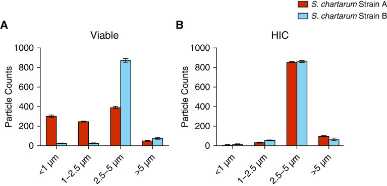
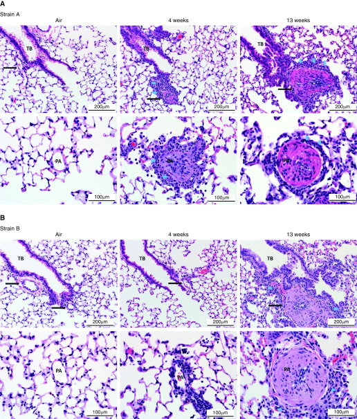
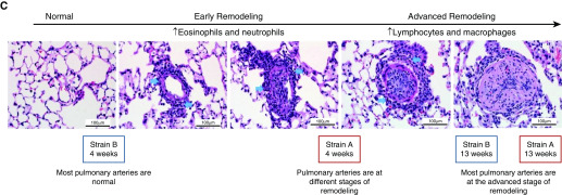
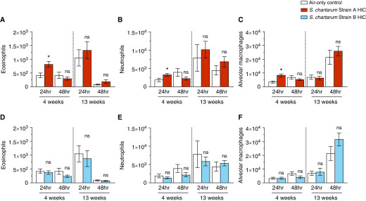
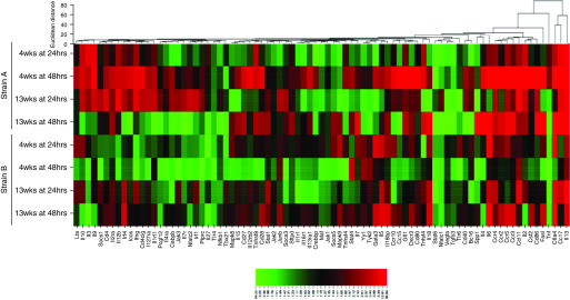
Stachybotrys chartarum is a fungal contaminant within the built environment and a respiratory health concern in the United States. The objective of this study was to characterize the mechanisms influencing pulmonary immune responses to repeatedly inhaled S. chartarum. Groups of B6C3F1/N mice repeatedly inhaled viable trichothecene-producing S. chartarum conidia (strain A or strain B), heat-inactivated conidia, or high-efficiency particulate absolute-filtered air twice per week for 4 and 13 weeks. Strain A was found to produce higher amounts of respirable fragments than strain B. Lung tissue, serum, and BAL fluid were collected at 24 and 48 hours after final exposure and processed for histology, flow cytometry, and RNA and proteomic analyses. At 4 weeks after exposure, a T-helper cell type 2-mediated response was observed. After 13 weeks, a mixed T-cell response was observed after exposure to strain A compared with a T-helper cell type 2-mediated response after strain B exposure. After exposure, both strains induced pulmonary arterial remodeling at 13 weeks; however, strain A-exposed mice progressed more quickly than strain B-exposed mice. BAL fluid was composed primarily of eosinophils, neutrophils, and macrophages. Both the immune response and the observed pulmonary arterial remodeling were supported by specific cellular, molecular, and proteomic profiles. The immunopathological responses occurred earlier in mice exposed to high fragment-producing strain A. The rather striking induction of pulmonary remodeling by S. chartarum appears to be related to the presence of fungal fragments during exposure.
Keywords: Stachybotrys chartarum fungal fragmentation; fungal exposure; pulmonary arterial remodeling.
Figures
Comment in
-
Killing Two Birds with One Stone: Mold-induced Pulmonary Immune Responses and Arterial Remodeling.Am J Respir Cell Mol Biol. 2020 May;62(5):537-538. doi: 10.1165/rcmb.2019-0386ED. Am J Respir Cell Mol Biol. 2020. PMID: 31693387 Free PMC article. No abstract available.
References
-
- World Health Organization (WHO) WHO guidelines for indoor air quality: dampness and mould. Geneva, Switzerland: WHO Regional Office for Europe; 2009. - PubMed
-
- Institute of Medicine. Damp indoor spaces and health. Washington, DC: National Academies Press; 2004. - PubMed
-
- Montaña E, Etzel RA, Allan T, Horgan TE, Dearborn DG. Environmental risk factors associated with pediatric idiopathic pulmonary hemorrhage and hemosiderosis in a Cleveland community. Pediatrics. 1997;99:e5. - PubMed
-
- Centers for Disease Control and Prevention (CDC) Acute pulmonary hemorrhage/hemosiderosis among infants: Cleveland, January 1993–November 1994. MMWR Morb Mortal Wkly Rep. 1994;43:881–883. - PubMed
-
- Centers for Disease Control and Prevention (CDC) Update: pulmonary hemorrhage/hemosiderosis among infants—Cleveland, Ohio, 1993–1996. MMWR Morb Mortal Wkly Rep. 2000;49:180–184. - PubMed
Publication types
MeSH terms
Substances
Grants and funding
LinkOut - more resources
Full Text Sources
