. 2019 Nov;8(11):1539-1552.
doi: 10.1530/EC-19-0442.

Leptin induces cell migration and invasion in a FAK-Src-dependent manner in breast cancer cells

Affiliations

Leptin induces cell migration and invasion in a FAK-Src-dependent manner in breast cancer cells

Juan Carlos Juárez-Cruz et al. Endocr Connect. 2019 Nov.

Abstract

Breast cancer is the most common invasive neoplasia, and the second leading cause of the cancer deaths in women worldwide. Mammary tumorigenesis is severely linked to obesity, one potential connection is leptin. Leptin is a hormone secreted by adipocytes, which contributes to the progression of breast cancer. Cell migration, metalloproteases secretion, and invasion are cellular processes associated with various stages of metastasis. These processes are regulated by the kinases FAK and Src. In this study, we utilized the breast cancer cell lines MCF7 and MDA-MB-231 to determine the effect of leptin on FAK and Src kinases activation, cell migration, metalloprotease secretion, and invasion. We found that leptin activates FAK and Src and induces the localization of FAK to the focal adhesions. Interestingly, leptin promotes the activation of FAK through a Src- and STAT3-dependent canonical pathway. Specific inhibitors of FAK, Src and STAT3 showed that the effect exerted by leptin in cell migration in breast cancer cells is dependent on these proteins. Moreover, we established that leptin promotes the secretion of the extracellular matrix remodelers, MMP-2 and MMP-9 and invasion in a FAK and Src-dependent manner. Our findings strongly suggest that leptin promotes the development of a more aggressive invasive phenotype in mammary cancer cells.

Keywords: FAK; Src; breast cancer; cell migration; invasion; leptin; metalloproteases.


Figures
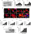
Figure 1
Figure 1
Activation and subcellular localization of FAK in leptin-stimulated breast cancer cell lines. (A) MDA-MB-231 and MCF7 cells were treated with 100 ng/mL and 200 ng/mL of leptin, respectively, for different times and cultures were lysed. Representative Western blots showing phosphorylated Y397 FAK and total FAK were detected using specific antibodies. The plots represent the densitometric and statistical analysis of the bands obtained by Western blot for MDA-MB-231 (B) and MCF7 (C); the values are the mean ± s.d. of at least three independent experiments and are expressed as changes with respect to the control (unstimulated cells). Asterisks denote comparisons made to unstimulated cells. *P < 0.05, **P < 0.01 by one-way ANOVA (Dunnett test). (D) Representative fluorescence microscopy images of MDA-MB-231 and MCF7 treated with or without 100 ng/mL leptin and 200 ng/mL at different time points. Anti-pY397 FAK staining is in green, TRITC-phalloidin is shown in red and DNA is stained with DAPI. Arrows indicate focal adhesions. (E) Quantification and statistical analyses of fluorescent signal of pY397 and stress fiber formation from fluorescence images obtained from MDA-MB-231 and MCF7. Asterisks denote comparisons made to unstimulated cells. *P < 0.05, **P < 0.01, ***P < 0.001 by one-way ANOVA (Dunnett test). (F) Representative Western blot of MDA-MB-231 cells treated with 100 ng/mL leptin at different time points. Phosphorylated Src was detected using an anti-pY418 as well as total Src. (G) Densitometric and statistical analysis of three independent Western blot analyses of phosphorylated Src at pY418. Data represents the mean ± s.d. and are expressed as changes with respect to the un-treated control. Statistical significance was established at ***P < 0.001 by Dunnett test.
Figure 2
Figure 2
Leptin-induced FAK activation occurs in a Src-dependent manner in MDA-MB-231 and MCF7 breast cancer cells. (A) Representative Western blot of FAK activation at Y397 in the presence or absence of 100 and 200 ng/mL leptin and 5 μM of the Src inhibitor SU6656 in MDA-MB-231 and MCF7 cells. Plots represent the densitometric statistical analyses for each cell type for FAK phosphorylation (B and C), and Src (D and E), respectively. (F) Representative Western blot of FAK activation at Y397 in the presence or absence of 100 ng/mL leptin and 5 μM of the Src inhibitor PP2 in MDA-MB-231 cells. Plots represent the densitometric statistical analyses for each cell type for FAK phosphorylation (G), and Src (H). Antibody against total FAK was used as loading control.
Figure 3
Figure 3
FAK/Src are required for migration of leptin-stimulated MDA-MB-231 and MCF7 breast cancer cells. Confluent monolayers of MDA-MB-231 cells were treated for 2 h with AraC to inhibit proliferation, and then pre-treated with 5 µM of the FAK inhibitor, PF-57322, or with 5 µM of the Src inhibitor, PP2, for 30 min. Wound-healing assays were performed by scratching the monolayer using a sterile pipette tip. Cells were washed and re-fed with DMEM supplemented with or without leptin. The progress of cell migration into the wound was registered at 0 (upper panel) and 48 h (lower panel). Representative light microscopy images and quantification of wounding assays in MDA-MB-231 (A and B) and MCF7 (C and D) breast cancer cell lines and effect of FAK inhibition in the wound closure. (E) Representative light microscopy images and quantification of wounding assays in MDA-MB-231 using SU6656, an Src inhibitor (F) and quantification of the wound closure and the effect of Src inhibition. Data are expressed as percentage of wound closure for each condition and represents the mean of three independent experiments ± s.d. The Newman–Keuls test compares all experimental conditions to determine statistically significant differences; **P < 0.01 by one-way ANOVA.
Figure 4
Figure 4
Leptin promotes cell migration and FAK activation independently of the canonical leptin receptor in a model for triple-negative breast cancer cells. (A) Representative Western blot of FAK activation at Y397 in the presence or absence of 100 ng/mL and 200 ng/mL leptin and 50 and 15 μM of the STAT3 inhibitor S3I201 in MDA-MB-231 and MCF7 cells. Plots represent the densitometric statistical analyses for FAK phosphorylation in MDA-MB-231 (B) and MCF7 cells (C). Confluent monolayers of MDA-MB-231 cells were treated for 2 h with AraC to inhibit proliferation, and then pre-treated with 50 µM of the STAT3 inhibitor, S3I201 for 24 h. Wound healing assays were performed by scratching the monolayer using a sterile pipette tip. Cells were washed and re-fed with DMEM supplemented with or without leptin. The progress of cell migration into the wound was registered at 0 (upper panel) and 48 h (lower panel). (D) Representative light microscopy images and (E) quantification of wounding assays in MDA-MB-231 breast cancer cells and effect of STAT inhibition in the wound closure. Data is expressed as percentage of wound closure for each condition and represents the mean of three independent experiments ± s.d. The Newman–Keuls test compares all experimental conditions to determine statistically significant differences; ***P < 0.001 by one-way ANOVA.
Figure 5
Figure 5
Leptin promotes the secretion of MMP-2 and MMP-9 in the MDA-MB-231 and MCF7 cancer cell lines. (A) Representative zymogram of the gelatinase activity of secreted MMP-2 and MMP-9 obtained from supernatants of MDA-MB-231 cells stimulated with increasing concentrations of leptin for 24 h. Quantification of the gelatinase activity of both secreted metalloproteases MMP-2 (B), MMP-9 (C) under each experimental condition. (D) Representative zymogram of the gelatinase activity of secreted MMP-2 and MMP-9 obtained from supernatants of MCF7 cells stimulated with increasing concentrations of leptin for 24 h. Quantification of the gelatinase activity of both secreted metalloproteases MMP-2 (E), MMP-9 (F) under each experimental condition. (G) Representative zymogram of the gelatinase activity of secreted MMP-2 and MMP-9 obtained from supernatants of MDA-MB-231 cells chronically stimulated with 100 ng/mL of leptin. Quantification of the gelatinase activity of both secreted metalloproteases MMP-2 (H), MMP-9 (I) under each experimental condition. Supernatant samples were collected at the indicated time points. Actin was used as loading control in all cases. Data represent the mean of three independent biological experiments ± s.d., and it is expressed as fold increase over the un-treated control. Statistical significance was established at *P < 0.05, **P < 0.01, ***P < 0.001 by Dunnett test.
Figure 6
Figure 6
Leptin promotes secretion of metalloproteases and increases an invasive phenotype of MDA-MB-231 cell line in a FAK-dependent manner. MDA-MB-231 cells were pre-treated for 30 min with 5 μM of the FAK-specific inhibitor, PF-573228. Then, the cells were stimulated without or with 100 ng/mL leptin for 24 h. (A) Representative zymogram of the gelatinase activity of MMP-2 and MMP-9 obtained from MDA-MB-231 cells supernatants, actin was used as loading control. Quantification of the activity of secreted MMP-2 (B) and MMP-9 (C) for each experimental condition, and it is expressed as fold increase over the un-treated control. Statistical significance was established at *P < 0.05, **P < 0.01, ***P < 0.001 by Dunnett test. (D) Representative light microscopy images of invasion assays for MDA-MB-231 cells. The cells were pre-treated with 5 μM PF-573228 and seeded at 1x10⁵ cells in Matrigel-coated chambers in the presence or absence of 100 ng/mL leptin for 48 h. (E) Cell invasion quantification represents the number of invading cells for each experimental condition. Data represents the mean ± s.d., and it is expressed as fold increase over the un-treated control. Statistical significance was established at *P < 0.05, **P < 0.01, by one-way ANOVA (Newman–Keuls test).
Figure 7
Figure 7
Leptin-induced secretion, activation of MMP-2 and MMP-9 and invasion in MDA-MB-231 cells is Src-dependent. (A) Representative zymogram MMP-2 and MMP-9 obtained from supernatants of MDA-MB-231 cells treated with 5 μM of the Src-specific inhibitor, PP2, and supplemented with or without 100 ng/mL leptin for 24 h. Quantification of the activity of secreted MMP-2 (B) and MMP-9 (C) for each experimental condition, and it is expressed as fold increase over the un-treated control. Statistical significance was established at P < 0.05, **P < 0.01, by one-way ANOVA (Newman–Keuls test). (D) Representative light microscopy images of invasion assays for MDA-MB-231 cells. The cells were pre-treated with 5 μM of PP2 and seeded at 1 × 105 cells in Matrigel-coated chambers in the presence or absence of 100 ng/mL leptin for 48 h. (E) Cell invasion quantification represents the number of invading cells for each experimental condition. Data represents the mean ± s.d., and it is expressed as fold increase over the un-treated control. Statistical significance was established at *P < 0.05, **P < 0.01, by one-way ANOVA (Newman–Keuls test).
Figure 8
Figure 8
Representative model of the MMP-2 and MMP-9 secretion induced by leptin in a cultured model for breast cancer. Leptin-dependent activation of FAK occurs via Src and an independent mechanism from the canonical ObR pathway. These kinases promote the secretion of MMPs, cell migration and invasion in cultured models for breast cancer cells.

Similar articles

Cited by

References

    1. Bray F, Ferlay J, Soerjomataram I, Siegel RL, Torre LA, Jemal A. Global cancer statistics 2018: GLOBOCAN estimates of incidence and mortality worldwide for 36 cancers in 185 countries. CA: A Cancer Journal for Clinicians 2018. 6 394–424. (10.3322/caac.21492) - DOI - PubMed
    1. Barone I, Giordano C, Bonofiglio D, Andò S, Catalano S. Leptin, obesity and breast cancer: progress to understanding the molecular connections. Current Opinion in Pharmacology 2016. 83–89. (10.1016/j.coph.2016.10.003) - DOI - PubMed
    1. Chaffer CL, Juan BPS, Lim E, Weinberg RA. EMT, cell plasticity and metastasis. Cancer Metastasis Reviews 2016. 645–654. (10.1007/s10555-016-9648-7) - DOI - PubMed
    1. Kaviani A, Neishaboury M, Mohammadzadeh N, Ansari-Damavandi M, Jamei K. Effects of obesity on presentation of breast cancer, lymph node metastasis and patient survival: a retrospective review. Asian Pacific Journal of Cancer Prevention 2013. 2225–2229. (10.7314/apjcp.2013.14.4.2225) - DOI - PubMed
    1. Iskander K, Farhour R, Ficek M, Ray A. Obesity-related complications: few biochemical phenomena with reference to tumorigenesis. Malaysian Journal of Pathology 2013. 1–15. - PubMed

LinkOut - more resources