. 2019 Oct 30;20(21):5414.
doi: 10.3390/ijms20215414.

Gender Differences in the Pharmacological Actions of Pegylated Glucagon-Like Peptide-1 on Endothelial Progenitor Cells and Angiogenic Precursor Cells in a Combination of Metabolic Disorders and Lung Emphysema

Affiliations

Gender Differences in the Pharmacological Actions of Pegylated Glucagon-Like Peptide-1 on Endothelial Progenitor Cells and Angiogenic Precursor Cells in a Combination of Metabolic Disorders and Lung Emphysema

Olga Victorovna Pershina et al. Int J Mol Sci. .

Abstract

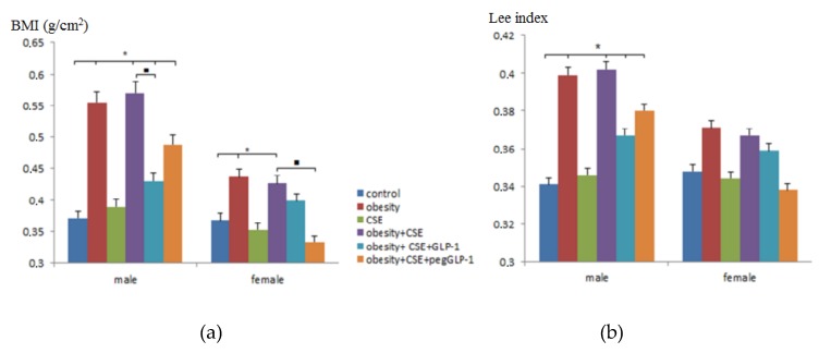
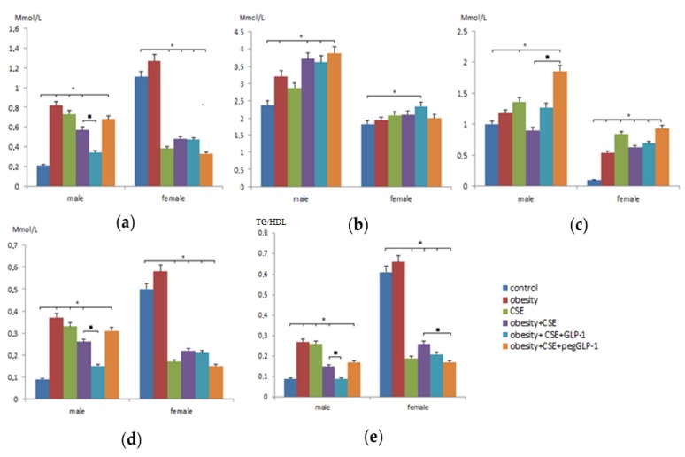
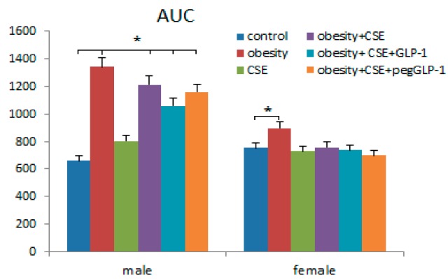
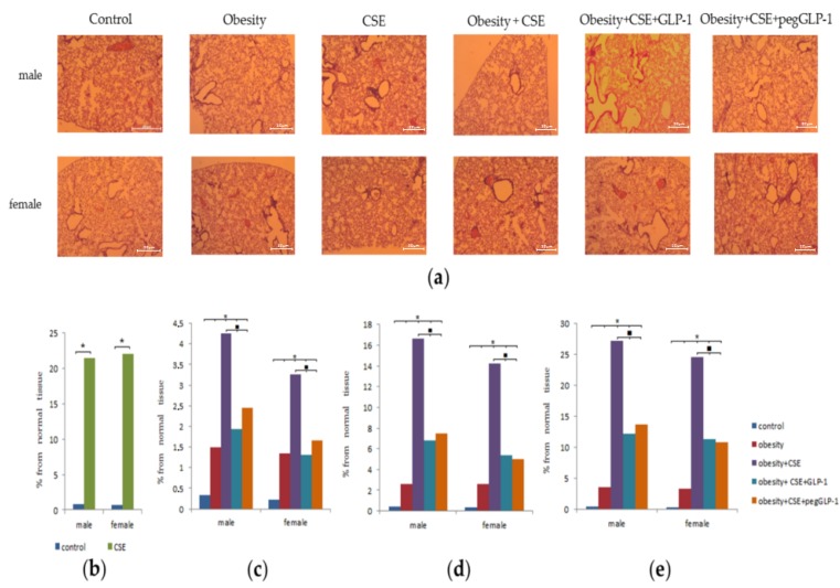
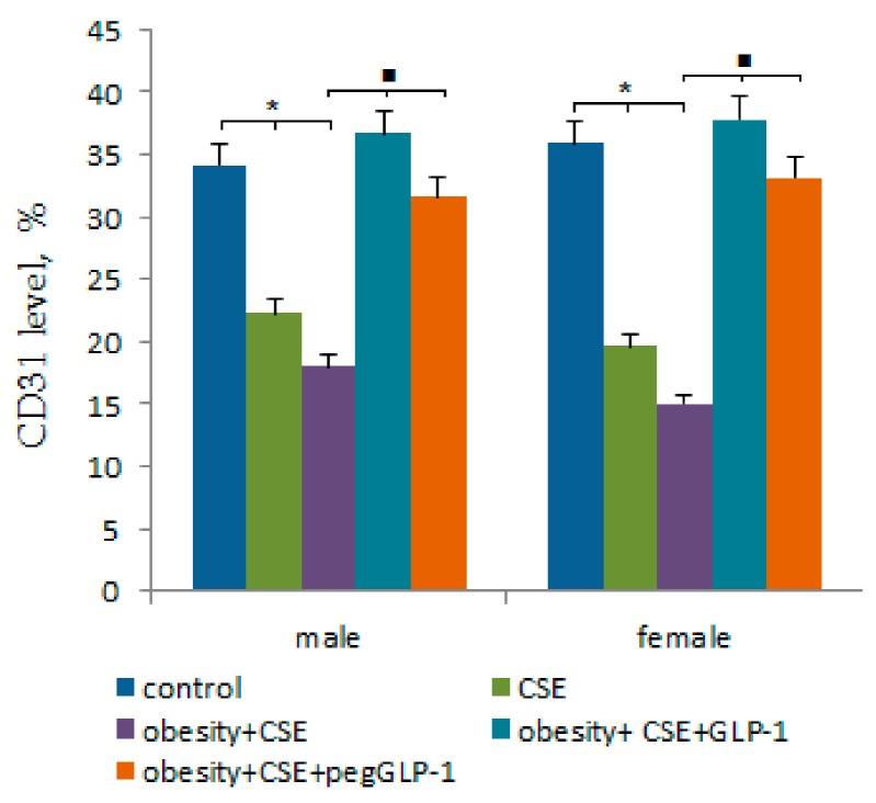
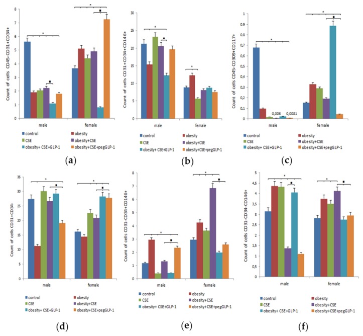
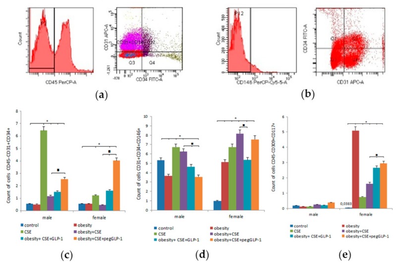
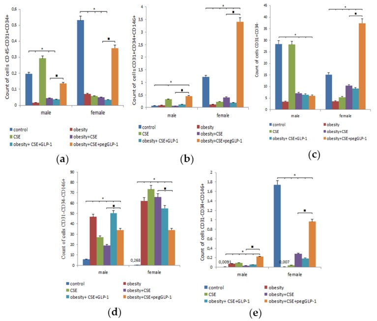
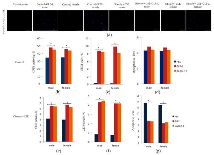
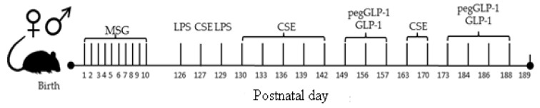
In clinical practice, the metabolic syndrome (MetS) is often associated with chronic obstructive pulmonary disease (COPD). Although gender differences in MetS are well documented, little is known about sex-specific differences in the pathogenesis of COPD, especially when combined with MetS. Consequently, it is not clear whether the same treatment regime has comparable efficacy in men and women diagnosed with MetS and COPD. In the present study, using sodium glutamate, lipopolysaccharide, and cigarette smoke extract, we simulated lipid metabolism disorders, obesity, hyperglycemia, and pulmonary emphysema (comorbidity) in male and female C57BL/6 mice. We assessed the gender-specific impact of lipid metabolism disorders and pulmonary emphysema on angiogenic precursor cells (endothelial progenitor cells (EPC), pericytes, vascular smooth muscle cells, cells of the lumen of the nascent vessel), as well as the biological effects of pegylated glucagon-like peptide 1 (pegGLP-1) in this experimental paradigm. Simulation of MetS/COPD comorbidity caused an accumulation of EPC (CD45-CD31+CD34+), pericytes, and vascular smooth muscle cells in the lungs of female mice. In contrast, the number of cells involved in the angiogenesis decreased in the lungs of male animals. PegGLP-1 had a positive effect on lipids and area under the curve (AUC), obesity, and prevented the development of pulmonary emphysema. The severity of these effects was stronger in males than in females. Furthermore, PegGLP-1 stimulated regeneration of pulmonary endothelium. At the same time, PegGLP-1 administration caused a mobilization of EPC (CD45-CD31+CD34+) into the bloodstream in females and migration of precursors of angiogenesis and vascular smooth muscle cells to the lungs in male animals. Gender differences in stimulatory action of pegGLP-1 on CD31+ endothelial lung cells in vitro were not observed. Based on these findings, we postulated that the cellular mechanism of in vivo regeneration of lung epithelium was at least partly gender-specific. Thus, we concluded that a pegGLP-1-based treatment regime for metabolic disorder and COPD should be further developed primarily for male patients.

Keywords: and endothelial regeneration; angiogenic precursor cells; dyslipidemia; endothelial progenitor cells; gender differences; hyperglycemia; obesity; pegylated glucagon-like peptide 1; pulmonary emphysema.


Conflict of interest statement

The authors declare no conflict of interest.

Figures

Figure 1
Figure 1
The effect of glucagon-like peptide-1 (GLP-1) and pegylated GLP-1 (pegGLP-1) on body mass index (BMI) and Lee index of male and female C57BL/6 mice on p189: (a) The BMI (g/cm2); (b) The Lee index. Groups: control—a control group from intact mice, obesity—mice with metabolic disorders (obesity and hyperglycemia), CSE—mice with lungs emphysema, obesity+CSE—mice with metabolic disorders (obesity and hyperglycemia) and lungs emphysema, obesity+CSE+GLP-1—mice with metabolic disorders (obesity and hyperglycemia) and lungs emphysema treated with GLP-1, obesity+CSE+pegGLP-1—mice with metabolic disorders (obesity and hyperglycemia) and lungs emphysema treated with pegGLP-1. Results are presented as the mean±SEM. *—significance of difference compared with control (p < 0.05); ■—significance of difference compared with the obesity+CSE group (p < 0.05). CSE, cigarette smoke extract.
Figure 2
Figure 2
Lipid profile measurements in the blood of female and male C57BL/6 mice on p189: (a) The level of triglycerides in serum (Mmol/l); (b) High-density lipoprotein level (Mmol/l); (c) Low-density lipoprotein level (Mmol/l); (d) Very low-density lipoprotein level (Mmol/l); (e) The ratio of triglycerides to high-density lipoproteins (TG/HDL). Groups: control—a control group from intact mice, obesity—mice with metabolic disorders (obesity and hyperglycemia), CSE—mice with lungs emphysema, obesity+CSE—mice with metabolic disorders (obesity and hyperglycemia) and lungs emphysema, obesity+CSE+GLP-1—mice with metabolic disorders (obesity and hyperglycemia) and lungs emphysema treated with GLP-1, obesity+CSE+pegGLP-1—mice with metabolic disorders (obesity and hyperglycemia) and lungs emphysema treated with pegGLP-1. *—significance of difference compared with control (p < 0.05); ■—significance of difference compared with the obesity+CSE group (p < 0.05).
Figure 3
Figure 3
The area under the curve (AUC) of female and male C57BL/6 mice during the glucose tolerance test (on p188). Groups: control—a control group from intact mice, obesity—mice with metabolic disorders (obesity and hyperglycemia), CSE-mice with lungs emphysema, obesity+CSE—mice with metabolic disorders (obesity and hyperglycemia) and lungs emphysema, obesity+CSE+GLP-1—mice with metabolic disorders (obesity and hyperglycemia) and lungs emphysema treated with GLP-1, obesity+CSE+pegGLP-1—mice with metabolic disorders (obesity and hyperglycemia) and lungs emphysema treated with pegGLP-1. *—significance of difference compared with control (p < 0.05).
Figure 4
Figure 4
Morphological study of lung obtained from male and female C57BL/6 mice (n = 6): (a) Photomicrographs of left lung sections (lower pulmonary field) (on p189). Tissues were stained with hematoxylin-eosin; (b) The area of emphysema-expanded lung tissue (lower pulmonary field) of mice from all groups (on p148); (c) The area of emphysema-expanded lung tissue (upper pulmonary field) of mice from all groups (on p); (d) The area of emphysema-expanded lung tissue (middle pulmonary field) of mice from all groups (on p189); (e) The area of emphysema-expanded lung tissue (lower middle pulmonary field) of mice from all groups (on the p189). Groups: control—a control group from intact mice, obesity—mice with metabolic disorders (obesity and hyperglycemia), CSE—mice with lungs emphysema, obesity+CSE—mice with metabolic disorders (obesity and hyperglycemia) and lungs emphysema, obesity+CSE+GLP-1–mice with metabolic disorders (obesity and hyperglycemia) and lungs emphysema treated with GLP-1, obesity+CSE+pegGLP-1—mice with metabolic disorders (obesity and hyperglycemia) and lungs emphysema treated with pegGLP-1. * p < 0.05 significance of difference compared with control group, ■—significance of difference compared with the obesity+CSE group (p < 0.05).
Figure 5
Figure 5
The relative content of cells expressing CD31 antigen in the lungs isolated from male and female C57BL/6 mice at the immunohistochemical staining for specific cellular marker: CD31 (on the p189). Groups: control—a control group from intact mice, obesity—mice with metabolic disorders (obesity and hyperglycemia), CSE—mice with lungs emphysema, obesity+CSE—mice with metabolic disorders (obesity and hyperglycemia) and lungs emphysema, obesity+CSE+GLP-1—mice with metabolic disorders (obesity and hyperglycemia) and lungs emphysema treated with GLP-1, obesity+CSE+pegGLP-1—mice with metabolic disorders (obesity and hyperglycemia) and lungs emphysema treated with pegGLP-1. *p < 0.05 significance of difference compared with control group, ■—significance of difference compared with the obesity+CSE group (p < 0.05).
Figure 6
Figure 6
Characterization of cell population isolated from the lung of male and female C57BL/6 mice on the p189. (a) The content of endothelial progenitor cells (EPC) (CD45CD31+CD34+); (b) The content of EPC (CD31+CD34+CD146+); (c) The content of angiogenesis precursors (CD45CD309+CD117+); (d) The content of cells in the lumen of the nascent vessel (CD31+CD34); (e) The content of vascular smooth muscle cells (CD31CD34+CD146+); (f) The content of pericytes (CD31CD34CD146+). Cells were analyzed by flow cytometry using antibodies for CD31, CD34, CD45, CD146, CD117, CD309 mice. Dot plots are representative of three independent experiments with the mean from three independent experiments. Groups: control—a control group from intact mice, obesity—mice with metabolic disorders (MD) (obesity and hyperglycemia), CSE—mice with lungs emphysema, obesity+CSE—mice with MD (obesity and hyperglycemia) and lungs emphysema, obesity+CSE+GLP-1—mice with MD (obesity and hyperglycemia) and lungs emphysema treated with GLP-1, obesity+CSE+pegGLP-1—mice with MD (obesity and hyperglycemia) and lungs emphysema treated with pegGLP-1. *—significance of difference compared with control (p < 0.05); ■—significance of difference compared with the obesity+CSE group (p < 0.05).
Figure 7
Figure 7
Characterization of cell population isolated from the bone marrow of male and female C57BL/6 mice on the p189. (a) Phenotype establishment and qualitative analysis of CD45 (PerCP), CD34 (FITC), and CD31 (APC) expression; (b) Phenotype establishment and qualitative analysis of CD34 (FITC), CD31 (APC), and CD146 (PerCP-Cy5.5) expression; (c) The content of EPC (CD45CD31+CD34+); (d) The content of EPC (CD31+CD34+CD146+); (e) The content of angiogenesis precursors (CD45CD309+CD117+). Cells were analyzed by flow cytometry using antibodies for CD45, CD31, CD34, CD146, CD117, CD309 mice. Dot plots are representative of three independent experiments with the mean from three independent experiments. Groups: control—a control group from intact mice, obesity—mice with MD (obesity and hyperglycemia), CSE—mice with lungs emphysema, obesity+CSE—mice with MD (obesity and hyperglycemia) and lungs emphysema, obesity+CSE+GLP-1—mice with MD (obesity and hyperglycemia) and lungs emphysema treated with GLP-1, obesity+CSE+pegGLP-1—mice with MD (obesity and hyperglycemia) and lungs emphysema treated with pegGLP-1. *—significance of difference compared with control (p < 0.05); ■—significance of difference compared with the obesity+CSE group (p < 0.05).
Figure 8
Figure 8
Characterization of cell population isolated from the blood of male and female C57BL/6 mice on the p189. (a) The content of EPC (CD45CD31+CD34+); (b) The content of EPC (CD31+CD34+CD146+); (c) The content of cells in the lumen of the nascent vessel (CD31+CD34); (d) The content of pericytes (CD31CD34CD146+); (e) The content of vascular smooth muscle cells (CD31CD34+CD146+). Cells were analyzed by flow cytometry using antibodies for CD45, CD31, CD34, CD146 mice. Dot plots are representative of three independent experiments with the mean from three independent experiments. Groups: control—a control group from intact mice, obesity—mice with MD (obesity and hyperglycemia), CSE—mice with lungs emphysema, obesity+CSE—mice with MD (obesity and hyperglycemia) and lungs emphysema, obesity+CSE+GLP-1—mice with MD (obesity and hyperglycemia) and lungs emphysema treated with GLP-1, obesity+CSE+pegGLP-1—mice with MD (obesity and hyperglycemia) and lungs emphysema treated with pegGLP-1. *—significance of difference compared with control (p < 0.05); ■—significance of difference compared with the obesity+CSE group (p < 0.05).
Figure 9
Figure 9
GLP-1 and pegGLP-1 treatment effects on CD31+ endothelial cells isolated from the lungs of male and female C57BL/6 mice in vitro: (a) Images of CD31+ cells stained with: Hoechst (blue) to identify cell nuclei; CD34 FITC (green) (Hoechst + CD34) composite image using all two colors. All scale bars are 100 µm; (bg) CD31+ endothelial cells from lung were precultured for 5 days, incubated with or without GLP-1 (10−7 M) or pegGLP-1 (10−7 M) for 24 h and then labeled with Hoechst, Carboxyfluorescein succinimidyl ester (CFSE) (b,e) CD34 FITC (c,f), Annexin V and 7-Aminoactinomycin D (7-AAD) (d,g) prior to fluorescence microscopic analysis. (b) CFSE activity after culture of cells isolated from the lung of intact mice; (c) the level of CD34+ cells after culture of cells isolated from the lung of intact mice; (d) the count of cells with apoptosis after culture of cells isolated from the lung of intact mice; (e) CFSE activity after culture of cells isolated from the lung of mice with MD and lung emphysema; (f) the level of CD34+ cells after culture of cells isolated from the lung of mice with MD and lung emphysema; (g) the count of cells with apoptosis after culture of cells isolated from the lung of mice with MD and lung emphysema. All data are expressed as mean ± SD, *—significance of difference compared with control (p < 0.05).
Figure 10
Figure 10
Schematic diagram of the experimental procedures.

References

    1. Lusis A.J., Attie A.D., Reue K. Metabolic syndrome: From epidemiology to systems biology. Nat. Rev. Genet. 2008;9:819–830. doi: 10.1038/nrg2468. - DOI - PMC - PubMed
    1. Mauvais-Jarvis F. Epidemiology of Gender Differences in Diabetes and Obesity. Adv. Exp. Med. Biol. 2017;1043:3–8. doi: 10.1007/978-3-319-70178-3_1. - DOI - PubMed
    1. Karastergiou K., Smith S.R., Greenberg A.S., Fried S.K. Sex differences in human adipose tissues e the biology of pear shape. Biol. Sex Differ. 2012;3:13. doi: 10.1186/2042-6410-3-13. - DOI - PMC - PubMed
    1. Mundi M.S., Koutsari C., Jensen M.D. Effects of increased free fatty acid availability on adipose tissue fatty acid storage in men. J. Clin. Endocrinol. Metab. 2014;99:E2635–E2642. doi: 10.1210/jc.2014-2690. - DOI - PMC - PubMed
    1. Karpe F., Pinnick K.E. Biology of upper-body and lower-body adipose tissue link to whole-body phenotypes. Nat. Rev. Endocrinol. 2015;11:90–100. doi: 10.1038/nrendo.2014.185. - DOI - PubMed

MeSH terms