. 2019 Oct 29;8(11):1334.
doi: 10.3390/cells8111334.

P65 Targets FGFR1 to Regulate the Survival of Ovarian Granulosa Cells

Affiliations

P65 Targets FGFR1 to Regulate the Survival of Ovarian Granulosa Cells

Xiaolong Yuan et al. Cells. .

Abstract

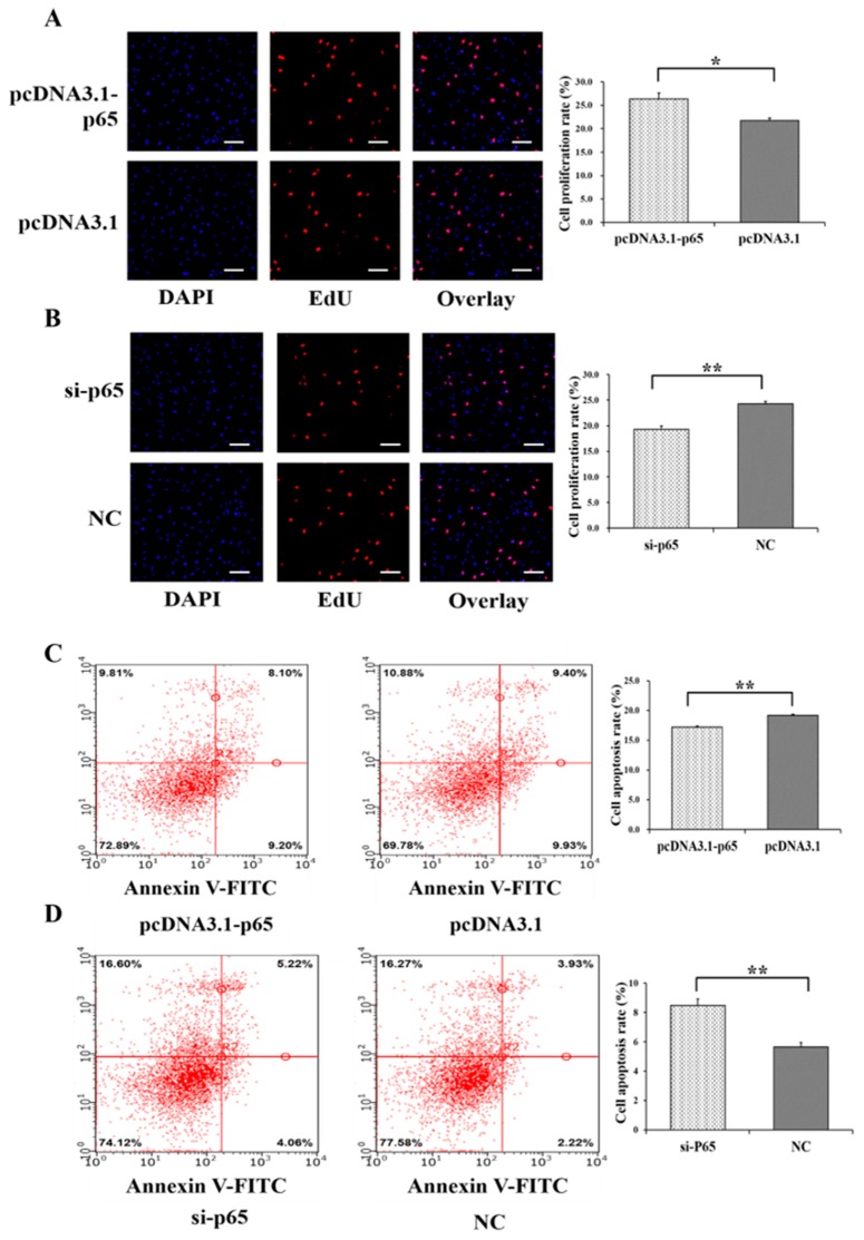
In female mammals, the abnormal apoptosis of ovarian granulosa cells (GCs) impairs follicular development and causes reproductive dysfunction. Many studies have indicated that the FGFR1 gene of the PI3K signaling pathway and the p65 subunit of the transcription factor NF-κB may regulate the proliferation and apoptosis of GCs involved in follicular development. However, little is known about whether p65 regulates the transcription of FGFR1, as well as the biological effects of p65 and FGFR1 on the survival of GCs and follicular development. In porcine follicles and GCs, we found that p65 and FGFR1 were exclusively expressed in the GCs of follicles, and the mRNA and protein levels of p65 and FGFR1 significantly increased from small to large follicles. Both p65 and FGFR1 were found to activate the PI3K signaling pathway, and the expressions of proliferation markers (PCNA and MKI67) and the anti-apoptotic gene BCL2 were significantly increased by p65 and FGFR1. Furthermore, both p65 and FGFR1 were observed to promote cell proliferation and inhibit the cell apoptosis of GCs, and p65 was confirmed to bind at the -348/-338 region of FGFR1 to positively regulate its transcription. Moreover, p65 was further found to enhance the pro-proliferation and anti-apoptotic effects of FGFR1. Taken together, p65 may target the -348/-338 region of FGFR1, promote the transcription of FGFR1, and enhance the pro-proliferation effect and anti-apoptotic effect of FGFR1 to facilitate the growth of follicles. This study will provide useful information for further investigations on the p65-mediated-FGFR1 signaling pathway during folliculogenesis in mammals.

Keywords: FGFR1; cell proliferation and apoptosis; ovarian granulosa cells; transcription factor p65.


Conflict of interest statement

The authors declare no conflict of interest.

Figures

Figure 1
Figure 1
Expression patterns of p65 and FGFR1 in follicles and granulosa cells (GCs). The representations of immunohistochemical results of p65 (A,B) and FGFR1 (C,D) in large and small follicles, respectively. The relative mRNA (E) and protein (F) levels of p65 in small and large follicles. The relative mRNA (G) and protein (H) levels of FGFR1 in small and large follicles. * indicates p < 0.05; ** indicates p < 0.01. Scale bars: 200 μm for 5× figures; 100 μm for 10× figures; 20 μm for 20× figures.
Figure 2
Figure 2
Biological effects of p65 on apoptosis and PI3K signaling pathway. (A) The mRNA level of p65 under different concentrations of pcDNA3.1-p65 plasmid; (B) the protein count of p65 was stimulated by 200 ng of pcDNA3.1-p65 plasmid; (C) the mRNA level of p65 with different concentrations of siRNAs; (D) the protein count of p65 was suppressed by 70 nM of si-p65. The relative mRNA expressions of genes from apoptosis signaling pathway after treatment by pcDNA3.1-p65 (E) and si-p65 (F); the relative mRNA expressions of genes from PI3K signaling pathway after treatment by pcDNA3.1-p65 (G) and si-p65 (H). * indicates p < 0.05; ** indicates p < 0.01. Data are represented as means ± SD. NC: negative control. Si-p65: specific siRNA for p65. PcDNA3.1-p65: pcDNA3.1 overexpression plasmid of p65.
Figure 3
Figure 3
Biological effects of p65 on cell proliferation and apoptosis of GCs. The cell proliferation rate of GCs after treatment by pcDNA3.1-p65 (A) and si-p65 (B). The cell apoptosis rate of GCs after treatment by pcDNA3.1-p65 (C) and si-p65 (D). * indicates p < 0.05; ** indicates p < 0.01. Data are represented as means ± SD. The scale bar represents 100 μm. NC: negative control. PcDNA3.1-p65: pcDNA3.1 overexpression plasmid of p65; si-p65: specific siRNA for p65.
Figure 4
Figure 4
p65 binds to the −348/−338 region of FGFR1 and positively regulate the expression of FGFR1. (A) The putative binding sites of p65 on the promoter of FGFR1; (B) PCR products of 5′ deletion fragments from promoter of FGFR1; (C) the relative luciferase activity of FGFR1 promoter after 5′ deletion of the putative binding sites of p65; (D) the relative luciferase activity of P6 with overexpression of p65; (E) the relative luciferase activity of P6 with interfering with expression of p65; (F) the relative luciferase activity of P6 after specific deletion of −348/−338; (G) confirmation of p65 binding at −348/−338 by chromatin immunoprecipitation (ChIP) assay. The relative mRNA (H) and protein (I) levels of FGFR1 stimulated by pcDNA3.1-p65; the relative mRNA (J) and protein (K) levels of FGFR1 inhibited by si-p65. * indicates p < 0.05; ** indicates p < 0.01. Data are represented as means ± SD. CAAT: CAAT box; TATA: TATA box: GC: GC box; M5000: DNA markers of 5000 bp. M500: DNA markers of 500 bp. Si-p65: specific siRNA for p65; NC: negative control. pcDNA3.1-p65: pcDNA3.1 overexpression plasmid of p65.
Figure 5
Figure 5
Biological effects of FGFR1 on apoptosis and PI3K signaling pathway. (A) The mRNA level of FGFR1 under different concentrations of pcDNA3.1-FGFR1 plasmid; (B) the FGFR1 protein count was stimulated by 200 ng of pcDNA3.1-FGFR1 plasmid; (C) the mRNA level of FGFR1 with different concentrations of siRNAs; (D) the FGFR1 protein count was suppressed by 70 nM of si-FGFR1. The relative mRNA expressions of genes from apoptosis signaling pathway after treatment by pcDNA3.1-FGFR1 (E) and si-FGFR1 (F); The relative mRNA expressions of genes from PI3K signaling pathway after treatment by pcDNA3.1-FGFR1 (G) and si-FGFR1 (H). * indicates p < 0.05; ** indicates p < 0.01. Data are represented as means ± SD. NC: negative control. Si-FGFR1: specific siRNA for FGFR1. PcDNA3.1-FGFR1: pcDNA3.1 overexpression plasmid of FGFR1.
Figure 6
Figure 6
Biological effects of FGFR1 on cell proliferation and apoptosis of GCs. The cell proliferation rate of GCs after treatment by pcDNA3.1-FGFR1 (A) and si-FGFR1 (B). The cell apoptosis rate of GCs after treatment by pcDNA3.1-FGFR1 (C) and si-FGFR1 (D). * indicates p < 0.05; ** indicates p < 0.01. Data are represented as means ± SD. The scale bar represents 100 μm. NC: negative control; PcDNA3.1-FGFR1: pcDNA3.1 overexpression plasmid of FGFR1; Si-FGFR1: specific siRNA for FGFR1.
Figure 7
Figure 7
p65 enhances the pro-proliferation and anti-apoptotic effects of FGFR1 in GCs. (A) PcDNA3.1-p65 increased the cell proliferation rate of pcDNA3.1-FGFR1 in GCs; (B) si-p65 decreased the cell proliferation rate of si-FGFR1 in GCs; (C) pcDNA3.1-p65 depressed the cell apoptosis rate of pcDNA3.1-FGFR1 in GCs; (D) si-p65 stimulated the cell apoptosis rate of si-FGFR1 in GCs. Data are represented as means ± SD. NC: negative control; PcDNA3.1-FGFR1: pcDNA3.1 overexpression plasmid of FGFR1; PcDNA3.1-p65: pcDNA3.1 overexpression plasmid of p65; si-FGFR1: specific siRNA for FGFR1; si-p65: specific siRNA for p65; NC: negative control; * indicates p < 0.05; ** indicates p < 0.01.

References

    1. Hunter M.G. Oocyte maturation and ovum quality in pigs. Rev. Reprod. 2000;5:122–130. doi: 10.1530/ror.0.0050122. - DOI - PubMed
    1. McGee E.A., Hsueh A.J. Initial and cyclic recruitment of ovarian follicles. Endocr. Rev. 2000;21:200–214. doi: 10.1210/er.21.2.200. - DOI - PubMed
    1. Soede N.M., Langendijk P., Kemp B. Reproductive cycles in pigs. Anim. Reprod. Sci. 2011;124:251–258. doi: 10.1016/j.anireprosci.2011.02.025. - DOI - PubMed
    1. Matsuda F., Inoue N., Manabe N., Ohkura S. Follicular growth and atresia in mammalian ovaries: Regulation by survival and death of granulosa cells. J. Reprod. Dev. 2012;58:44–50. doi: 10.1262/jrd.2011-012. - DOI - PubMed
    1. Manabe N., Goto Y., Matsuda-Minehata F., Inoue N., Maeda A., Sakamaki K., Miyano T. Regulation mechanism of selective atresia in porcine follicles: Regulation of granulosa cell apoptosis during atresia. J. Reprod. Dev. 2004;50:493–514. doi: 10.1262/jrd.50.493. - DOI - PubMed

Publication types

Substances

LinkOut - more resources