Review
. 2019 Oct 29;8(4):82.
doi: 10.3390/biology8040082.

Autophagy in Cancer Cell Death

Affiliations
Review

Autophagy in Cancer Cell Death

Benedikt Linder et al. Biology (Basel). .

Abstract

Autophagy has important functions in maintaining energy metabolism under conditions of starvation and to alleviate stress by removal of damaged and potentially harmful cellular components. Therefore, autophagy represents a pro-survival stress response in the majority of cases. However, the role of autophagy in cell survival and cell death decisions is highly dependent on its extent, duration, and on the respective cellular context. An alternative pro-death function of autophagy has been consistently observed in different settings, in particular, in developmental cell death of lower organisms and in drug-induced cancer cell death. This cell death is referred to as autophagic cell death (ACD) or autophagy-dependent cell death (ADCD), a type of cellular demise that may act as a backup cell death program in apoptosis-deficient tumors. This pro-death function of autophagy may be exerted either via non-selective bulk autophagy or excessive (lethal) removal of mitochondria via selective mitophagy, opening new avenues for the therapeutic exploitation of autophagy/mitophagy in cancer treatment.

Keywords: AT-101; autophagy; cancer; cell death; gossypol; mitochondria; mitophagy.


Conflict of interest statement

The authors declare no conflict of interest. The funders had no role in the design of the study; in the collection, analyses, or interpretation of data; in the writing of the manuscript, or in the decision to publish the results.

Figures

Figure 1
Figure 1
Molecular basis of mammalian autophagy. Autophagy is a multistep process involving several key ATG proteins and signaling complexes. It requires the formation of double-membrane-containing autophagosomes that sequester proteins, lipids, organelles or invasive microbes and fuse with lysosomes for digestion of content by acidic hydrolases. ULK1, a protein kinase serving as the central initiator of autophagy, is inhibited by the mTORC1 complex that contains mTOR. AMPK serves as a nutrient sensor and negative regulator of mTORC1. Autophagosome biogenesis starts with the formation of an initiation membrane that is derived either from the endoplasmatic reticulum (ER) or from several other cellular membrane sources. Vesicle nucleation is promoted by the BECN1/Vps34 core complex containing the lipid kinase Vps34. Vesicle elongation is regulated by the two ubiquitin-like conjugation systems (UBLs) ATG12-UBL and LC3-UBL that cooperate to catalyze the conjugation of phosphatidylethanolamine (PE) to LC3 and facilitate the conversion of cytosolic LC3-I into a membrane-associated LC3-II that is translocated to the autophagosomal membrane. Following vesicle closure, mature autophagosomes fuse with lysosomes to generate autolysosomes that digest the autophagosomal content by lysosomal proteases for cellular recycling [5]. This figure was created using Servier Medical Art templates, which are licensed under a Creative Commons Attribution 3.0 Unported License; https://smart.servier.com.
Figure 2
Figure 2
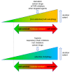
Context-dependent cell responses to bulk autophagy and selective mitophagy. Both autophagy and mitophagy can either promote or inhibit cell death in cancer cells. This response is highly dependent on the cell type, the trigger of auto-/mitophagy, its duration, and its extent. Accordingly, excessive autophagy can lead to cell death, however too little autophagy/mitophagy (marked by dotted line) can also be detrimental to the cells due to an impaired quality control/removal of harmful cellular material.
Figure 3
Figure 3
AT-101/gossypol as a trigger of lethal mitophagy. AT-101 causes mitochondrial damage including mitochondrial permeability transition pore (mPTP) opening, loss of membrane potentials, and decreased oxygen consumption. Damaged mitochondria are selectively degraded via mitophagy. In parallel, AT-101 induces the mitophagy receptors BNIP3 and BNIP3L/NIX, as well as the mitophagy-inducer HMOX1, which will further facilitate the extent of mitophagy. This excessive mitophagy finally leads to the demise of the cancer cell(s). This figure was created using Servier Medical Art templates, which are licensed under a Creative Commons Attribution 3.0 Unported License; https://smart.servier.com.

References

    1. Hengartner M.O. The biochemistry of apoptosis. Nature. 2000;407:770–776. doi: 10.1038/35037710. - DOI - PubMed
    1. Kroemer G., Martin S.J. Caspase-independent cell death. Nat. Med. 2005;11:725–730. doi: 10.1038/nm1263. - DOI - PubMed
    1. Galluzzi L., Vitale I., Aaronson S.A., Abrams J.M., Adam D., Agostinis P., Alnemri E.S., Altucci L., Amelio I., Andrews D.W., et al. Molecular mechanisms of cell death: Recommendations of the Nomenclature Committee on Cell Death 2018. Cell Death Differ. 2018;25:486–541. doi: 10.1038/s41418-017-0012-4. - DOI - PMC - PubMed
    1. Nikoletopoulou V., Markaki M., Palikaras K., Tavernarakis N. Crosstalk between apoptosis, necrosis and autophagy. Biochim. Biophys. Acta. 2013;1833:3448–3459. doi: 10.1016/j.bbamcr.2013.06.001. - DOI - PubMed
    1. Marino G., Niso-Santano M., Baehrecke E.H., Kroemer G. Self-consumption: The interplay of autophagy and apoptosis. Nat. Rev. Mol. Cell. Biol. 2014;15:81–94. doi: 10.1038/nrm3735. - DOI - PMC - PubMed

LinkOut - more resources