

Review
doi: 10.1016/j.path.2019.08.006.
Epub 2019 Sep 27.
Parathyroid Pathology
Affiliations
- PMID: 31672291
- PMCID: PMC7395581
- DOI: 10.1016/j.path.2019.08.006
Item in Clipboard
Review
Parathyroid Pathology
Surg Pathol Clin.
2019 Dec.
Abstract
Proliferative pathologic lesions of parathyroid glands encompass a spectrum of entities ranging from benign hyperplastic processes to malignant neoplasia. This review article outlines the pathophysiologic classification of parathyroid disorders and describes histologic, immunohistochemical, and molecular features that can be assessed to render accurate diagnoses.
Keywords: Parafibromin; Parathyroid; Parathyroid adenoma; Parathyroid carcinoma; Primary hyperplasia.
Copyright © 2019 Elsevier Inc. All rights reserved.
Conflict of interest statement
Conflict of interest: There are no conflicts of interest to report by any of the authors.
Figures
Normal parathyroid gland composed mainly of chief cells and adipocytes with thin fibrous septa dividing gland into lobules (hematoxylineosin [H&E], original magnification ×400).
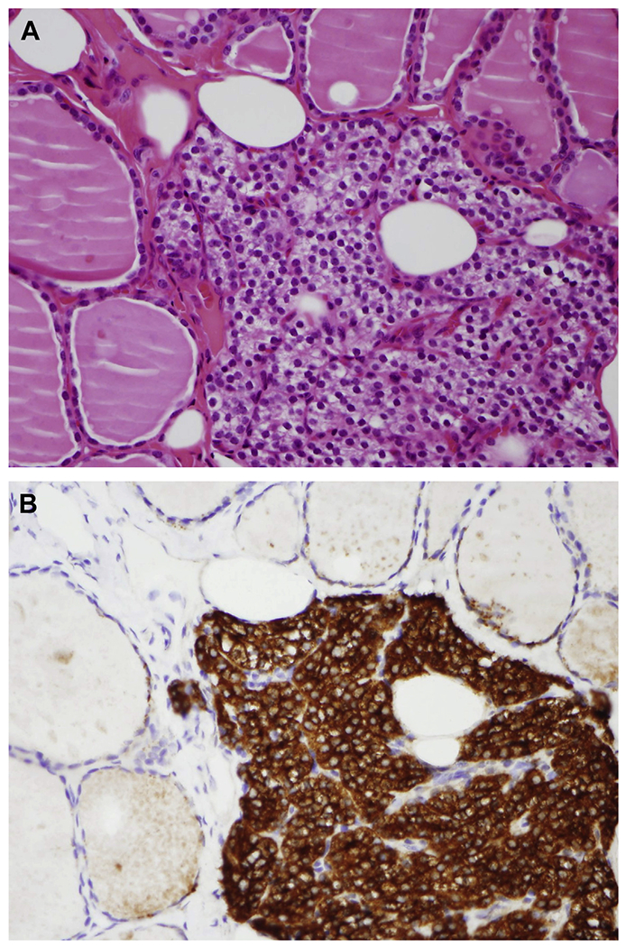
(A) Incidental finding of ectopic intrathyroidal parathyroid tissue (H&E, original magnification ×400). (B) Parathyroid hormone (PTH) by immunohistochemistry highlights parathyroid chief cells, allowing proper identification (PTH, original magnification ×400).
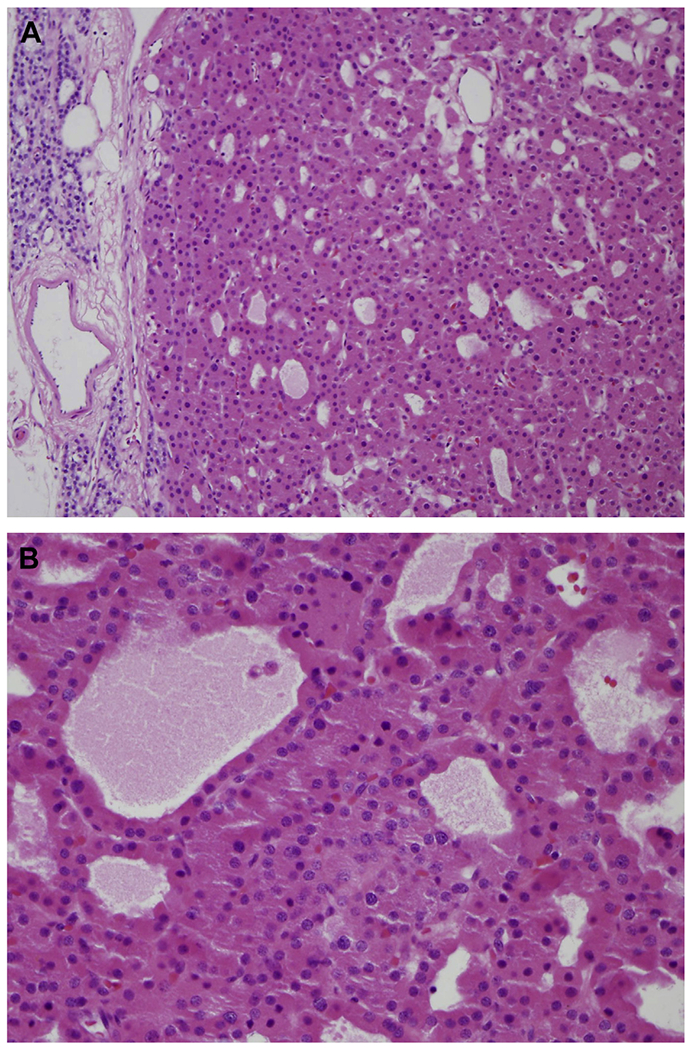
(A) Parathyroid hyperplasia is characterized by chief cell proliferation involving all 4 glands (H&E, original magnification ×200). (B) Nodules of oncocytic cells are common (H&E, original magnification ×400).
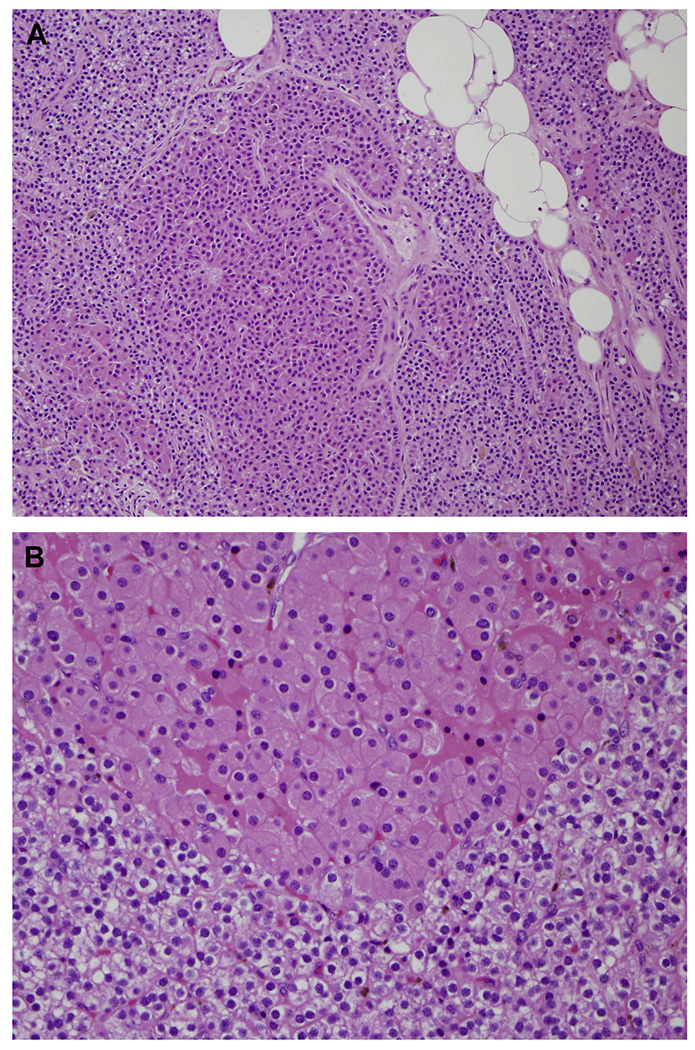
(A) Oncocytic PA with residual normocellular parathyroid tissue identified outside the adenoma capsule (H&E, original magnification ×200). (B) Microcystic or cystic architecture may be observed inside the adenoma (H&E, original magnification ×400).
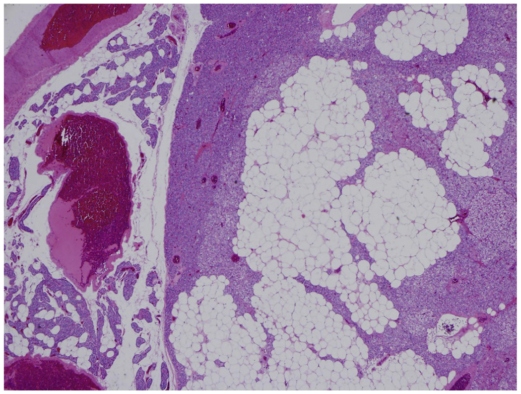
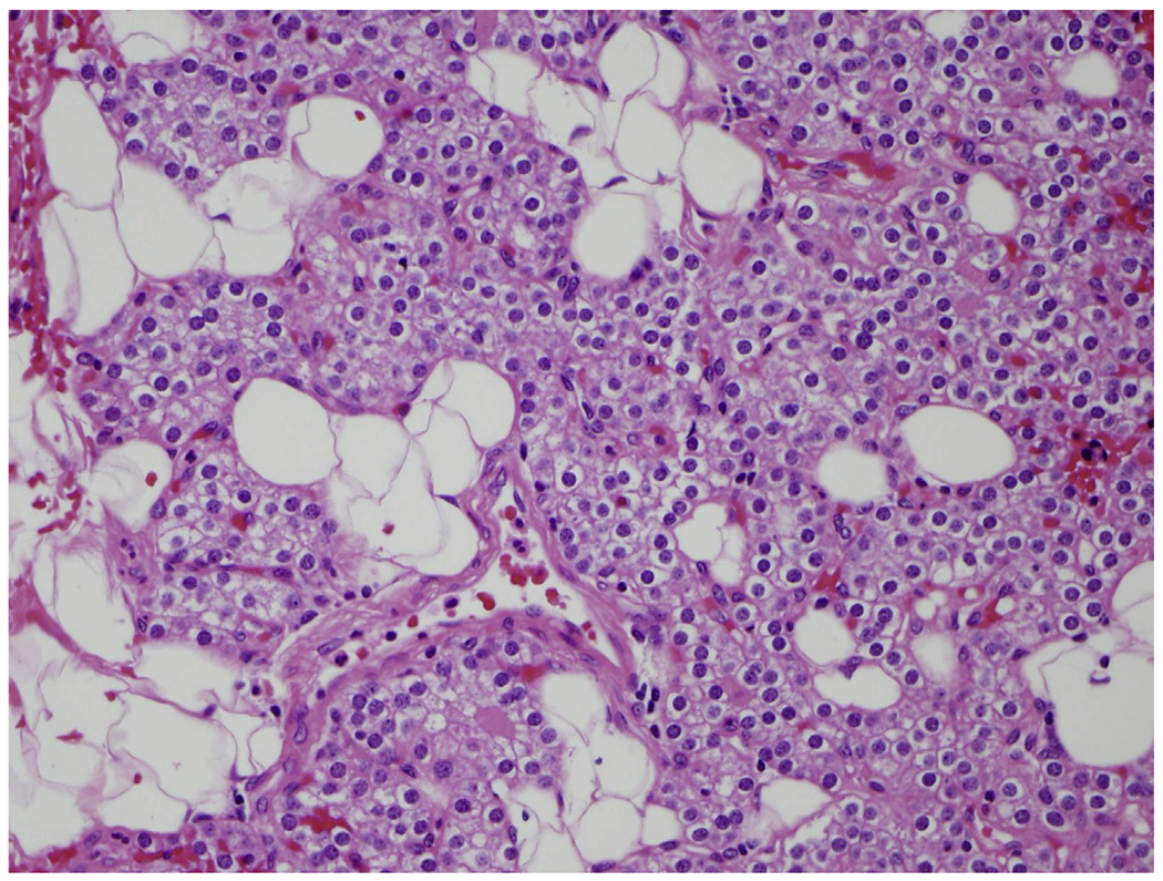
Lipoadenoma is a rare variant of PA composed of mature adipose tissue and chief cells (H&E, original magnification ×200).
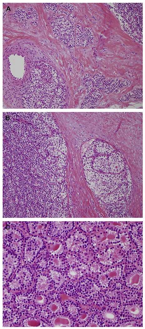
(A) Thick fibrotic capsule with parathyroid cells infiltrating surrounding soft tissue associated with (B) true vascular invasion are key features of PC (H&E, original magnification ×200). (C) Microcystic growth pattern in PC with unremarkable bland cytologic features (H&E, original magnification ×400).
References
-
- Mallik S, Aggarwal P, Singh I, et al. A study on development and morphogenesis of parathyroid gland in the developing human embryo. J Med Soc 2017;31(3):195–200.
-
- Grevellec A, Tucker AS. The pharyngeal pouches and clefts: development, evolution, structure and derivatives. Semin Cell Dev Biol 2010;21(3): 325–32. - PubMed
-
- Akerstrom G, Malmaeus J, Bergstrom R. Surgical anatomy of human parathyroid glands. Surgery 1984;95(1):14–21. - PubMed
-
- Fancy T, Gallagher D, Hornig JD. Surgical anatomy of the thyroid and parathyroid glands. Otolaryngol Clin North Am 2010;43(2):221–7. - PubMed
-
- Åkerström G, Rudberg C, Grimelius L, et al. Histologic parathyroid abnormalities in an autopsy series. Hum Pathol 1986;17(5):520–7. - PubMed
Publication types
MeSH terms
Grants and funding
LinkOut - more resources
Full Text Sources
