. 2019 Dec;40(12):2066-2072.
doi: 10.3174/ajnr.A6291. Epub 2019 Oct 31.

Angiographic Analysis of Natural Anastomoses between the Posterior and Anterior Cerebral Arteries in Moyamoya Disease and Syndrome

Affiliations

Angiographic Analysis of Natural Anastomoses between the Posterior and Anterior Cerebral Arteries in Moyamoya Disease and Syndrome

S Bonasia et al. AJNR Am J Neuroradiol. 2019 Dec.

Abstract

Background and purpose: Moyamoya disease is a chronic neurovascular steno-occlusive disease of the internal carotid artery and its main branches, associated with the development of compensatory vascular collaterals. Literature is lacking about the precise description of these compensatory vascular systems. Usually, the posterior circulation is less affected, and its vascular flow could compensate the hypoperfusion of the ICA territories. The aim of this study was to describe these natural connections between the posterior cerebral artery and the anterior cerebral artery necessary to compensate the lack of perfusion of the anterior cerebral artery territories in the Moyamoya population.

Materials and methods: All patients treated for Moyamoya disease from 2004 to 2018 in 4 neurosurgical centers with available cerebral digital subtraction angiography were included. Forty patients (80 hemispheres) with the diagnosis of Moyamoya disease were evaluated. The presence of anastomoses between the posterior cerebral artery and the anterior cerebral artery was found in 31 hemispheres (38.7%).

Results: Among these 31 hemispheres presenting with posterior cerebral artery-anterior cerebral artery anastomoses, the most frequently encountered collaterals were branches from the posterior callosal artery (20%) and the posterior choroidal arteries (20%). Another possible connection found was pio-pial anastomosis between cortical branches of the posterior cerebral artery and the anterior cerebral artery (15%). We also proposed a 4-grade classification based on the competence of these anastomoses to supply retrogradely the territories of the anterior cerebral artery.

Conclusions: We found 3 different types of anastomoses between the anterior and posterior circulations, with different abilities to compensate the anterior circulation. Their development depends on the perfusion needs of the territories of the anterior cerebral artery and can provide the retrograde refilling of the anterior cerebral artery branches.


Figures

Fig 1.
Fig 1.
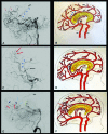
The 3 types of PCA-ACA collaterals are shown by angiograms with corresponding graphic illustrations. The blue triangles (A and B) indicate the anastomosis between the posterior pericallosal artery and the anterior pericallosal artery (type I). The red arrows indicate the contribution of pio-pial connections to the anastomosis. C and D, Type II collateral, between the medial posterior choroidal artery (MPChoA) and the anterior pericallosal artery (APA) (blue triangles). The MPChoA turns first anteriorly and then backward around the splenium of the corpus callosum to reach the APA. Pio-pial connections are also visible in the angiogram (red arrows). Type III collaterals are visible (E and F), where the dark blue arrows indicate pio-pial or leptomeningeal connections between cortical branches from the PCA (red arrows) and cortical branches from the ACA (light blue arrows). The green triangles show the adjunctive presence of posterior pericallosal artery (PPA)-APA connections. POA indicates parieto-occipital artery; CMA, calloso-marginal artery; BA, basilar artery.
Fig 2.
Fig 2.
Diagram representation of the distribution of Suzuki scores and PCA-ACA anastomosis depending on the age group.
Fig 3.
Fig 3.
A–D, The angiograms and graphic illustrations show the capacity to compensate the ACA territories by the PCA-ACA anastomoses in case of proximal ICA stenosis (black arrow), through a 4-grade classification. In grade I (A and B), the collaterals refill just the first part of the ACA, without seeing any cortical branch. The angiogram on the left shows that the contribution to the refilling is made by the posterior pericallosal artery (blue star) and by pio-pial connections (green star). C and D, The retrograde flow reaches a larger part of the ACA, also highlighting a cortical branch of the ACA (grade II). A double contribution from the pio-pial connection (green star) and medial posterior choroidal artery (blue star) is visible in the angiogram. Grade III (E and F) consists of the retrograde refilling of 2 or 3 ACA branches (red arrows in E), which, in this case, are supported by a medial posterior choroidal artery–anterior pericallosal artery anastomosis. In grade IV, almost all the ACA territory is retrogradely refilled (G and H). The green and blue stars indicate the 2 main connections that compensate the ACA territories hypoperfusion.

Comment in

  • Reply.
    Bonasia S, Robert T. Bonasia S, et al. AJNR Am J Neuroradiol. 2020 Jun;41(6):E42. doi: 10.3174/ajnr.A6503. Epub 2020 Apr 2. AJNR Am J Neuroradiol. 2020. PMID: 32241768 Free PMC article. No abstract available.
  • The Significance of Natural Anastomoses among Intracranial Vessels in Moyamoya Disease.
    Yu J, Zhang J, Chen J. Yu J, et al. AJNR Am J Neuroradiol. 2020 Jun;41(6):E41. doi: 10.3174/ajnr.A6502. Epub 2020 Apr 2. AJNR Am J Neuroradiol. 2020. PMID: 32241773 Free PMC article. No abstract available.

References

    1. Bang OY, Fujimura M, Kim SK.. The pathophysiology of Moyamoya disease: an update. J Stroke 2016;18:12–20 10.5853/jos.2015.01760 - DOI - PMC - PubMed
    1. Burke GM, Burke AM, Sherma AK.. Moyamoya disease: a summary. Neurosurg Focus 2009;26:E11 10.3171/2009.1.FOCUS08310 - DOI - PubMed
    1. Scott RM, Smith ER.. Moyamoya disease and moyamoya syndrome. N Engl J Med 2009;360:1226–37 10.1056/NEJMra0804622 - DOI - PubMed
    1. Kim JS. Moyamoya disease: epidemiology, clinical features, and diagnosis. J Stroke 2016;18:2–11 10.5853/jos.2015.01627 - DOI - PMC - PubMed
    1. Kwag HJ, Jeong DW, Lee SH, et al. . Intracranial hemodynamic changes during adult moyamoya disease progression. J Clin Neurol 2008;4:67–74 10.3988/jcn.2008.4.2.67 - DOI - PMC - PubMed

LinkOut - more resources