. 2020 Jan 1;80(1):102-115.
doi: 10.1158/0008-5472.CAN-19-1957. Epub 2019 Oct 31.

Targeting Novel Sodium Iodide Symporter Interactors ADP-Ribosylation Factor 4 and Valosin-Containing Protein Enhances Radioiodine Uptake

Affiliations

Targeting Novel Sodium Iodide Symporter Interactors ADP-Ribosylation Factor 4 and Valosin-Containing Protein Enhances Radioiodine Uptake

Alice Fletcher et al. Cancer Res. .

Abstract

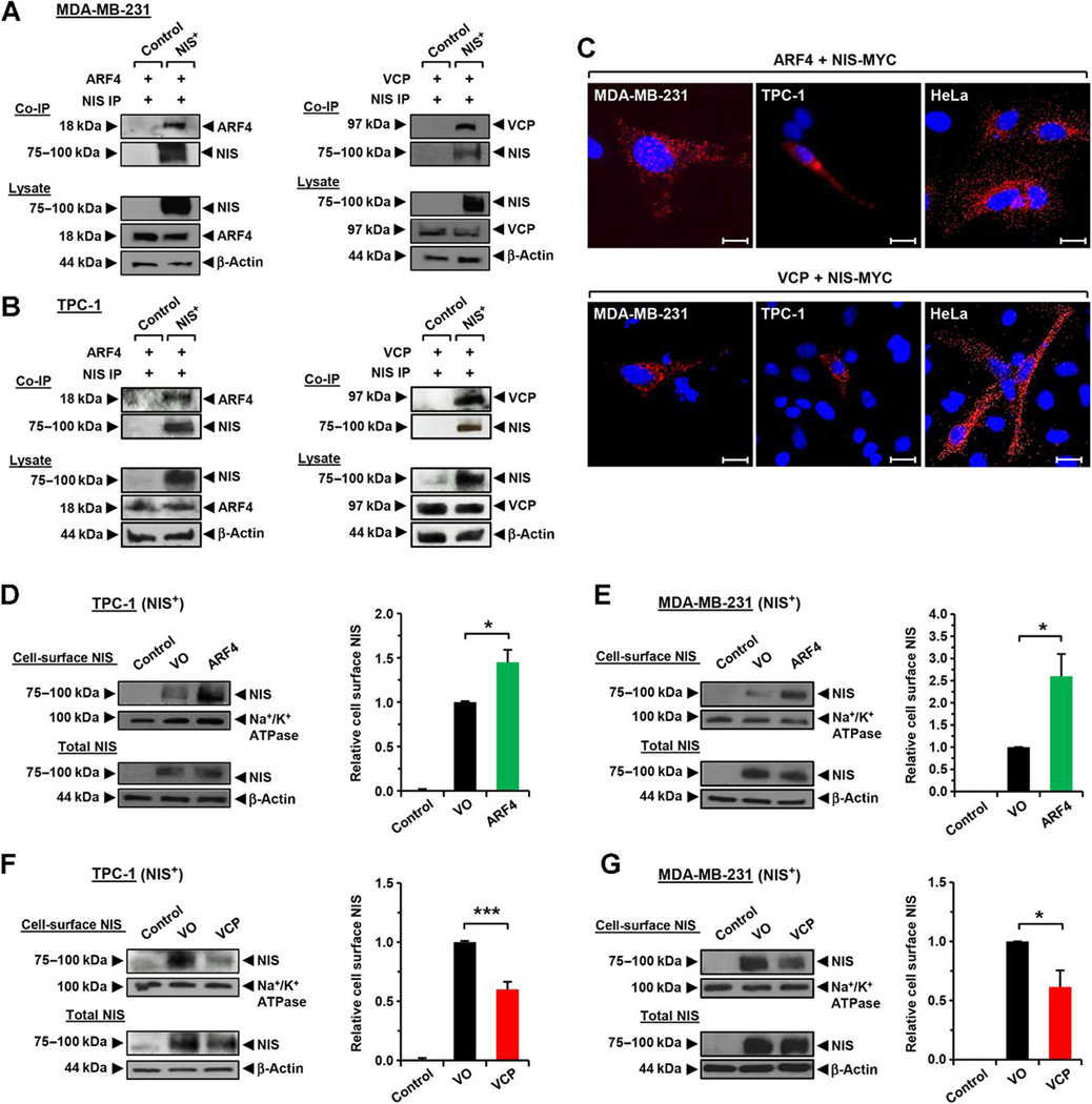
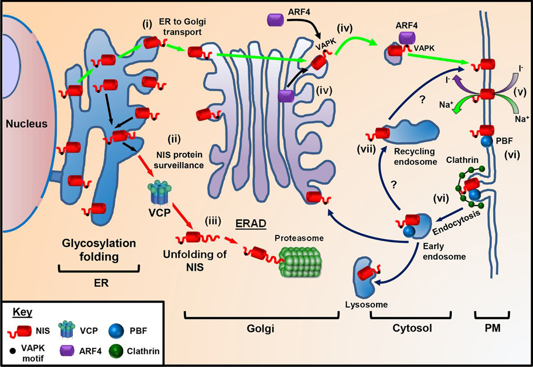
The sodium iodide symporter (NIS) is required for iodide uptake, which facilitates thyroid hormone biosynthesis. NIS has been exploited for over 75 years in ablative radioiodine (RAI) treatment of thyroid cancer, where its ability to transport radioisotopes depends on its localization to the plasma membrane. The advent of NIS-based in vivo imaging and theranostic strategies in other malignancies and disease modalities has recently increased the clinical importance of NIS. However, NIS trafficking remains ill-defined. Here, we used tandem mass spectrometry followed by coimmunoprecipitation and proximity ligation assays to identify and validate two key nodes-ADP-ribosylation factor 4 (ARF4) and valosin-containing protein (VCP)-controlling NIS trafficking. Using cell-surface biotinylation assays and highly inclined and laminated optical sheet microscopy, we demonstrated that ARF4 enhanced NIS vesicular trafficking from the Golgi to the plasma membrane, whereas VCP-a principal component of endoplasmic reticulum (ER)-associated degradation-governed NIS proteolysis. Gene expression analysis indicated VCP expression was particularly induced in aggressive thyroid cancers and in patients who had poorer outcomes following RAI treatment. Two repurposed FDA-approved VCP inhibitors abrogated VCP-mediated repression of NIS function, resulting in significantly increased NIS at the cell-surface and markedly increased RAI uptake in mouse and human thyroid models. Collectively, these discoveries delineate NIS trafficking and highlight the new possibility of systemically enhancing RAI therapy in patients using FDA-approved drugs. SIGNIFICANCE: These findings show that ARF4 and VCP are involved in NIS trafficking to the plasma membrane and highlight the possible therapeutic role of VCP inhibitors in enhancing radioiodine effectiveness in radioiodine-refractory thyroid cancer.


Conflict of interest statement

Disclosure of Potential Conflicts of Interest

No potential conflicts of interest were disclosed.

Figures

Figure 1.
Figure 1.
Identification of ARF4 and VCP as regulators of NIS activity. A, Western blot analysis of whole-cell lysate and PM fraction in MDA-MB-231 (NIS+) cells used in MS/MS. B, Top hits for putative NIS interactors identified by MS/MS (peptides ≥ 6). C and D, Western blot analysis and RAI uptake of MDA-MB-231 (NIS+) cells transfected with esiRNA specific for indicated NIS interactors. NT, nontransfected cells. E and F, Western blot analysis and RAI uptake in MDA-MB-231 (NIS+) cells, TPC-1 (NIS+) cells, and human primary thyrocytes transfected with ARF4 siRNA (E) or ARF4 (F). G and H, Same as E and F, but cells transfected with VCP siRNA (G) or VCP (H). NS, not significant; *, P < 0.05; **, P < 0.01; ***, P < 0.001.
Figure 2.
Figure 2.
ARF4 and VCP bind NIS in vitro and modulate PM NIS. A, Co-IP assays in MDA-MB-231 (NIS+) cells showing specific interaction between NIS and ARF4 (left) or VCP (right). B, Same as A, but in TPC-1 (NIS+) cells. C, PLA demonstrating specific interaction between NIS-MYC and ARF4 (top) or VCP (bottom) in MDA-MB-231, TPC-1, and HeLa cells. Red fluorescent spots, specific interactions. Blue, DAPI nuclear staining. Magnification, ×100. Scale bars, 10 μm. D and E, Western blot analysis of NIS protein levels at the PM relative to Na+/K+ ATPase following the CSBA in TPC-1 (NIS+) cells (D) and MDA-MB-231 (NIS+) cells (E) after ARF4 transfection. F and G, Same as D and E, but after VCP transfection. *, P < 0.05; ***, P < 0.001.
Figure 3.
Figure 3.
Involvement of ARF4 in trafficking NIS at the PM. A and B, HILO microscopy images demonstrating trafficking of ARF4-dsRED (red), NIS-GFP (green), and colocalization (yellow) to the PM in HeLa cells. Video capture times (hr:min:sec) are indicated. PM regions in framed areas are magnified in bottom right, which highlight the movement of ARF4 and NIS (white and orange arrowheads; see bottom). Scale bars, 10 μm. C, Representative images of NIS-GFP movement patterns tracked using ImageJ software. Scale bars, 10 μm. D, Box-whisker plot of velocity (μm/sec) and distance traveled (μm) of NIS-GFP in HeLa cells transfected with ARF4 (n = 475) or VO (n = 339). E, RAI uptake in TPC-1 (NIS+) and MDA-MB-231 (NIS+) cells transfected with ARF4 and treated with dynasore for 1 hour prior to addition of 125I. F, Western blot analysis of NIS expression levels in TPC-1 (NIS+) cells as described in E. G, RAI uptake in TPC-1 cells transfected with ARF4, as well as WT NIS, 574AAAK577-mutant NIS, or 475ALAS478-mutant NIS. H, Representative co-IP assay for ARF4 with WT or mutant NIS. NS, not significant; *, P < 0.05; ***, P < 0.001.
Figure 4.
Figure 4.
Inhibition of VCP enhances NIS function. A, RAI uptake of MDA-MB-231 (NIS+) and TPC-1 (NIS+) cells treated with ES-1 for 24 hours. B, Same as A, but cells treated with NMS-873. C, Western blot analysis following ES-1 or NMS-873 treatment in TPC-1 (NIS+) cells. D, PLA showing specific interaction (red fluorescent spots) between NIS-MYC and VCP in TPC-1 cells treated with ES-1. Blue, DAPI nuclear stain. Magnification, ×100. Scale bars, 10 μm. E, Co-IP assays demonstrating interaction of VCP and NIS in TPC-1 (NIS+) cells treated with ES-1 or NMS-873. F and G, RAI uptake and relative NIS protein levels in parental TPC-1 cells treated with ES-1, NMS-873, or DMSO. H, Time course of RAI uptake (top) and relative NIS protein levels (bottom) in TPC-1 (NIS+) cells treated with 2.5 μmol/L ES-1. I, Same as H, but cells treated with 5 μmol/L NMS-873. J, RAI uptake and Western blot analysis in TPC-1 (NIS+) cells transfected with VCP or Scr siRNA, then treated with ES-1. K, RAI uptake and Western blot analysis in MDA-MB-231 (NIS+) cells, TPC-1 (NIS+) cells, and human primary thyrocytes transfected with WT VCP or QQ VCP mutant. NS, not significant; *, P < 0.05; **, P < 0.01; ***, P < 0.001.
Figure 5.
Figure 5.
VCP inhibitors increase PM NIS expression and function. A, TPC-1 (NIS+) cells treated with VCP inhibitors (ebastine, clotrimazole, and astemizole). B and C, Western blot (B) and RAI uptake (C) analyses of VCP expression following VCP-siRNA depletion and treatment with 0.5 μmol/L ebastine (top), 0.25 μmol/L clotrimazole (middle), 0.25 μmol/L astemizole (bottom) in TPC-1 (NIS+) cells. D and E, Cell-surface biotinylation assay analysis of NIS protein levels at the PM relative to Na+/K+ ATPase in the TPC-1 (NIS+) cell line after treatment with 0.5 μmol/L ebastine, 0.25 μmol/L clotrimazole, or 0.25 μmol/L astemizole. F, Western blot analysis of VCP expression in TPC-1 (NIS+) cells following treatment with 0.5 μmol/L ebastine, 0.25 μmol/L clotrimazole, or 0.25 μmol/L astemizole. G, Schematic indicating protocol for obtaining mouse thyrocytes. H and I, RAI uptake (H) and Western blot analysis (I) in mouse thyrocytes treated with 0.5 μmol/L ebastine (n = 8) or 0.25 μmol/L clotrimazole (n = 8). J, RAI uptake in human thyrocytes treated as described with ebastine or clotrimazole at varying of doses of VCP inhibitors due to previous evidence of variability in VCP inhibitor sensitivity (50). *, P < 0.05; **, P < 0.01.
Figure 6.
Figure 6.
VCP and ARF4 expression is associated with poorer survival and response to RAI. A, ARF4 (left) and VCP (right) expression in normal thyroid and PTC in the THCA TCGA data set. B, VCP expression in normal thyroid (n = 12), PDTC (n = 17), and ATC (n = 20). C, ARF4 (left) and VCP (right) expression in PTC with BRAF-like (n = 272) or RAS-like genetic signatures (n = 119). D, ARF4 expression in PTC with indicated genetic alterations. E, Frequency (%) of indicated genetic alterations in PTC with low (Q1Q2) versus high ARF4 (Q3Q4) expression. F and G, Same as D and E, but for VCP expression. H–J, DFS for THCA with high (Q3Q4; >12.71) versus low (Q1Q2; <12.71) VCP expression for the entire PTC cohort (H), RAI-treated patients (I) and non–RAI-treated patients (J). K, Hazard ratios ±95% CI for patients stratified on median VCP and ARF4 tumoral expression in THCA with the indicated treatment and genetic signature or alteration. L and M, DFS for THCA with high (Q3Q4; >11.84) versus low (Q1Q2; <11.84) ARF4 expression for RAI-treated PTC patients (L) and non–RAI-treated PTC patients (M). NS, not significant; *, P < 0.05; **, P < 0.01; ***, P < 0.001.
Figure 7.
Figure 7.
Putative model of NIS trafficking. NIS maintains a delicate balance between protein synthesis, folding, assembly, trafficking, and degradation. (i) We propose NIS is glycosylated in the ER and upon correct folding transported to the Golgi. (ii) Protein surveillance pathways exist that target NIS for ERAD. As VCP does not require ATPase activity to inhibit NIS function, it is likely VCP acts to unfold NIS (iii) prior to proteasomal degradation. (iv) ARF4 recognizes the VAPK motif in the NIS C-terminus and promotes vesicular trafficking to the PM, where NIS is active (v). (vi) PBF has a YARF endocytosis motif and acts to bind and internalize NIS away from the PM in a clathrin-dependent process. (vii) Although inhibition of recycling by dynasore suggests that ARF4 shuttles NIS to the PM, other proteins must promote recycling of NIS to the PM as with most PM transporters.

References

    1. Haugen BR, Alexander EK, Bible KC, Doherty GM, Mandel SJ, Nikiforov YE, et al. 2015 American thyroid association management guidelines for adult patients with thyroid nodules and differentiated thyroid cancer: the American Thyroid Association guidelines task force on thyroid nodules and differentiated thyroid cancer. Thyroid 2016;26:1–133. - PMC - PubMed
    1. Schlumberger M, Brose M, Elisei R, Leboulleux S, Luster M, Pitoia F, et al. Definition and management of radioactive iodine-refractory differentiated thyroid cancer. Lancet Diabetes Endocrinol 2014;2:356–8. - PubMed
    1. Spitzweg C, Bible KC, Hofbauer LC, Morris JC. Advanced radioiodine-refractory differentiated thyroid cancer: the sodium iodide symporter and other emerging therapeutic targets. Lancet Diabetes Endocrinol 2014;10:830–42. - PubMed
    1. La Vecchia C, Malvezzi M, Bosetti C, Garavello W, Bertuccio P, Levi F, et al. Thyroid cancer mortality and incidence: a global overview. Int J Cancer 2015; 136:2187–95. - PubMed
    1. Dai G, Levy O, Carrasco N. Cloning and characterization of the thyroid iodide transporter. Nature 1996;379:458–60. - PubMed

Publication types

MeSH terms