. 2019 Nov 1;4(21):e129224.
doi: 10.1172/jci.insight.129224.

Progenitor cell combination normalizes retinal vascular development in the oxygen-induced retinopathy (OIR) model

Affiliations

Progenitor cell combination normalizes retinal vascular development in the oxygen-induced retinopathy (OIR) model

Sergio Li Calzi et al. JCI Insight. .

Abstract

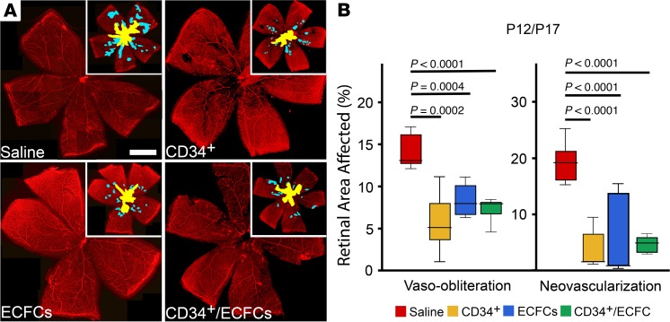
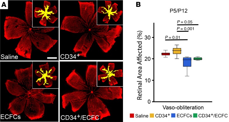
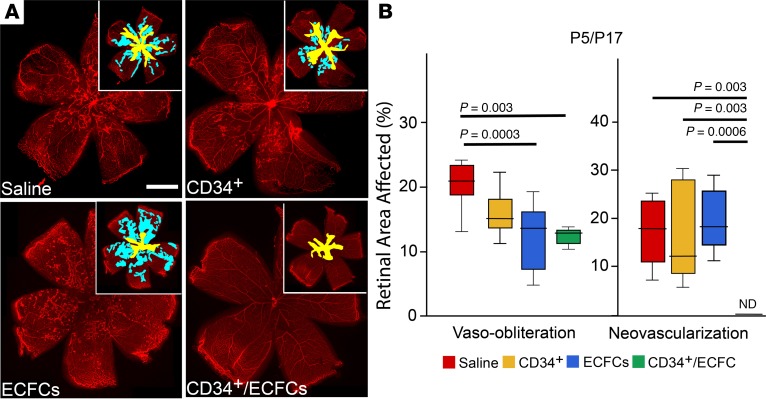
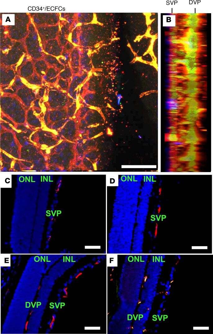
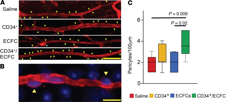
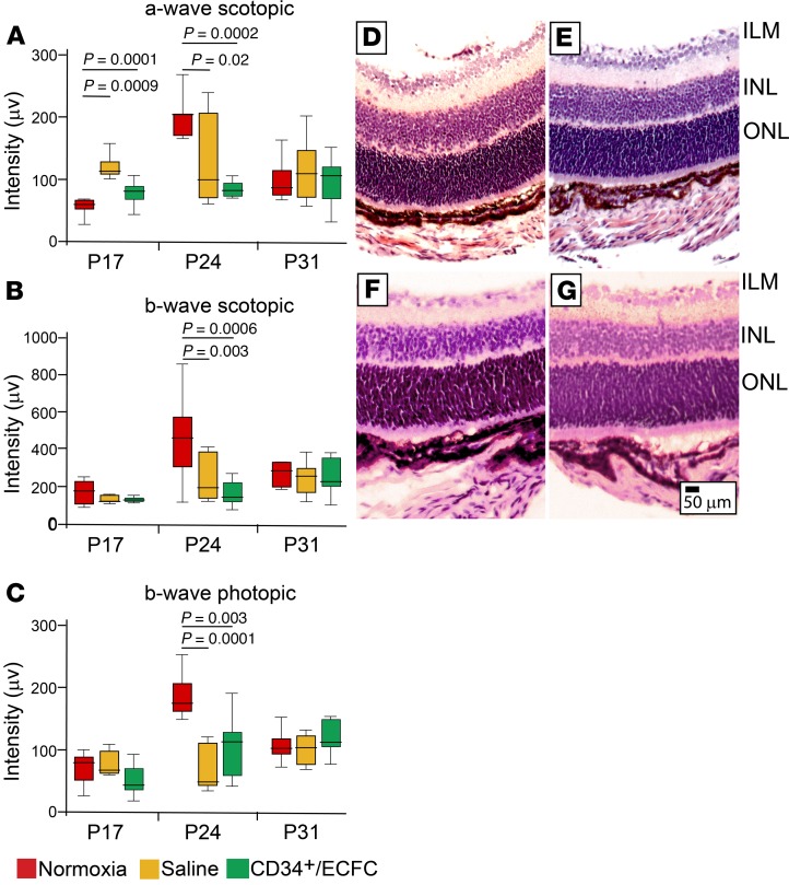
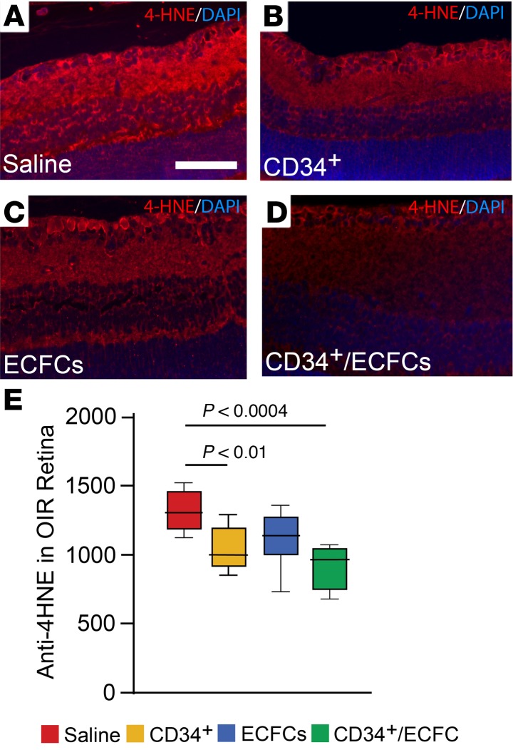
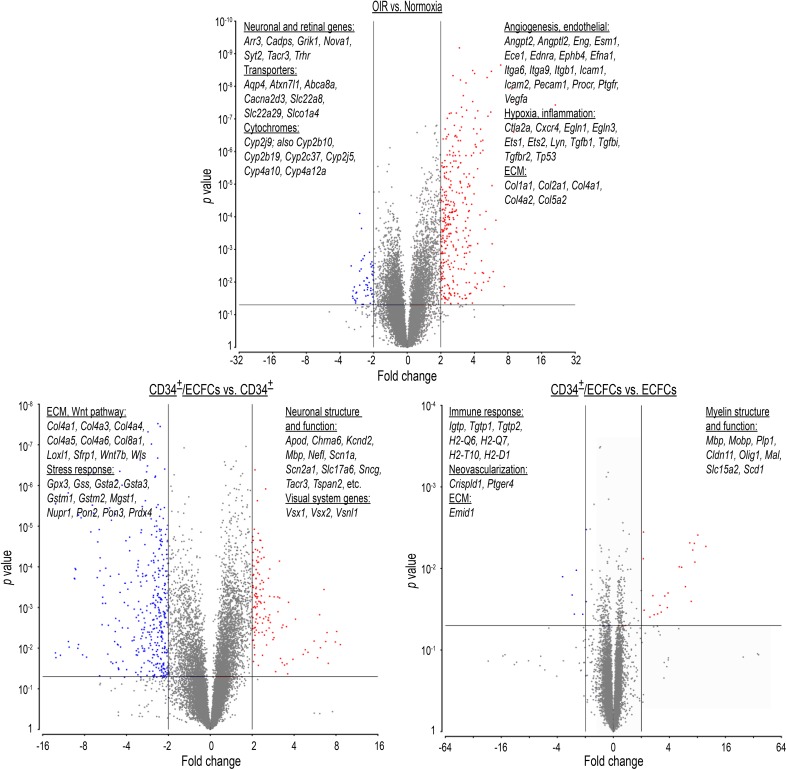
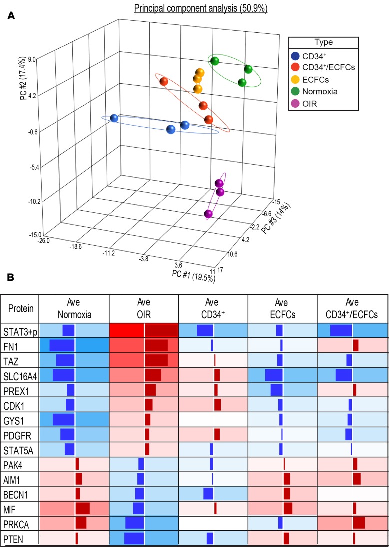
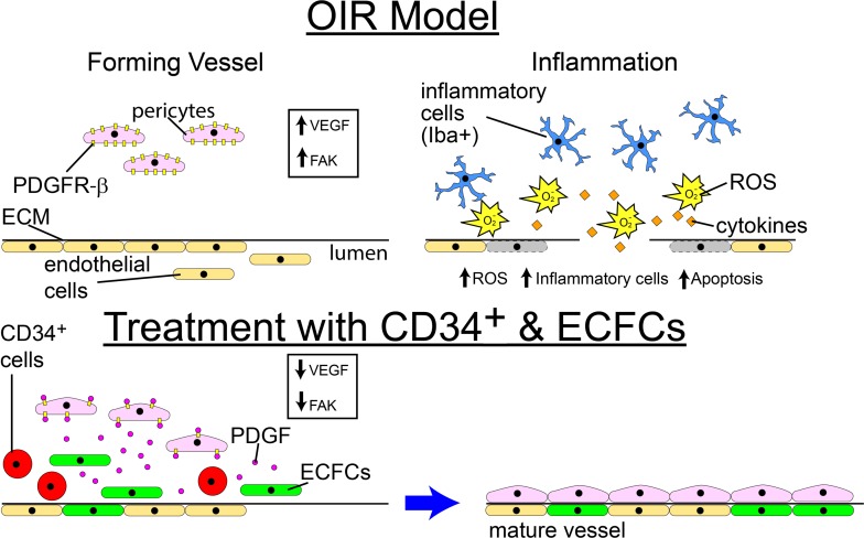
Retinopathy of prematurity (ROP) is a disorder of the developing retina of preterm infants. ROP can lead to blindness because of abnormal angiogenesis that is the result of suspended vascular development and vaso-obliteration leading to severe retinal stress and hypoxia. We tested the hypothesis that the use of the human progenitor cell combination, bone marrow-derived CD34+ cells and vascular wall-derived endothelial colony-forming cells (ECFCs), would synergistically protect the developing retinal vasculature in a mouse model of ROP, called oxygen-induced retinopathy (OIR). CD34+ cells alone, ECFCs alone, or the combination thereof were injected intravitreally at either P5 or P12 and pups were euthanized at P17. Retinas from OIR mice injected with ECFCs or the combined treatment revealed formation of the deep vascular plexus (DVP) while still in hyperoxia, with normal-appearing connections between the superficial vascular plexus (SVP) and the DVP. In addition, the combination of cells completely prevented aberrant retinal neovascularization and was more effective anatomically and functionally at rescuing the ischemia phenotype than either cell type alone. We show that the beneficial effects of the cell combination are the result of their ability to orchestrate an acceleration of vascular development and more rapid ensheathment of pericytes on the developing vessels. Lastly, our proteomic and transcriptomic data sets reveal pathways altered by the dual cell therapy, including many involved in neuroretinal maintenance, and principal component analysis (PCA) showed that cell therapy restored OIR retinas to a state that was closely associated with age-matched normal retinas. Together, these data herein support the use of dual cell therapy as a promising preventive treatment for the development of ROP in premature infants.

Keywords: Adult stem cells; Angiogenesis; Stem cells; hypoxia.


Conflict of interest statement

Conflict of interest: The authors have declared that no conflict of interest exists.

Figures

Figure 1
Figure 1. Reparative responses in the retina following injection of CD34+ cells, ECFCs, or the combination of both cell types at P12 and euthanized at P17.
(A) Flat-mounted retinas from OIR pups injected on P12 and euthanized on P17, stained for collagen IV to visualize the retinal vessels. Insets: areas of vaso-obliteration (yellow) and neovascularization (blue) are shown. (B) Summary of quantification of P12/P17 vaso-obliteration and neovascularization areas. Scale bar: 100 μm. All data were assessed using 1-way ANOVA. When the results were significant, we determined which means differed from each other using Tukey’s multiple-comparisons test. Values are expressed as percentage of total retina ± SEM, n = 10–12 mice for each data set. Only significant comparisons are shown in the figures.
Figure 2
Figure 2. Reparative responses in the retina following injection of CD34+ cells, ECFCs, or the combination of both cell types at P5 and euthanized at P12.
(A) Flat-mounted retinas from OIR pups injected on P5 and euthanized on P12 stained for collagen IV to visualize the retinal vessels. Insets: areas of vaso-obliteration (yellow) are shown. (B) Summary of quantification of P5/P12 vaso-obliteration areas. In this group, there is no neovascularization because mice were euthanized before the beginning of the neovascular phase of the model. Scale bar: 100 μm. All data were assessed using 1-way ANOVA. When the results were significant, we determined which means differed from each other using Tukey’s multiple-comparisons test. Values are expressed as percentage of total retina ± SEM, n = 10–12 mice for each data set. Only significant comparisons are shown in the figures.
Figure 3
Figure 3. Reparative responses in the retina following injection of CD34+ cells, ECFCs, or the combination of both cell types at P5 and euthanized at P17.
(A) Flat-mounted retinas from OIR pups injected on P5 and euthanized on P17 stained for collagen IV to visualize the retinal vessels. Insets: areas of vaso-obliteration (yellow) and neovascularization (blue) are shown. (B) Summary of quantification of P5/P17 vaso-obliteration and neovascularization areas. Scale bar: 100 μm. All data were assessed using 1-way ANOVA. When the results were significant, we determined which means differed from each other using Tukey’s multiple-comparisons test. Values are expressed as percentage of total retina ± SEM, n = 10–12 mice for each data set. Only significant comparisons are shown in the figures.
Figure 4
Figure 4. Injection of ECFCs on P5 stimulates the development of the deep vascular plexus in the OIR pups prior to return to normoxic conditions on P12.
(A) Representative projection of Z-series confocal images from flat-mounted retinas from OIR pups injected with the combination of CD34+ cells and ECFCs on P5 and euthanized on P12. Retinas were stained for collagen IV (red), histone H1 (blue), and GFP (green). (B) Lateral view of A. (CF) Paraffin cross sections of OIR retinas from pups treated with saline (C), CD34+ cells (D), ECFCs (E), or the combination of ECFCs and CD34+ cells (F). Sections were stained with mouse antibody to CD31 (red) and DAPI (blue). DVP, deep vascular plexus; INL, inner nuclear layer; ONL, outer nuclear layer; SVP, superficial vascular plexus. Scale bar (all panels): 50 μm.
Figure 5
Figure 5. The combination of ECFCs and CD34+ cells increases pericyte ensheathment in the OIR model.
(A) Representative fluorescence images showing retinal blood vessels stained for collagen IV (red). Pericytes (yellow arrowheads) are easily recognizable as they appear as areas that are “bulging out” of the blood vessels. Blue arrowhead: small vascular tuft. Scale bar: 50 μm. (B) Representative fluorescence high magnification image showing that collagen IV (red) allows the identification of pericytes (yellow arrowheads). Scale bar: 10 μm. Blue stain is DAPI. (C) Quantification of pericytes in retina blood vessels of OIR mouse pups injected at P5 and analyzed at P17. Values are expressed as average/100 μm ± SEM. All data were assessed using 1-way ANOVA. When the results were significant, we determined which means differed from each other using Tukey’s multiple-comparisons test. Only significant comparisons are shown in the figures. Vehicle, n = 9; CD34, n = 12; ECFC, n = 7; ECFC/CD34, n = 6.
Figure 6
Figure 6. Combined treatment with CD34+ cells and ECFCs has no effect on neural retinal function in OIR mice.
(A) Scotopic a- and (B) scotopic b-waves and (C) photopic b-wave intensities were performed at P17, P24, and P31 in control (normoxia) mice, OIR mice injected with saline, or OIR mice injected with CD34+/ECFCs at P5. At P17, the combination of CD34+ cells and ECFCs resulted in a normalization of the a-wave. At P24, normoxia (control) mice showed significantly increased intensities of scotopic a- and b-waves and photopic b-wave compared with saline-injected or CD34+/ECFCs-injected OIR mice. Intensity measurements returned to baseline by P31 in all 3 experimental cohorts. All data were assessed using 1-way ANOVA. When the results were significant, we determined which means differed from each other using Tukey’s multiple-comparisons test. Only significant comparisons are shown in the figures. (DG) H&E-stained ocular sections. (D) Saline-injected pups. (E) CD34+ cell–injected pups. (F) ECFC-injected pups. (G) CD34+/ECFC-injected pups. Scale bar: 50 μm. ILM, inner limiting membrane; INL, inner nuclear layer; ONL, outer nuclear layer.
Figure 7
Figure 7. Combined treatment with CD34+ cells and ECFCs reduces oxidative stress.
OIR mice were injected at P5 with either CD34+ cells, ECFCs, or a combination of the 2 cells. (AD) 4-Hydroxynonenal (4-HNE) detection in the retinas. (A) Saline-injected pups. (B) CD34+ cell–injected pups. (C) ECFC-injected pups. (D) CD34+/ECFC-injected pups. CD34+ cell treatment and the cell combination have the strongest effect on reduction of retinal oxidative stress. (E) Summary of quantification of all the tissue sections analyzed. All data were assessed using 1-way ANOVA. When the results were significant, we determined which means differed from each other using Tukey’s multiple-comparisons test. Only significant comparisons are shown in the figures.
Figure 8
Figure 8. Microarray analyses on whole retinal mRNA.
Validation study: P17 OIR retinas were compared with age-matched normoxia control retinas. A total of 894 genes were identified with significantly altered expression. A total of 640 genes were upregulated and included pathways involved in (i) angiogenesis, (ii) inflammation, (iii) extracellular matrix (ECM) remodeling, and (iv) endothelial-specific pathways, while 254 were downregulated and included (i) transporters and channels, (ii) neuronal-specific pathways, and (iii) cytochrome P450 genes.
Figure 9
Figure 9. Retinal protein expression differences in OIR mice treated with dual cell therapy.
(A) Principal component analysis (PCA) from proteomic data sets demonstrates closer association of cell therapy–treated OIR retinas with control (normoxia) retinas. (B) Proteomic analysis of contralateral OIR retinas from the transcriptomic data set treated with CD34+, ECFCs, or CD34+/ECFCs. Shown are the proteins that were significantly changed in the OIR model (P < 0.05), and whose expression was reversed by either cell treatment. Some of these proteins are discussed in the text (STAT3, PDGFR, STAT5A, AIM1, BECN1, PTEN, PRKCA, MIF, PAK4). Data represent NormLog2-Median Centered expression data, averaged (Ave) per set. Red, upregulated; blue, downregulated. In each case, the intensity of the color and the length of the column is proportional to the level of expression. Stat3+p, phosphorylated STAT3.
Figure 10
Figure 10. Schematic showing proposed mechanism for the beneficial effect of CD34+ cell and ECFC combination.
The combination of ECFCs and CD34+ cells may represent an improved strategy compared with either cell alone when used for prevention of ROP by reducing oxidative injury and inflammation, promoting pericyte ensheathment, and targeting critical proteins such as PDGF, FAK, STAT-pY705, PTEN, and PKCα-pS657.

References

    1. Alon T, Hemo I, Itin A, Pe’er J, Stone J, Keshet E. Vascular endothelial growth factor acts as a survival factor for newly formed retinal vessels and has implications for retinopathy of prematurity. Nat Med. 1995;1(10):1024–1028. doi: 10.1038/nm1095-1024. - DOI - PubMed
    1. Hartnett ME, Penn JS. Mechanisms and management of retinopathy of prematurity. N Engl J Med. 2012;367(26):2515–2526. doi: 10.1056/NEJMra1208129. - DOI - PMC - PubMed
    1. Hellström A, Smith LE, Dammann O. Retinopathy of prematurity. Lancet. 2013;382(9902):1445–1457. doi: 10.1016/S0140-6736(13)60178-6. - DOI - PMC - PubMed
    1. Selvam S, Kumar T, Fruttiger M. Retinal vasculature development in health and disease. Prog Retin Eye Res. 2018;63:1–19. doi: 10.1016/j.preteyeres.2017.11.001. - DOI - PubMed
    1. Connor KM, et al. Quantification of oxygen-induced retinopathy in the mouse: a model of vessel loss, vessel regrowth and pathological angiogenesis. Nat Protoc. 2009;4(11):1565–1573. doi: 10.1038/nprot.2009.187. - DOI - PMC - PubMed

Publication types

MeSH terms