. 2019 Oct 31;9(1):15778.
doi: 10.1038/s41598-019-52215-4.

Molecular signatures of retinal ganglion cells revealed through single cell profiling

Affiliations

Molecular signatures of retinal ganglion cells revealed through single cell profiling

Lauren A Laboissonniere et al. Sci Rep. .

Abstract

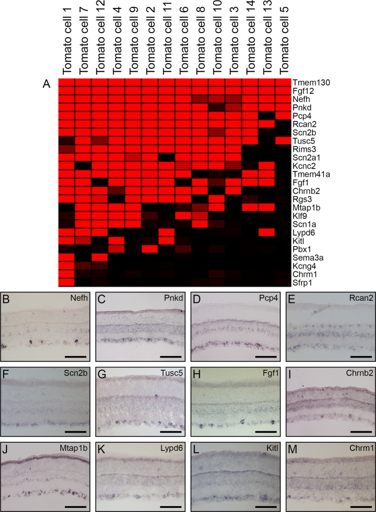
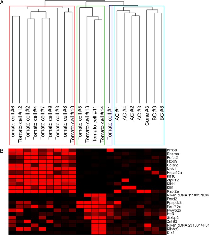
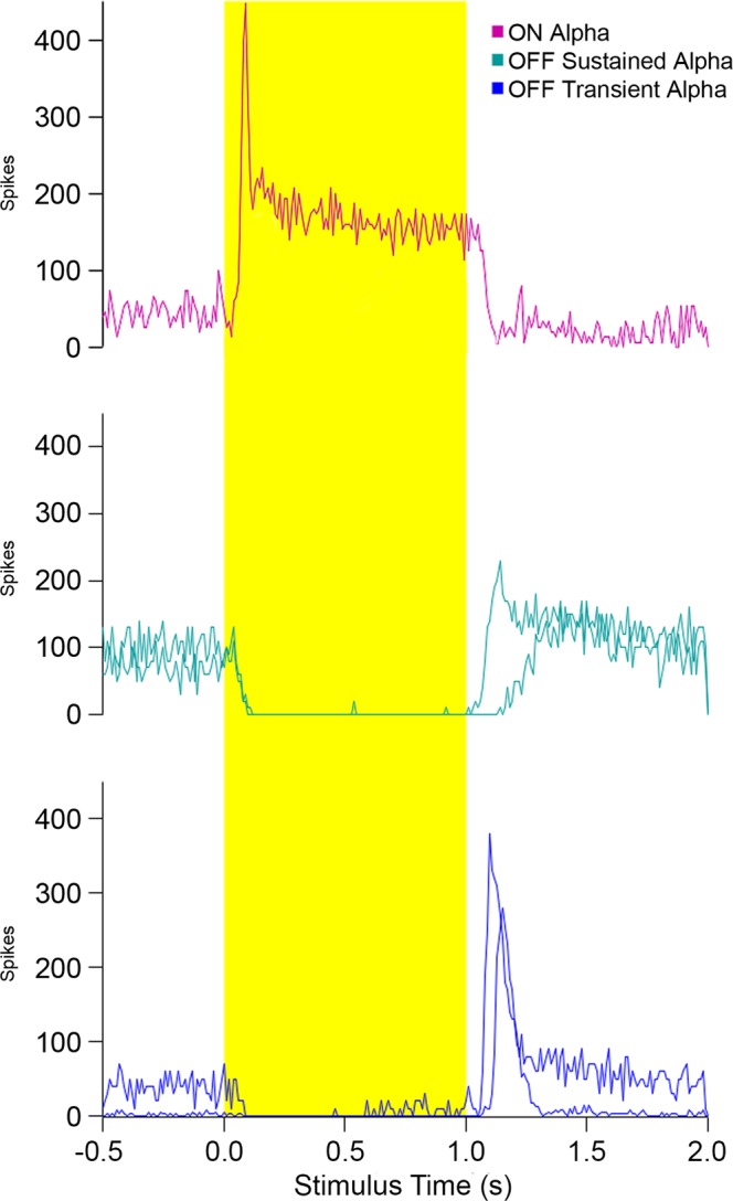
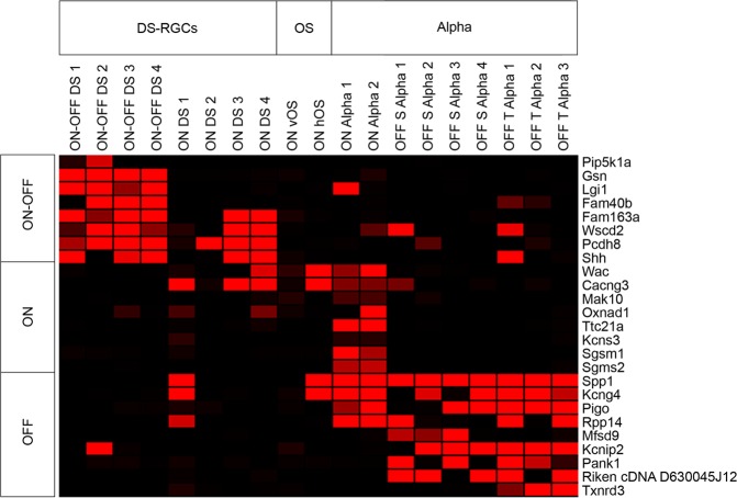
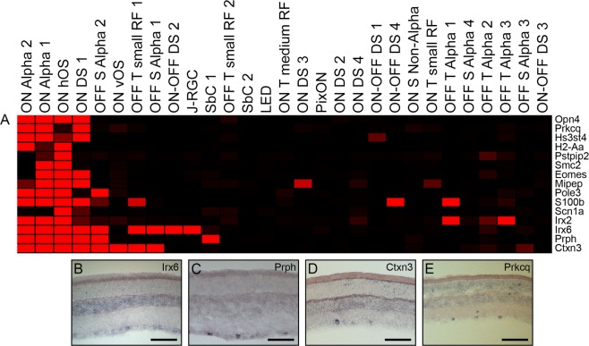
Retinal ganglion cells can be classified into more than 40 distinct subtypes, whether by functional classification or transcriptomics. The examination of these subtypes in relation to their physiology, projection patterns, and circuitry would be greatly facilitated through the identification of specific molecular identifiers for the generation of transgenic mice. Advances in single cell transcriptomic profiling have enabled the identification of molecular signatures for cellular subtypes that are only rarely found. Therefore, we used single cell profiling combined with hierarchical clustering and correlate analyses to identify genes expressed in distinct populations of Parvalbumin-expressing cells and functionally classified RGCs. RGCs were manually isolated based either upon fluorescence or physiological distinction through cell-attached recordings. Microarray hybridization and RNA-Sequencing were employed for the characterization of transcriptomes and in situ hybridization was utilized to further characterize gene candidate expression. Gene candidates were identified based upon cluster correlation, as well as expression specificity within physiologically distinct classes of RGCs. Further, we identified Prph, Ctxn3, and Prkcq as potential candidates for ipRGC classification in the murine retina. The use of these genes, or one of the other newly identified subset markers, for the generation of a transgenic mouse would enable future studies of RGC-subtype specific function, wiring, and projection.


Conflict of interest statement

The authors declare no competing interests.

Figures

Figure 1
Figure 1
Retinal ganglion cell subset markers revealed through transcriptome profiling of tdTomato+ cells. Fourteen tdTomato+ cells were hybridized to Affymetrix microarrays and the resulting data was extracted and normalized by MAS5 software. The genes expressed in these cells were visualized on a heatmap created with Genesis software, where red signal indicates high expression of the gene in a particular cell, and black signal indicates the absence of expression. Subset genes were identified based on their expression in the majority of the tdTomato+ cells (A) and were examined through in situ hybridization (BM). Those examined include: Nefh (B), Pnkd (C), Pcp4 (D), Rcan2 (E), Scn2b (F), Tusc5 (G), Fgf1 (H), Chrn2b (I), Mtap1b (J), Lypd6 (K), Kitl (L), and Chrm1 (M). Scale bars represent 100 µm.
Figure 2
Figure 2
tdTomato cells cluster into distinct groups. The tdTomato+ cells were clustered using Pearson correlation with average linkage and 3 separate clusters of RGCs were identified (A). A heatmap showing the genes expressed highly by cells in those clusters (B), with red signal indicating high expression of the gene in a particular cell, and black signal indicating the absence of expression.
Figure 3
Figure 3
Spike trains of alpha RGCs following whole cell patch clamping. The spike trains of representative alpha RGCs were examined over a 2-second interval during which light was shone upon the cells’ receptive fields. On Alpha, Off Sustained Alpha, and Off Transient Alpha cells are shown.
Figure 4
Figure 4
Markers of physiologically distinct RGCs identified. Three major groups of RGCs were classified: DS-RGCs, OS, and Alpha RGCs. The genes expressed by the cells belonging to these three major classifications are displayed on the heatmap. Clear distinctions were observed between cells with a preference for light during On-Off transitions, On alone, and Off alone.
Figure 5
Figure 5
Potential genetic markers of ipRGCs. The expression of Opn4-correlated genes in the categorized RGCs was determined by Pearson correlation and visualized by heatmap (A). Opn4+ cell candidate markers were investigated by in situ hybridization (BE). Genes examined included Irx6 (B), Prph (C), Ctxn3 (D), and Prkcq (E). Scale bars represent 100 µm.
Figure 6
Figure 6
Voltage-gated channel genes detected in subsets of RGCs. The expression of potassium and sodium channel subunits were queried in our tdTomato+ (A) and physiologically characterized RGCs (B) and displayed by heatmap. Four of these ion channel genes were detected by ISH: Kcnc2 (C), Kcnab2 (D), Scn2a1 (E), and Scn1a (F). Voltage-gated channel genes were then explored for expression among scRNA-seq data of 8 physiologically characterized cells and visualized on a heatmap (G). Scn4b expression was further examined by section ISH (H) and flat-mount ISH (I). Scale bars represent 100 µm.

References

    1. Sanes JR, Masland RH. The Types of Retinal Ganglion Cells: Current Status and Implications for Neuronal Classification. Annu. Rev. Neurosci. 2015;38:221–246. doi: 10.1146/annurev-neuro-071714-034120. - DOI - PubMed
    1. Baden T, et al. The functional diversity of retinal ganglion cells in the mouse. Nature. 2016;529:345–350. doi: 10.1038/nature16468. - DOI - PMC - PubMed
    1. Rheaume BA, et al. Single cell transcriptome profiling of retinal ganglion cells identifies cellular subtypes. Nat. Commun. 2018;9:2759. doi: 10.1038/s41467-018-05134-3. - DOI - PMC - PubMed
    1. Stevens CF. Neuronal diversity: Too many cell types for comfort? Curr. Biol. 1998;8:708–710. doi: 10.1016/S0960-9822(98)70454-3. - DOI - PubMed
    1. London A, Benhar I, Schwartz M. The retina as a window to the brain-from eye research to CNS disorders. Nat. Rev. Neurol. 2013;9:44–53. doi: 10.1038/nrneurol.2012.227. - DOI - PubMed