. 2019 Oct 31;9(1):15765.
doi: 10.1038/s41598-019-52323-1.

Tubular Deficiency of Heterogeneous Nuclear Ribonucleoprotein F Elevates Systolic Blood Pressure and Induces Glycosuria in Mice

Affiliations

Tubular Deficiency of Heterogeneous Nuclear Ribonucleoprotein F Elevates Systolic Blood Pressure and Induces Glycosuria in Mice

Chao-Sheng Lo et al. Sci Rep. .

Abstract

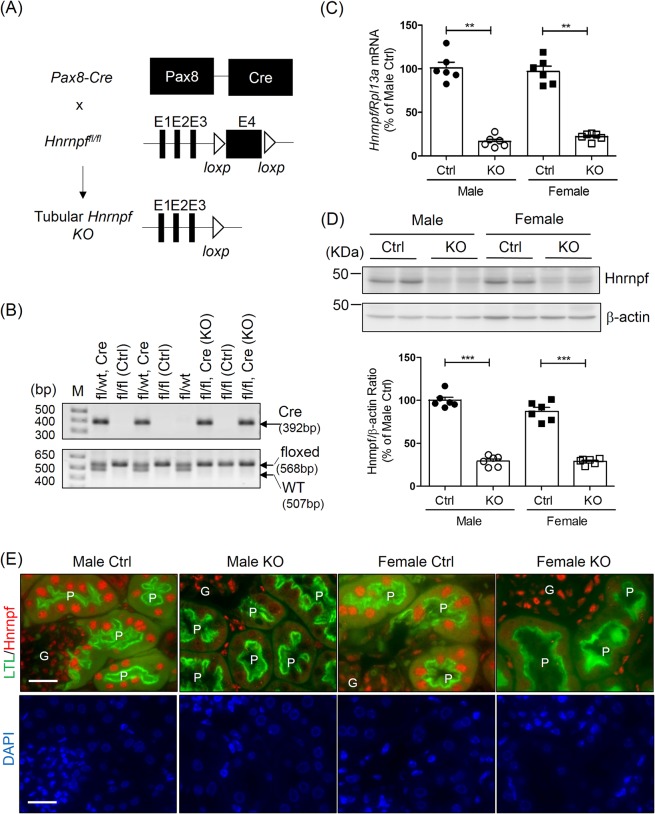
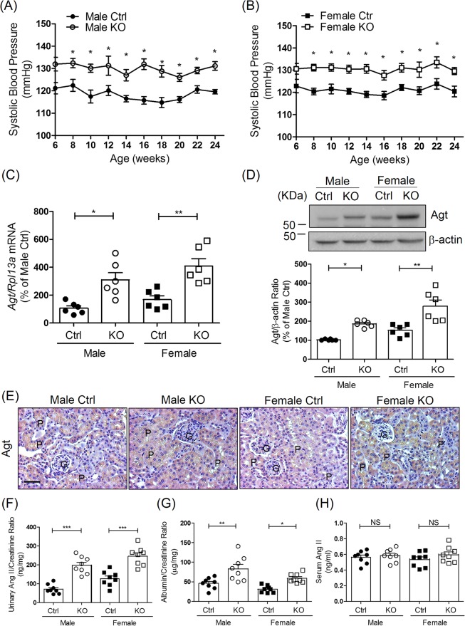
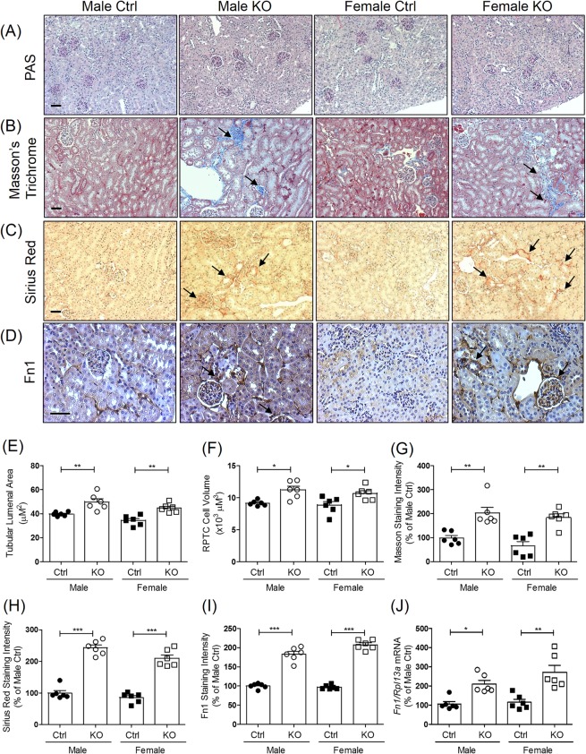
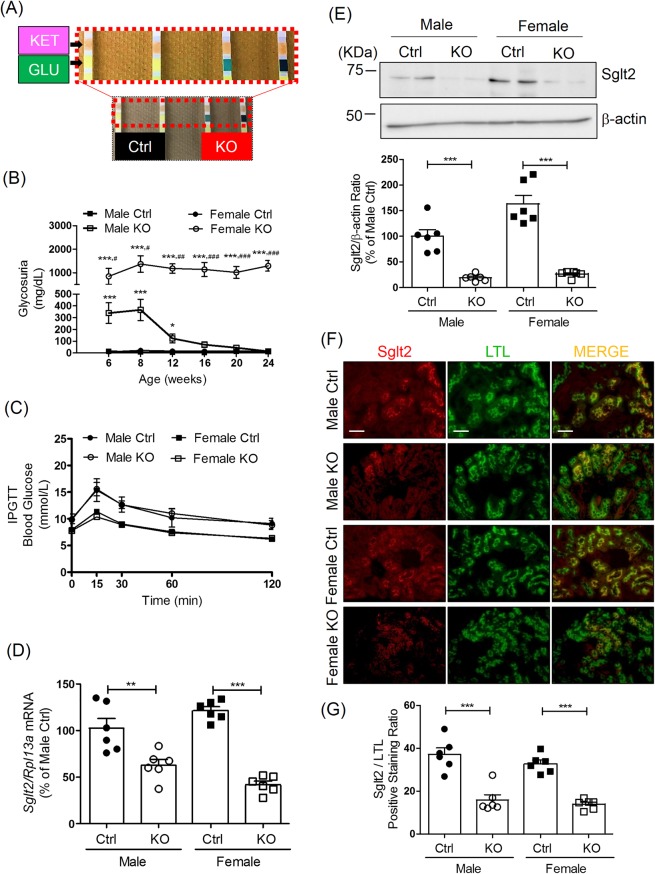
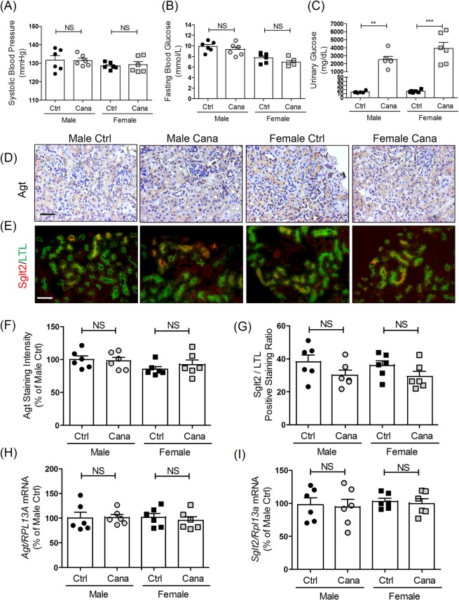
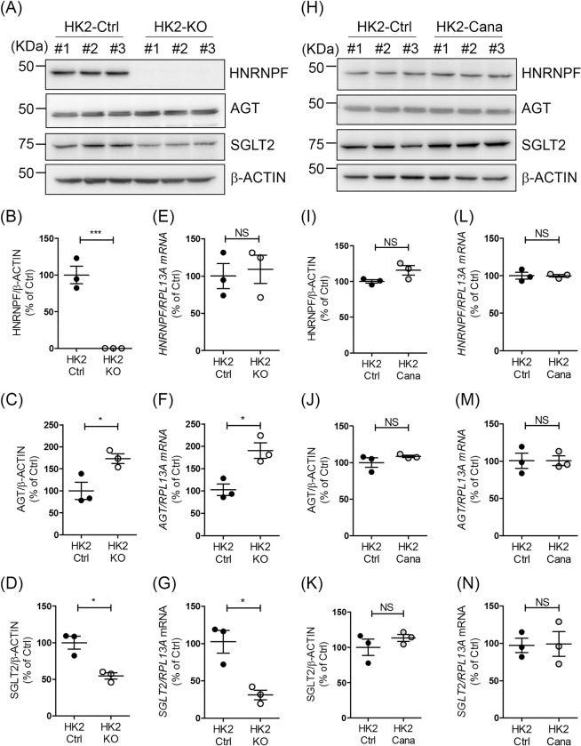
We reported previously that overexpression of heterogeneous nuclear ribonucleoprotein F (Hnrnpf) in renal proximal tubular cells (RPTCs) suppresses angiotensinogen (Agt) expression, and attenuates systemic hypertension and renal injury in diabetic Hnrnpf-transgenic (Tg) mice. We thus hypothesized that deletion of Hnrnpf in the renal proximal tubules (RPT) of mice would worsen systemic hypertension and kidney injury, perhaps revealing novel mechanism(s). Tubule-specific Hnrnpf knockout (KO) mice were generated by crossbreeding Pax8-Cre mice with floxed Hnrnpf mice on a C57BL/6 background. Both male and female KO mice exhibited elevated systolic blood pressure, increased urinary albumin/creatinine ratio, tubulo-interstitial fibrosis and glycosuria without changes in blood glucose or glomerular filtration rate compared with control littermates. However, glycosuria disappeared in male KO mice at the age of 12 weeks, while female KO mice had persistent glycosuria. Agt expression was elevated, whereas sodium-glucose co-transporter 2 (Sglt2) expression was down-regulated in RPTs of both male and female KO mice as compared to control littermates. In vitro, KO of HNRNPF in human RPTCs (HK-2) by CRISPR gRNA up-regulated AGT and down-regulated SGLT2 expression. The Sglt2 inhibitor canagliflozin treatment had no effect on Agt and Sglt2 expression in HK-2 and in RPTCs of wild-type mice but induced glycosuria. Our results demonstrate that Hnrnpf plays a role in the development of hypertension and glycosuria through modulation of renal Agt and Sglt2 expression in mice, respectively.


Conflict of interest statement

The authors declare no competing interests.

Figures

Figure 1
Figure 1
Generation of tubular Hnrnpf KO mice. (A) Schematic diagram describing the strategy of generating tubular Hnrnpf gene knockout mice. Exon 4 (E4) of the Hnrnpf gene is deleted; arrowheads: loxP sites. (B) Genotyping identification, the PCR bands of Cre (392 bp), floxed (568 bp) and wild-type (507 bp) alleles of Hnrnpf are indicated. Genotyping of representative litters are indicated; fl, Hnrnpf floxed; Control (Ctrl) (genotype: fl/fl) and KO (genotype: fl/fl, Cre). (C) Quantitative Hnrnpf mRNA expression level in male and female Ctrl and KO 24 week-old mice. **P < 0.01, KO versus Ctrl; n = 6 per group. (D) Representative WB and quantification of Hnrnpf protein expression in male and female Ctrl and KO 24 week-old mice. ***P < 0.005, KO versus Ctrl; n = 6 per group. (E) Immunostaining for Hnrnpf (red color) and a proximal tubular marker (lotus tetragonolobus lectin, LTL)(green color) in Ctrl and KO mice (original magnification ×600). DAPI staining (blue color) for cellular nucleus. Scale bars = 20 μm. G, glomerulus; P, proximal tubule.
Figure 2
Figure 2
Systolic blood pressure (SBP) and intrarenal angiotensinogen (Agt) expression in tubular Hnrnpf KO mice. (A) Longitudinal average SBP measurement (performed two or three times per mouse per week in the morning without fasting) in (A) male and (B) female mice. Baseline SBP was measured daily over a 5-day period before initiation of actual measurement at week 6. Values are means ± SEM, n = 10 for each group. *P < 0.05, KO versus Ctrl. (C) Agt mRNA levels in male and female Ctrl and KO mice at the age of 24 weeks. *P < 0.05, **P < 0.01, n = 6 per group, KO versus Ctrl. (D) Representative WB of Agt protein expression and quantitation of Agt expression in Ctrl and KO groups from 24 week-old male and female mice. *P < 0.05, **P < 0.01, n = 6 per group, KO versus Ctrl. (E) Representative immunostaining for Agt in Ctrl and KO mice (original magnification ×200). Scale bars = 50 μm. G, Glomerulus; P, proximal tubule. (F) Urinary Ang II, (G) ACR and (H) serum Ang II levels at week 24 in Ctrl and KO mice. Urinary Ang II and albumin levels were normalized with urinary creatinine levels. Values are mean ± SEM, n = 8 per group. *P < 0.05, **P < 0.01 and ***P < 0.005; KO versus Ctrl.
Figure 3
Figure 3
Tubulo-interstitial fibrosis in mouse kidneys. (A) Representative image of Periodic acid-Schiff (PAS) staining, (B) Masson’s trichrome staining, (C) Sirius Red staining and (D) fibronectin-1 (Fn1) immunostaining (original magnification ×100) in kidneys from male and female Ctrl and KO mice at the age of 24 weeks. (G) Glomerulus; P, proximal tubule. Scale bars = 50 μm. Semi-quantitation of tubule lumenal area (E), RPTC volume (F), Masson’s trichrome staining (G), Sirius Red staining (H) and Fn1 immunostaining (I) of Ctrl and KO mice at the age of 24 weeks. RT-qPCR of Fn1(J) in freshly RPTs from male and female Ctrl and KO mice. Values are means ± SEM, n = 6. *P < 0.05, **P < 0.01, ***P < 0.005; KO versus Ctrl.
Figure 4
Figure 4
Glycosuria and Sglt2 expression in Hnrnpf KO mice. (A) Urinary glucose in Ctrl and KO mice detected by dipstick test at the age of 6 weeks. (B) Longitudinal urinary glucose levels in male and female KO mice and Ctrl from the age of 6 weeks to 24 weeks measured by glucose colorimetric kit. Values are means ± SEM, n = 6. **P < 0.01; ***P < 0.005. KO versus Ctrl. ##P < 0.01; ###P < 0.005, female KO versus male KO. IPGTT test in male and female (C) Ctrl and KO mice at the age of 23 weeks. (D) Ratio of Sglt2/Rpl13a mRNA expression quantified by RT-qPCR and (E) Representative WB of Sglt2 protein expression in male and female mouse RPTs at the age of 24 weeks. Values are means ± SEM, n = 6. ***P < 0.005; KO versus Ctrl. (F) Double immunostaining of Sglt2 and LTL (magnification x100) and semi-quantification of Sglt2/LTL immunostaining ratio (G) in male and female Ctrl and KO mouse kidneys at the age of 24 weeks. Values are Sglt2/LTL positive staining ratio, n = 6. ***P < 0.005, KO versus Ctrl.
Figure 5
Figure 5
Effect of canagliflozin treatment on blood and urinary glucose levels, AGT and SGLT2 expression in mice. (A) SBP, (B) blood glucose and (C) urinary glucose levels after 4 weeks of treatment with or without canagliflozin in adult male and female wild-type (WT) mice. Values are means ± SEM, n = 6. **P < 0.01, ***P < 0.005; canagliflozin-treated versus non-treated mice. Immunostaining for Agt (D) and immunoflourescent staining of Sglt2 (E) in the kidneys of WT male and female mice with or without 4 weeks of canagliflozin treatment. Magnification x 200. Scale bars = 50 μm. Semi-quantification of Agt (F) and Sglt2 (G) immunostaining in male and female WT mouse kidneys after 4 weeks of treatment with or without canagliflozin. RT-qPCR of Agt (H) and Sglt2 (I) expression in isolated RPTs of WT male and female mice with or without 4 weeks of canagliflozin treatment. Values are means ± SEM, n = 6. NS, not significant; canagliflozin treated versus non-treated mice.
Figure 6
Figure 6
AGT and SGLT2 expression in HK-2 with or without HNRNPF KO. (A) WB, (BD) semi-quantitation of WB and (EG) RT-qPCR of HNRNPF, AGT, SGLT2 and β-ACTIN in different clones of HK-2 Ctrl and HK-2 with HNRNPF KO by CRISPR gRNA. Values are means ± SEM, n = 3. *P < 0.05, **P < 0.01; HK-2-HNRNPF KO versus HK-2 Ctrl. (H) WB, (IK) semi-quantitation of WB and (L)(M)(N) RT-qPCR of HNRNPF, AGT, SGLT2 and β-ACTIN of expression in HK-2 with or without canagliflozin (Cana) (0.5 mM) treatment for 24 hours. Values are means ± SEM, n = 3. NS, not significant. HK-2-Cana versus HK-2 Ctrl.

Similar articles

Cited by

References

    1. Ingelfinger JR, Zuo WM, Fon EA, Ellison KE, Dzau VJ. In situ hybridization evidence for angiotensinogen messenger RNA in the rat proximal tubule. An hypothesis for the intrarenal renin angiotensin system. The Journal of clinical investigation. 1990;85:417–423. doi: 10.1172/jci114454. - DOI - PMC - PubMed
    1. Gomez RA, et al. Renin and angiotensinogen gene expression in maturing rat kidney. The American journal of physiology. 1988;254:F582–587. doi: 10.1152/ajpcell.1988.254.4.C582. - DOI - PubMed
    1. Kamiyama M, Farragut KM, Garner MK, Navar LG, Kobori H. Divergent localization of angiotensinogen mRNA and protein in proximal tubule segments of normal rat kidney. Journal of hypertension. 2012;30:2365–2372. doi: 10.1097/HJH.0b013e3283598eed. - DOI - PMC - PubMed
    1. Tang SS, et al. Temperature-sensitive SV40 immortalized rat proximal tubule cell line has functional renin-angiotensin system. Am J Physiol. 1995;268:F435–446. doi: 10.1152/ajprenal.1995.268.3.F435. - DOI - PubMed
    1. Siragy HM, Carey RM. Role of the intrarenal renin-angiotensin-aldosterone system in chronic kidney disease. American journal of nephrology. 2010;31:541–550. doi: 10.1159/000313363. - DOI - PMC - PubMed

Publication types

MeSH terms

Substances