. 2019 Oct 31;10(1):4978.
doi: 10.1038/s41467-019-12979-9.

Molecular architecture of softwood revealed by solid-state NMR

Affiliations

Molecular architecture of softwood revealed by solid-state NMR

Oliver M Terrett et al. Nat Commun. .

Abstract

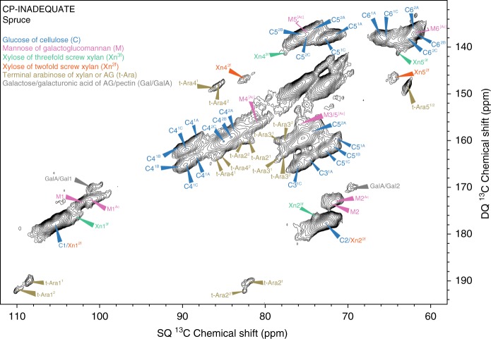
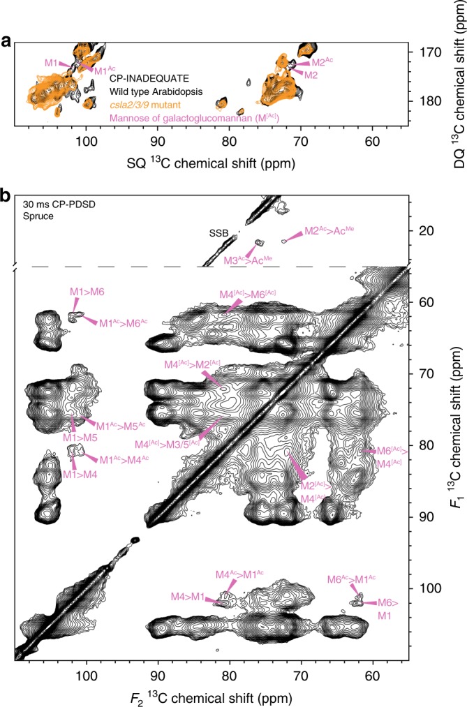
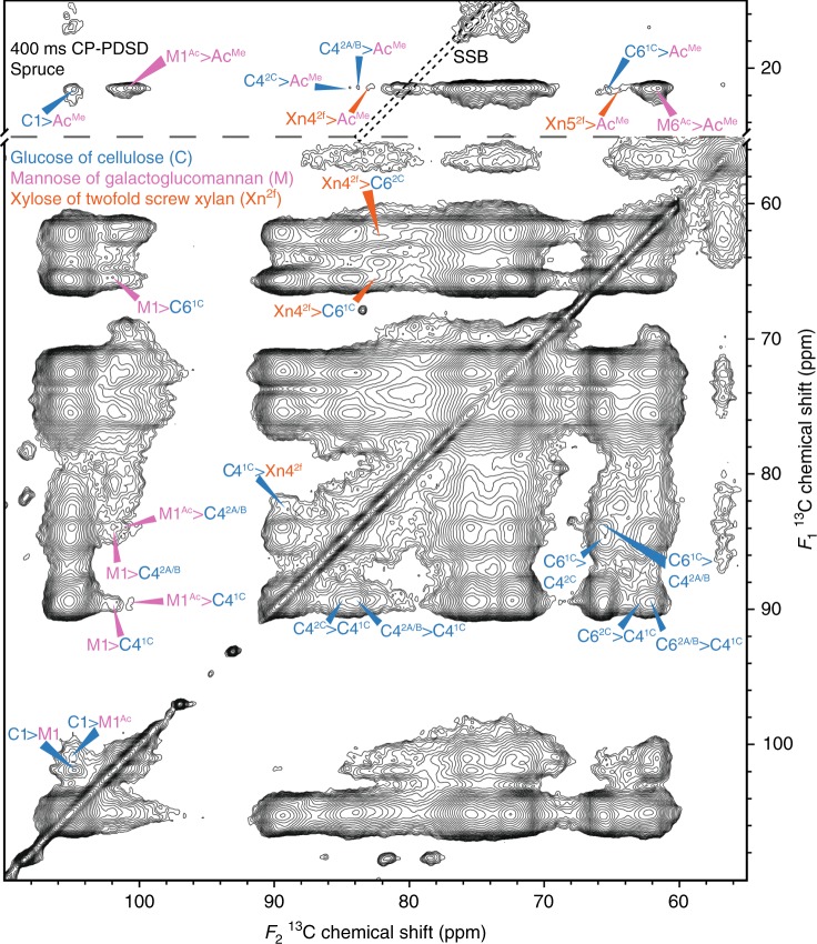
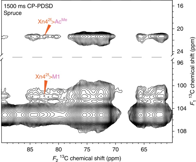
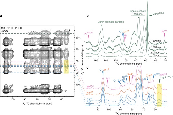
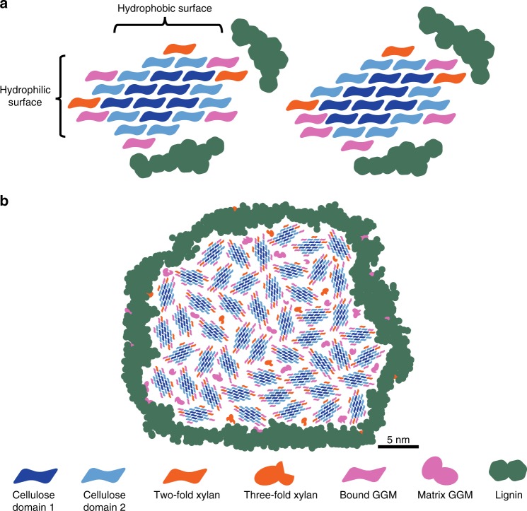
Economically important softwood from conifers is mainly composed of the polysaccharides cellulose, galactoglucomannan and xylan, and the phenolic polymer, lignin. The interactions between these polymers lead to wood mechanical strength and must be overcome in biorefining. Here, we use 13C multidimensional solid-state NMR to analyse the polymer interactions in never-dried cell walls of the softwood, spruce. In contrast to some earlier softwood cell wall models, most of the xylan binds to cellulose in the two-fold screw conformation. Moreover, galactoglucomannan alters its conformation by intimately binding to the surface of cellulose microfibrils in a semi-crystalline fashion. Some galactoglucomannan and xylan bind to the same cellulose microfibrils, and lignin is associated with both of these cellulose-bound polysaccharides. We propose a model of softwood molecular architecture which explains the origin of the different cellulose environments observed in the NMR experiments. Our model will assist strategies for improving wood usage in a sustainable bioeconomy.


Conflict of interest statement

The authors declare no competing interests.

Figures

Fig. 1
Fig. 1
An overview of immobile polysaccharides in spruce cell walls. The carbohydrate region of a refocussed CP-INADEQUATE 13C MAS NMR spectrum of 13C enriched spruce wood. Most carbons in the major polysaccharides cellulose, galactoglucomannan (GGM) and xylan are labelled, as are the terminal arabinose of xylan or arabinogalactans (AG) and galactose/galacturonic acid residues of arabinogalactans or pectin. The two terminal arabinose residues are labelled 1 and 2 to differentiate them. For cellulose, the environments have been split into two groups, domain 1 and 2, as in. Here, each cellulose domain is further resolved into environments. For GGM, the acetylated mannosyl residue carbons are labelled Ac, where they share the same chemical shift as the unacetylated mannose carbons, they are labelled [Ac]. The positions of GGM carbons 5–6 are obscured by cellulose peaks in the same region in the INADEQUATE spectrum but can be determined from a 30 ms PDSD experiment. Their positions, see full assignments in Supplementary Table 2, are labelled with an unfilled arrow. The region with single quantum (SQ) = 70–80 ppm and double quantum (DQ) = 142–152 ppm is unlabelled due to overlapping peaks from multiple polysaccharides
Fig. 2
Fig. 2
Assigning the chemical shifts of GGM in spruce. a The carbon 1-2 region of 13C CP-INADEQUATE MAS NMR spectra of wild type Arabidopsis and the csla2/3/9 mutant. The GGM carbons 1 and 2 are labelled. The wild type spectrum was previously published in (Grantham et al., 2017). b The acetate methyl (AcMe) and carbohydrate regions of a 30 ms mixing time 13C CP-PDSD MAS NMR spectra of spruce is shown. The intramolecular cross-peaks of two GGM mannose backbone residues are labelled. For GGM, the acetylated mannosyl residue carbons are labelled Ac; where they share the same chemical shift as the unacetylated mannose carbons they are labelled [Ac]. Spinning side bands are marked SSB
Fig. 3
Fig. 3
Xylan and GGM are a similar distance from the cellulose microfibril surface as glucan chains are from each other. The acetate methyl and carbohydrate regions of a 400 ms mixing time CP-PDSD 13C MAS NMR spectrum of spruce is shown. Cross-peaks between the different cellulose domains are labelled on the right hand side of the diagonal line in the carbohydrate region. Cross-peaks between cellulose and xylan/GGM are labelled on the left hand side of the diagonal line in the carbohydrate region and in the acetate methyl region of the spectra. Spinning side bands are surrounded by black dotted lines and are marked SSB
Fig. 4
Fig. 4
Xylan and GGM bind to the same cellulose microfibrils. Acetate methyl and carbon 1 region of a 1500 ms mixing time 13C CP-PDSD MAS NMR spectrum of spruce. Cross-peaks between xylan and GGM are labelled
Fig. 5
Fig. 5
Lignin is more intimately associated with GGM and xylan than cellulose in spruce. a The carbohydrate region of a 13C MAS NMR 1500 ms mixing time CP-PDSD spectrum is shown, with lines drawn to mark where the 1D slices are derived from the 2D spectrum. b The lignin methoxyl (56.5 ppm) slice extracted from 2D CP-PDSD spectra at 100, 400, 1000 and 1500 ms mixing times are overlaid. The different spectra are normalised to the self-peak at 56.5 ppm. Cross-peaks between the lignin methoxyl and GGM acetate methyl and cellulose/xylan and GGM carbon 1 are labelled. c The carbon 4 slices of GGM (80.4 ppm), xylan (82.4 ppm) and three cellulose (2B = 83.7, 2C = 84.5, 1C = 89.5 ppm) sub-domains from the 1500 ms CP-PDSD spectrum are shown. Cross-peaks between the polysaccharides and the lignin methoxyl are labelled. All spectra are normalised so that the self-peak of carbon 4 is the same height. A translucent yellow box highlights the cross-peak to the lignin methoxyl
Fig. 6
Fig. 6
a Possible models of the molecular architecture of softwood. The ratio of polysaccharide chains is based on the integrals of carbon 4–6 (carbon 4–5 for xylan) cross-peaks in the 30 ms CP-PDSD MAS NMR spectrum (see Fig. 2b) and the monosaccharide analysis. Both acetylated and unacetylated GGM are quantified together, as are the sub-domains of cellulose 1A–C and 2A–C. On the left, only xylan is shown as being able to convert domain 2 (C4 ≈ 84 ppm) to domain 1 (C4 ≈ 89 ppm) upon binding to the hydrophilic surface of the microfibril. On the right, binding of both xylan and some GGM to the hydrophilic surface can change domain 2 to domain 1. Lignin is shown mostly associated with itself, but is close to GGM, xylan and domain 2 cellulose. The cellulose microfibrils are taken to have 18 glucan chains with a 2, 3, 4, 4, 3, 2 habit to match the measured cellulose domain 1 to domain 2 ratio upon xylan binding. b Model of spruce cell wall macrofibril. Groups of cellulose microfibrils with bound GGM and xylan form macrofibrils in spruce cell walls. In addition to cellulose-bound xylan and GGM macrofibrils may contain some three-fold xylan and matrix GGM. Lignin is localised to the surface of the polysaccharide core of the macrofibril and interacts predominantly with GGM, xylan and cellulose domain 2. Size bar (5 nm) is provided for Fig. 6b) and is based on measurements presented in the literature,

References

    1. Bar-On YM, Phillips R, Milo R. The biomass distribution on Earth. Proc. Natl Acad. Sci USA. 2018;115:6506–6511. doi: 10.1073/pnas.1711842115. - DOI - PMC - PubMed
    1. Ramage MH, et al. The wood from the trees: the use of timber in construction. Renew. Sustain. Energy Rev. 2017;68:333–359. doi: 10.1016/j.rser.2016.09.107. - DOI
    1. Bradshaw CJA, Warkentin IG, Sodhi NS. Urgent preservation of boreal carbon stocks and biodiversity. Trends Ecol. Evol. 2009;24:541–548. doi: 10.1016/j.tree.2009.03.019. - DOI - PubMed
    1. Del Lungo, A., Ball, J. & Carle, J. Global planted forests thematic study: results and analysis, Planted Forests and Trees Working Paper 38, Food and Agricultural Organisation of the United Nations. (2006).
    1. Scheller HV, Ulvskov P. Hemicelluloses. Annu. Rev. Plant Biol. 2010;61:263–289. doi: 10.1146/annurev-arplant-042809-112315. - DOI - PubMed

Publication types