

Reticular Dysgenesis and Mitochondriopathy Induced by Adenylate Kinase 2 Deficiency with Atypical Presentation
- PMID: 31673062
- PMCID: PMC6823482
- DOI: 10.1038/s41598-019-51922-2
Reticular Dysgenesis and Mitochondriopathy Induced by Adenylate Kinase 2 Deficiency with Atypical Presentation
Abstract
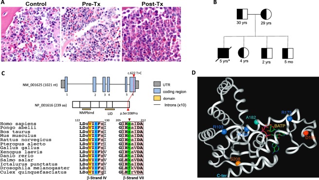
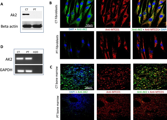
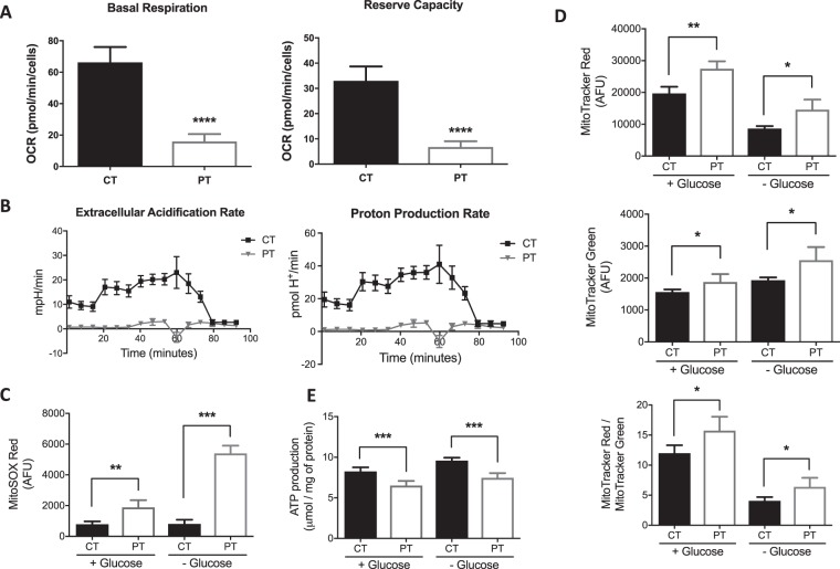
Reticular dysgenesis is an autosomal recessive form of severe combined immunodeficiency (SCID) that usually manifests in newborns. It is a unique example of an immune deficiency that is linked to dysfunctional mitochondrial energy metabolism and caused by adenylate kinase 2 (AK2) deficiency. It is characterized by an early differentiation arrest in the myeloid lineage, impaired lymphoid maturation, and sensorineural hearing loss. In this study, a novel AK2 homozygous mutation, c.622 T > C [p.Ser208Pro], was identified in an Old Order Amish patient through whole exome sequencing. Functional studies showed that the patient's cells have no detectable AK2 protein, as well as low oxygen consumption rate (OCR), extracellular acidification rate (ECAR) and proton production rate (PPR). An increased production of reactive oxygen species, mitochondrial membrane permeability, and mitochondrial mass, and decreased ATP production, were also observed. The results confirm the pathogenicity of the AK2 mutation and demonstrate that reticular dysgenesis should be considered in Amish individuals presenting with immune deficiency. We also describe other pathophysiological aspects of AK2 deficiency not previously reported.
Conflict of interest statement
The authors declare no competing interests.
Figures
Similar articles
-
Reticular dysgenesis (aleukocytosis) is caused by mutations in the gene encoding mitochondrial adenylate kinase 2.Nat Genet. 2009 Jan;41(1):101-5. doi: 10.1038/ng.265. Epub 2008 Nov 30. Nat Genet. 2009. PMID: 19043417
-
Recent advances in understanding the pathogenesis and management of reticular dysgenesis.Br J Haematol. 2018 Mar;180(5):644-653. doi: 10.1111/bjh.15045. Epub 2017 Dec 21. Br J Haematol. 2018. PMID: 29270983 Review.
-
AK2 deficiency compromises the mitochondrial energy metabolism required for differentiation of human neutrophil and lymphoid lineages.Cell Death Dis. 2015 Aug 13;6(8):e1856. doi: 10.1038/cddis.2015.211. Cell Death Dis. 2015. PMID: 26270350 Free PMC article.
-
Human adenylate kinase 2 deficiency causes a profound hematopoietic defect associated with sensorineural deafness.Nat Genet. 2009 Jan;41(1):106-11. doi: 10.1038/ng.278. Epub 2008 Nov 30. Nat Genet. 2009. PMID: 19043416 Free PMC article.
-
Adenylate kinase and AMP signaling networks: metabolic monitoring, signal communication and body energy sensing.Int J Mol Sci. 2009 Apr 17;10(4):1729-1772. doi: 10.3390/ijms10041729. Int J Mol Sci. 2009. PMID: 19468337 Free PMC article. Review.
Cited by
-
Human coilin interacting nuclear ATPase protein in cancer: uncovering new insights into pathogenesis and therapy.Am J Transl Res. 2020 Jul 15;12(7):4051-4058. eCollection 2020. Am J Transl Res. 2020. PMID: 32774758 Free PMC article.
-
Mesenchymal stem cell energy deficit and oxidative stress contribute to osteopenia in the Pahenu2 classical PKU mouse.Mol Genet Metab. 2021 Mar;132(3):173-179. doi: 10.1016/j.ymgme.2021.01.014. Epub 2021 Feb 11. Mol Genet Metab. 2021. PMID: 33602601 Free PMC article.
-
The Influence of Blue Light Exposure on Reconstructed 3-Dimensional Skin Model: Molecular Changes and Gene Expression Profile.JID Innov. 2023 Dec 5;4(2):100252. doi: 10.1016/j.xjidi.2023.100252. eCollection 2024 Mar. JID Innov. 2023. PMID: 38328595 Free PMC article.
-
Adenylate Kinase and Metabolic Signaling in Cancer Cells.Front Oncol. 2020 May 19;10:660. doi: 10.3389/fonc.2020.00660. eCollection 2020. Front Oncol. 2020. PMID: 32509571 Free PMC article. Review.
-
Ancestral retrovirus envelope protein ERVWE1 upregulates circ_0001810, a potential biomarker for schizophrenia, and induces neuronal mitochondrial dysfunction via activating AK2.Cell Biosci. 2024 Nov 14;14(1):138. doi: 10.1186/s13578-024-01318-1. Cell Biosci. 2024. PMID: 39543767 Free PMC article.
References
Publication types
MeSH terms
Substances
Supplementary concepts
Grants and funding
LinkOut - more resources
Full Text Sources
Miscellaneous
