Review
. 2020 Feb;16(2):99-111.
doi: 10.1038/s41581-019-0216-6. Epub 2019 Oct 31.

NAD+ homeostasis in renal health and disease

Affiliations
Review

NAD+ homeostasis in renal health and disease

Kenneth M Ralto et al. Nat Rev Nephrol. 2020 Feb.

Abstract

The mammalian kidney relies on abundant mitochondria in the renal tubule to generate sufficient ATP to provide the energy required for constant reclamation of solutes from crude blood filtrate. The highly metabolically active cells of the renal tubule also pair their energetic needs to the regulation of diverse cellular processes, including energy generation, antioxidant responses, autophagy and mitochondrial quality control. Nicotinamide adenine dinucleotide (NAD+) is essential not only for the harvesting of energy from substrates but also for an array of regulatory reactions that determine cellular health. In acute kidney injury (AKI), substantial decreases in the levels of NAD+ impair energy generation and, ultimately, the core kidney function of selective solute transport. Conversely, augmentation of NAD+ may protect the kidney tubule against diverse acute stressors. For example, NAD+ augmentation can ameliorate experimental AKI triggered by ischaemia-reperfusion, toxic injury and systemic inflammation. NAD+-dependent maintenance of renal tubular metabolic health may also attenuate long-term profibrotic responses that could lead to chronic kidney disease. Further understanding of the genetic, environmental and nutritional factors that influence NAD+ biosynthesis and renal resilience may lead to novel approaches for the prevention and treatment of kidney disease.


Conflict of interest statement

S.M.P. is an inventor of patent filings from Beth Israel Deaconess Medical Center. He holds equity in Raksana, serves on the Scientific Advisory Board of Aerpio and has received consulting fees from Cytokinetics, Astellas, Janssen, Mission Therapeutics and Aerpio. K.M.R. and E.P.R. declare no competing interests.

Figures

Fig. 1
Fig. 1. Overview of NAD+ metabolism
In highly metabolically active mammalian cells such as the renal tubular epithelium, nicotinamide adenine dinucleotide (NAD+) acts as a hub that coordinates various aspects of metabolism. Addition of a hydride ion (H-) (an electron pair) to NAD+ yields its reduced form, NADH. Reduction of NAD+ to NADH is required for glycolysis, fatty acid oxidation and the tricarboxylic acid cycle (also known as the citric acid cycle and Krebs cycle). Oxidation of NADH (donation of the electron pair) moves high-energy electrons from fuel substrates to Complex I of the mitochondrial electron transport chain (ETC). Sequential oxidation–reduction reactions in the ETC generate a proton motive force that is utilized by mitochondrial ATP synthase to generate ATP (not shown). Oxidation of NADH is also required to generate lactic acid from pyruvate and for the desaturation of polyunsaturated fatty acids (PUFAs). NAD+ can also be phosphorylated to form NADP+. Oxidation–reduction reactions involving NADP+ and its reduced form, NADPH, facilitate defence against oxidative stress by serving as a cofactor for thioredoxin-dependent reduction of disulfides and as a cofactor for glutathione reductase, which reduces the disulfide form of glutathione to its antioxidant sulfhydryl form. Reduction of NADP+ also occurs in the pentose phosphate pathway, which generates chemical building blocks for sugars and nucleic acids. NADPH serves as a substrate for NADPH oxidase enzymes (NOXs) that create second messengers of free radicals. NAD+ can also be cleaved by poly(ADP-ribose) polymerases (PARPs), sirtuins and the cyclic ADP-ribose (ADPR) synthetases CD38 and CD157 to generate nicotinamide (Nam) and ADPR.
Fig. 2
Fig. 2. Pathways of NAD+ biosynthesis
Dietary nutrients are converted to nicotinamide adenine dinucleotide (NAD+) through discrete endogenous biosynthetic routes. The de novo pathway converts tryptophan to NAD+ via a series of enzymatic steps. Indoleamine-2,3-dioxygenase (IDO) is a rate-limiting enzyme that catalyses the first step in this pathway, the conversion of tryptophan to N-formylkynurenine (not shown). Quinolinate phosphoribosyltransferase (QPRT) catalyses the conversion of quinolinic acid to nicotinate mononucleotide (NAMN). This bottleneck step commits the pathway to NAD+ synthesis. The Preiss–Handler pathway converts the acid form of vitamin B3, nicotinic acid (also known as niacin), to NAMN. This conversion is catalysed by nicotinate phosphoribosyltransferase (NAPRT). NAMN is converted to NAD+ by the sequential action of nicotinamide mononucleotide adenylyl transferases (NMNATs) and NAD synthetase (NADSYN). The salvage pathway converts the base analogue of vitamin B3, nicotinamide (Nam; also known as niacinamide), to NAD+ via the rate-limiting enzyme Nam phosphoribosyltransferase (NAMPT). Nam mononucleotide (NMN) is a measurable intermediate in this pathway that can also be obtained through supplements and synthesized from the dietary precursor nicotinamide riboside. The salvage pathway is so named because NAD+ is ‘reclaimed’ from the Nam product of enzymes that consume NAD+ to generate ADP-ribose (ADPR) or cyclic ADPR (cADPR). NAD+ is converted to NAD+ phosphate (NADP+) by NAD+ kinases (NADKs). Hydride transfer from NADH to NADP+ by nicotinamide nucleotide transhydrogenase (NNT) also generates NADPH. NRKs, nicotinamide riboside kinases.
Fig. 3
Fig. 3. NAD+-consuming enzymes.
Nicotinamide adenine dinucleotide (NAD+)-consuming enzymes comprise sirtuins (part a), poly(ADP-ribose) polymerases (PARPs) (part b) and the cyclic ADP-ribose (cADPR) synthetases CD38 and CD157 (part c). Sirtuins deacetylate or deacylate proteins by cleaving NAD+ to nicotinamide (Nam) and ADPR, which accepts the acetyl or acyl group (AC) from the target protein. PARPs utilize NAD+ cleavage to attach single or multiple ADPR subunits to target proteins. CD38 and CD157 cleave NAD+ to form cADPR and Nam.
Fig. 4
Fig. 4. The de novo NAD+ biosynthesis pathway.
The de novo nicotinamide adenine dinucleotide (NAD+) biosynthesis pathway (also known as the kynurenine pathway or tryptophan–kynurenine pathway) consists of eight steps that convert dietary tryptophan to NAD+. Tryptophan has multiple metabolic fates that compete with its conversion to N-formylkynurenine, which is catalysed by indoleamine 2,3-dioxygenase (IDO) or Trp 2,3-dioxygenase (TDO). N-formylkynurenine is hydrolysed to kynurenine by arylformamidase (AFMID). Kynurenine is hydroxylated to 3-hydroxy-kynurenine by kynurenine 3-monooxygenase (KMO). 3-Hydroxy-kynurenine is a major branch point in the pathway as this metabolite can be converted to xanthurenate or other molecules. Kynureninase (KYNU) catalyses the formation of 3-hydroxyanthranilic acid, which is converted to α-amino-β-carboxymuconate-ε-semialdehyde (ACMS) by 3-hydroxyanthranilate 3,4-dioxygenase (HAAO). ACMS can either spontaneously cyclize to form quinolinic acid or can be decarboxylated by ACMS decarboxylase (ACMSD) to form picolinic acid. The only known fate of quinolinic acid is conversion to nicotinate mononucleotide (NAMN) by quinolinate phosphoribosyltransferase (QPRT). Quinolinic acid is therefore the first metabolite in the pathway that is committed to NAD+ biosynthesis. Nicotinamide mononucleotide adenylyltransferase (NMNAT) catalyses the final step to create NAD+.
Fig. 5
Fig. 5. Glycolytic NAD+ recycling and lipid accumulation in AKI.
Nicotinamide adenine dinucleotide (NAD+) is reduced to NADH via glycolysis in the cytosol and via the tricarboxylic acid (TCA) cycle and β-oxidation of fatty acids (FAO) in mitochondria. Under aerobic conditions, NAD+ is regenerated as electrons are transported to the mitochondrial electron transport chain (ETC) to drive oxidative phosphorylation, which produces ATP. During acute kidney injury (AKI), mitochondrial respiration and function are impaired, glycolysis increases to meet cellular energy demands and cytosolic NAD+ recycling is required. Lactate dehydrogenase (LDH), which converts pyruvate to lactate, has a major role in cytosolic NAD+ recycling. Desaturation of polyunsaturated fatty acids (PUFAs) to form highly unsaturated fatty acids (HUFAs) is an additional mechanism of glycolytic NAD+ recycling that is mediated by delta-5 and delta-6 fatty acid desaturases (D5D and D6D). D5D and D6D are located in the endoplasmic reticulum (ER) membrane and have cytosol-facing catalytic domains; they are highly expressed in the kidney and liver. Experimental AKI is associated with >50–100-fold increases in the levels of HUFA-containing triglycerides and cellular lipid accumulation in renal tubular cells.
Fig. 6
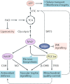
Fig. 6. Potential renoprotective actions of NAD+.
Nicotinamide adenine dinucleotide (NAD+) is required for distinct reactions in glycolysis, fatty acid oxidation (FAO) and the tricarboxylic acid (TCA) cycle that lead to the generation of ATP via the electron transport chain (ETC) in mitochondria. In healthy renal tubular epithelial cells, this ATP is utilized to provide the energy required for key functions, such as solute transport and maintenance of membrane integrity. Efficient FAO also prevents the potentially toxic accumulation of storage fats. NAD+ might also exert renoprotective effects via its interactions with sirtuins and PPARγ co-activator 1α (PGC1α). Sirtuin 1 (SIRT1) activates PGC1α via NAD+-dependent deacetylation and PGC1α in turn promotes NAD+ biosynthesis via the de novo pathway by co-ordinately upregulating the expression of the genes encoding enzymes in this pathway. PGC1α promotes mitochondrial quality control and ATP production via interrelated process that include mitochondrial biogenesis and the induction of mitophagy via transcription factor EB (TFEB). SIRT3 also utilizes NAD+ to directly promote healthy mitochondrial function and SIRT1 may limit stress signalling through the pro-apoptotic Jun N-terminal kinase (JNK) pathway. In addition to its effects on mitochondria, PGC1α signalling can induce the production of vascular trophic molecules, such as VEGF. In tubular epithelial cells, a PGC1α-dependent product of FAO, β-hydroxybutyrate (β-OHB), may signal the production of vasodilator prostaglandins that can maintain renal blood flow during conditions such as shock that would otherwise promote renal ischaemia. Phosphorylation of NAD+ to NADP+ may potentiate defence against oxidant stress induced by inflammation, toxins, or ischaemia–reperfusion injury by promoting the reduction of glutathione (GSH) and through the vasodilator nitric oxide (NO•). Mitochondrial quality control may also limit the burden of free radicals emanating from injured mitochondria.

References

    1. Poyan Mehr A, et al. De novo NAD+ biosynthetic impairment in acute kidney injury in humans. Nat. Med. 2018;24:1351–1359. doi: 10.1038/s41591-018-0138-z. - DOI - PMC - PubMed
    1. Evans J, Wang TC, Heyes MP, Markey SP. LC/MS analysis of NAD biosynthesis using stable isotope pyridine precursors. Anal. Biochem. 2002;306:197–203. doi: 10.1006/abio.2002.5715. - DOI - PubMed
    1. Revollo JR, Grimm AA, Imai S. The NAD biosynthesis pathway mediated by nicotinamide phosphoribosyltransferase regulates Sir2 activity in mammalian cells. J. Biol. Chem. 2004;279:50754–50763. doi: 10.1074/jbc.M408388200. - DOI - PubMed
    1. Grozio A, et al. Slc12a8 is a nicotinamide mononucleotide transporter. Nat. Metab. 2019;1:47–57. doi: 10.1038/s42255-018-0009-4. - DOI - PMC - PubMed
    1. Carpenter KJ. The relationship of pellagra to corn and the low availability of niacin in cereals. Experientia Suppl. 1983;44:197–222. doi: 10.1007/978-3-0348-6540-1_12. - DOI - PubMed