. 2019 Jul 22;10(35):8164-8170.
doi: 10.1039/c9sc01495k. eCollection 2019 Sep 21.

Seconds-resolved pharmacokinetic measurements of the chemotherapeutic irinotecan in situ in the living body

Affiliations

Seconds-resolved pharmacokinetic measurements of the chemotherapeutic irinotecan in situ in the living body

Andrea Idili et al. Chem Sci. .

Abstract

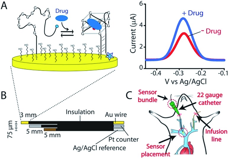
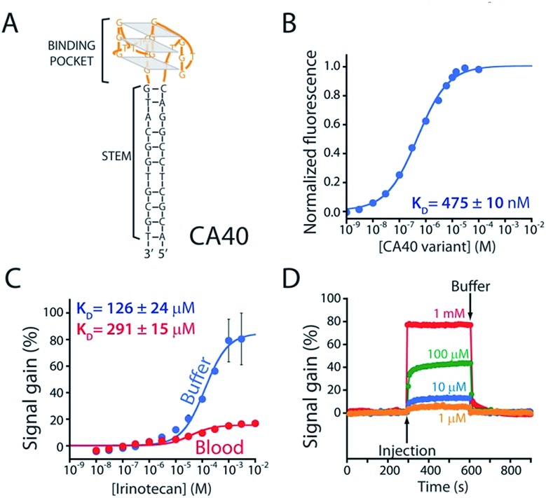
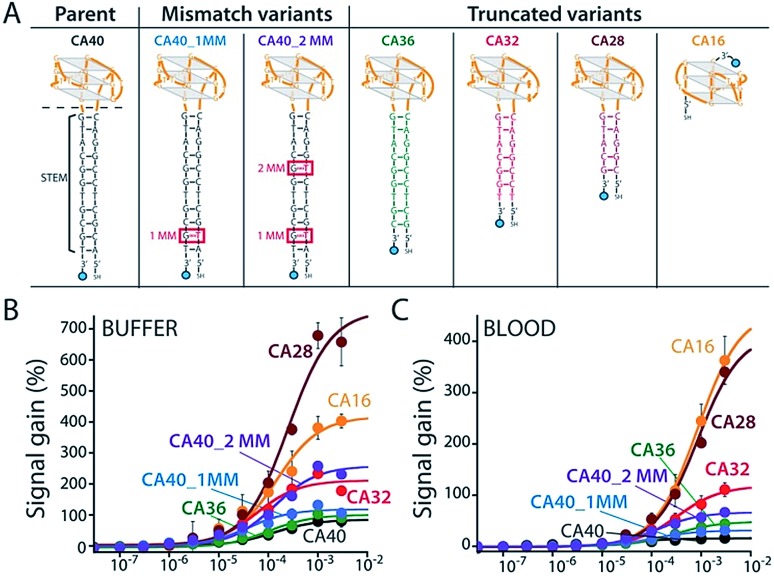
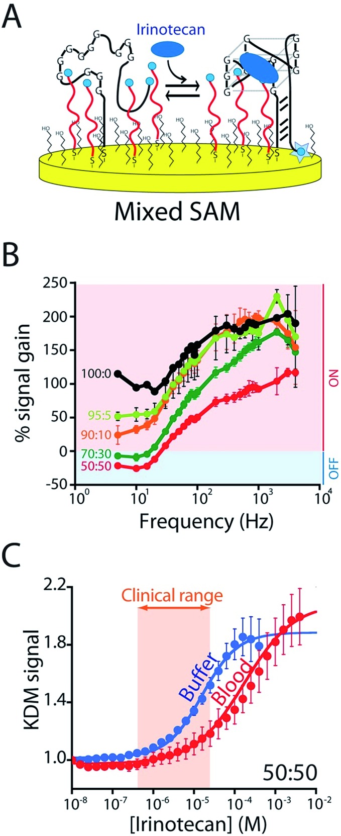
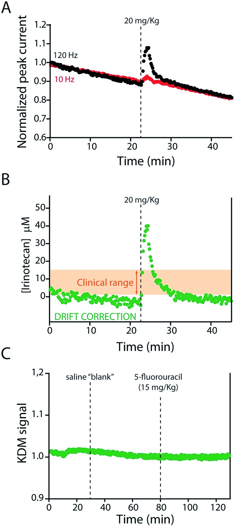
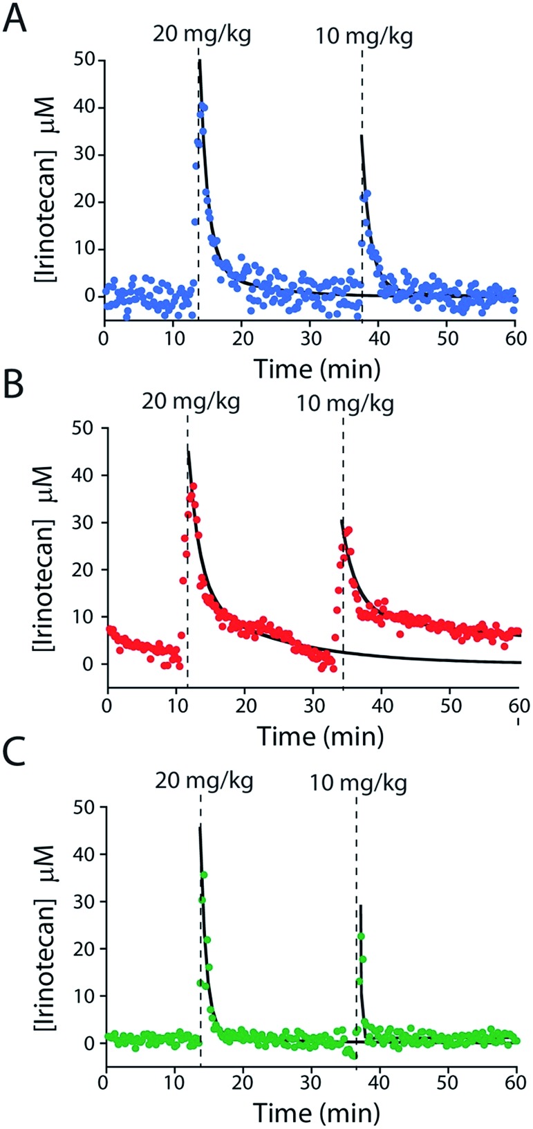
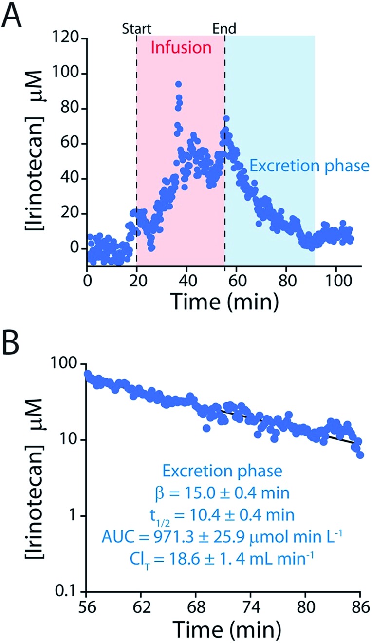
The ability to measure drugs in the body rapidly and in real time would advance both our understanding of pharmacokinetics and our ability to optimally dose and deliver pharmacological therapies. To this end, we are developing electrochemical aptamer-based (E-AB) sensors, a seconds-resolved platform technology that, as critical for performing measurements in vivo, is reagentless, reversible, and selective enough to work when placed directly in bodily fluids. Here we describe the development of an E-AB sensor against irinotecan, a member of the camptothecin family of cancer chemotherapeutics, and its adaptation to in vivo sensing. To achieve this we first re-engineered (via truncation) a previously reported DNA aptamer against the camptothecins to support high-gain E-AB signaling. We then co-deposited the modified aptamer with an unstructured, redox-reporter-modified DNA sequence whose output was independent of target concentration, rendering the sensor's signal gain a sufficiently strong function of square-wave frequency to support kinetic-differential-measurement drift correction. The resultant, 200 μm-diameter, 3 mm-long sensor achieves 20 s-resolved, multi-hour measurements of plasma irinotecan when emplaced in the jugular veins of live rats, thus providing an unprecedentedly high-precision view into the pharmacokinetics of this class of chemotherapeutics.


Figures

Fig. 1
Fig. 1. Indwelling E-AB sensors supporting the high-frequency measurements of plasma irinotecan levels in situ in the living body. (A) Electrochemical aptamer-based (E-AB) sensors consist of a redox-reporter-modified aptamer covalently attached to a gold electrode via an alkane-thiol self-assembled monolayer. In the absence of its specific target, the aptamer is partially or entirely unfolded (left). Target binding induces a conformational change, altering the efficiency with which the redox reporter (here a methylene blue molecule) approaches the electrode and thus altering electron transfer and (right) the signal observed upon voltammetric (here square-wave) interrogation. (B) In the completed sensor a 75 μm gold-wire working electrode is bundled with same-diameter platinum counter and a silver/silver-chloride reference electrodes, creating a device small enough and flexible enough to (C) be emplaced via a 22-gauge guide catheter in one of the external jugular veins of a live rat.
Fig. 2
Fig. 2. (A) The parent aptamer (CA40) is predicted to fold into a G-quadruplex, which is thought to be the target-binding site,, flanked by a 12-base-pair stem. (B) Exploiting the intrinsic fluorescence of irinotecan (Fig. S1†) we find that, when free in solution, the aptamer exhibits a dissociation constant of 475 nM. (C) When redox-reporter-modified and anchored to the sensor's interrogating electrode, however, its affinity and signal gain are reduced significantly, particularly when deployed in undiluted whole blood. (D) The E-AB sensor nevertheless rapidly responds to when challenged (here in buffer) with irinotecan. Binding curves in panel C employed a square-wave frequency of 120 Hz. The kinetic experiments in panel D employed a square-wave frequency of 500 Hz and a repetition rate of 0.2 Hz.
Fig. 3
Fig. 3. We reengineered the parent aptamer to produce higher-gain E-AB signaling. (A) We did so by destabilizing the aptamer's stem-loop (thus increasing the population of unfolded molecules poised to respond to target) via either introduction of one (CA40_1MM) or two (CA40_2MM) mismatches or via truncation (CA36, CA32, CA28, CA16) of the stem. (B) When challenged in a simple buffered solution all of the re-engineered variants exhibited higher gain than that of the parent aptamer (see ESI Table 1†), with the most destabilized (CA40_2MM, CA32, CA28, CA16) producing the greatest signal gain. (C) When tested in whole blood their gain and affinity are reduced, but the best performing nevertheless still support high-gain E-AB sensing.
Fig. 4
Fig. 4. To correct the drift of seen during in vivo deployment we modified the E-AB sensor so that it better supports “Kinetic Differential Measurements” (KDM). (A) KDM requires that the gain of an E-AB sensor be a strong function of square wave frequency. To induce this we co-deposited the aptamer with a redox-reporter-modified linear DNA sequence that does not respond to target. (B) The signal gain (relative signal change between no target and saturating target – i.e., 100 μM) of the original E-AB sensor (100 : 0 black curve) is a relatively minor function of square-wave frequency. Upon co-deposition with increasing amounts of the linear-strand (to a maximum ratio of 50 : 50, red curve) we observe increasingly strong frequency dependence, albeit with a corresponding reduction in the maximum gain. (C) A sensor fabricated using a 50 : 50 mixture of the two strands and employing KDM drift correction (here the difference in the relative signals seen at 10 and 120 Hz) responds to target over the clinically-relevant range (0.5 μM to 15 μM; 0.06 and 10 μg mL–1), in both buffer and in undiluted whole blood.
Fig. 5
Fig. 5. The KDM-corrected E-AB sensor supports real time, seconds-resolved measurements of plasma irinotecan levels., (A) In the absence of KDM signals collected at high (120 Hz) and low (10 Hz) frequencies both drift significantly, but because they drift in concert (B) taking their difference (KDM) produces a stable baseline. (C) As expected, control injections of either a saline “blank” or a second chemotherapeutic (5-fluorouracil, which is often co-administered with irinotecan), do not produce any measurable sensor response.
Fig. 6
Fig. 6. E-AB sensors support the measurements of plasma drug levels with unprecedented time resolution, providing a high-precision window into, for example, intra-subject pharmacokinetic variability. Shown are plasma irinotecan levels following multiple intravenous injections into three live rats (A–C). The black lines represent the fit of each injection dataset to a two-compartment pharmacokinetic model.
Fig. 7
Fig. 7. (A) To determine irinotecan's pharmacokinetics with more precision we performed an intravenous injection at a much higher dose (60 mg kg–1). (B) The higher peak concentrations reached in this experiment lead to longer measurement runs, in turn improving the precision of our estimates of the relevant pharmacokinetic parameters.

References

    1. Hamburg M. A., Collins F. S. N. Engl. J. Med. 2010;363:301–304. - PubMed
    1. Reuben D. B., Tinetti M. E. N. Engl. J. Med. 2012;366:777–779. - PubMed
    1. Sanavio B., Krol S. Front. Bioeng. Biotechnol. 2015;3:20. - PMC - PubMed
    1. Tucker G. T. Pharm. Res. 2017;34:1539–1543. - PMC - PubMed
    1. Arroyo-Curras N., Ortega G., Copp D. A., Ploense K. L., Plaxco Z. A., Kippin T. E., Hespanha J. P., Plaxco K. W. ACS Pharmacol. Transl. Sci. 2018;1:110–118. - PMC - PubMed