. 2019 Oct 31;114(6):47.
doi: 10.1007/s00395-019-0758-6.

Glucocorticoids preserve the t-tubular system in ventricular cardiomyocytes by upregulation of autophagic flux

Affiliations

Glucocorticoids preserve the t-tubular system in ventricular cardiomyocytes by upregulation of autophagic flux

Thomas Seidel et al. Basic Res Cardiol. .

Abstract

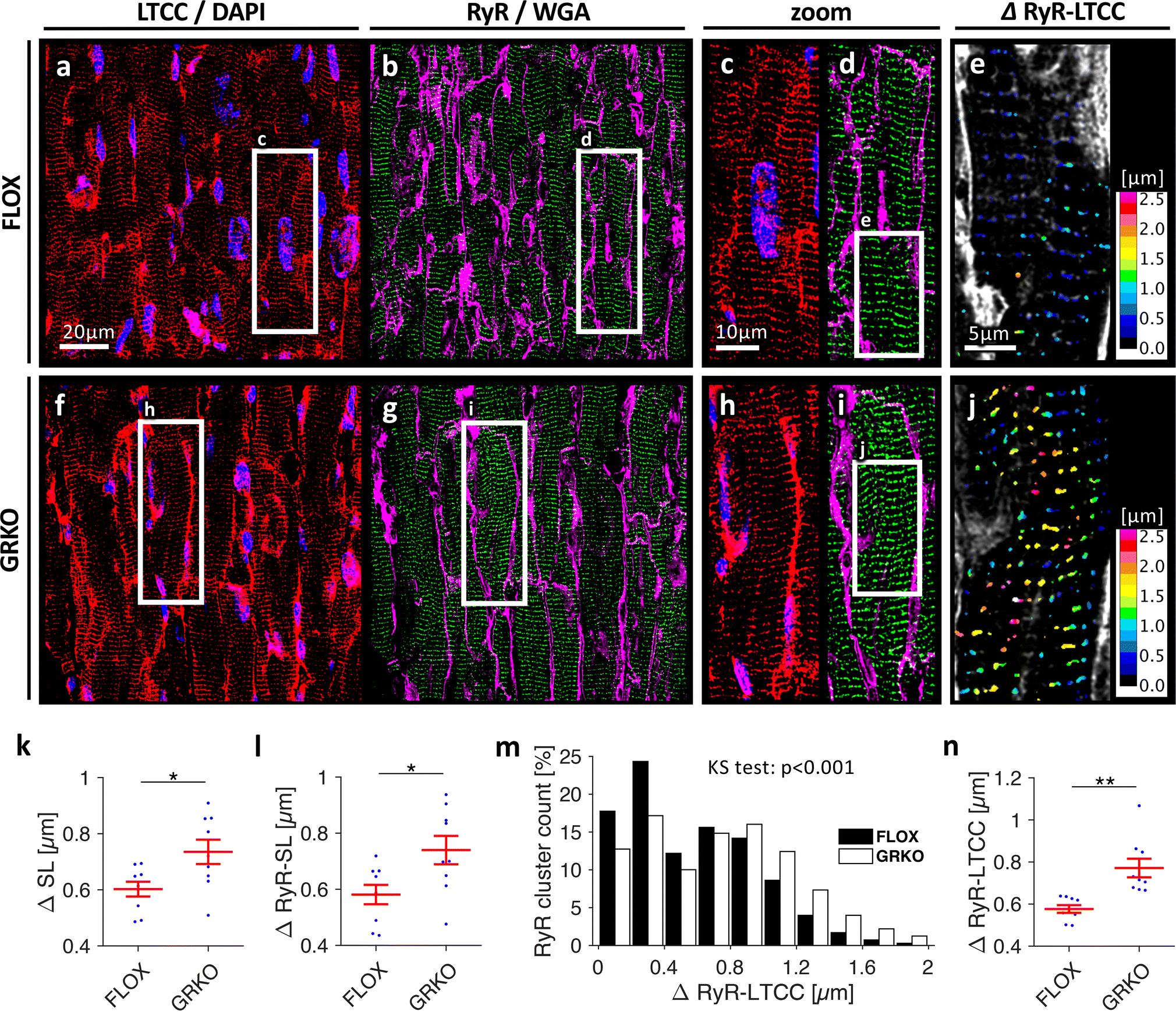
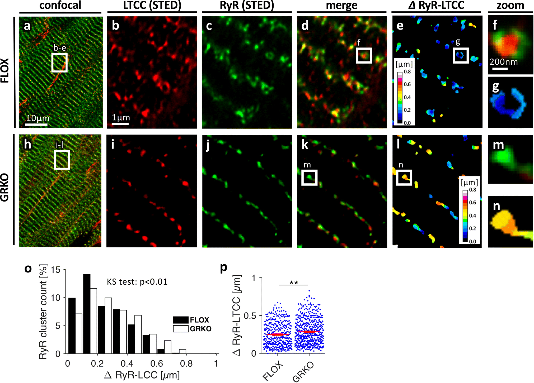
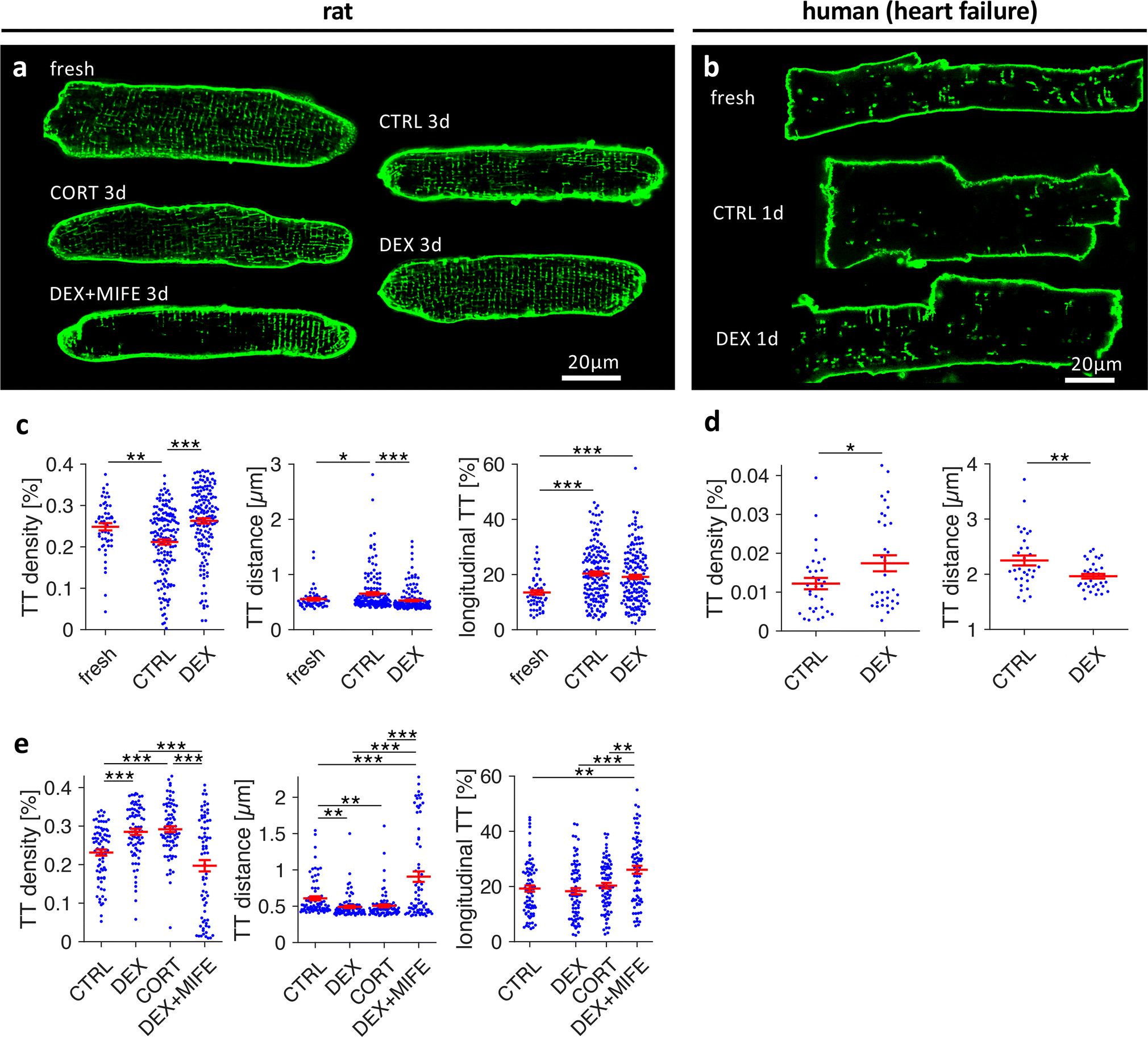
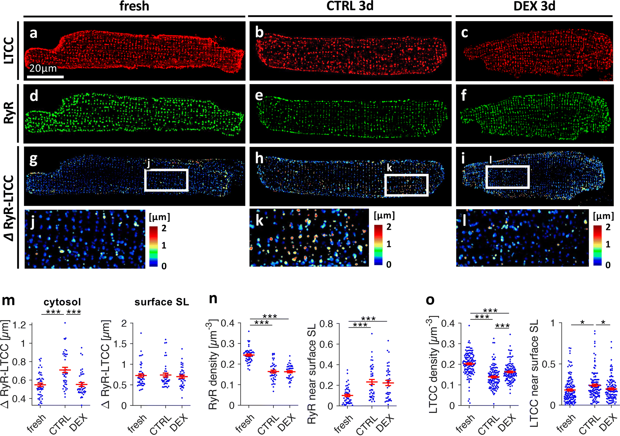
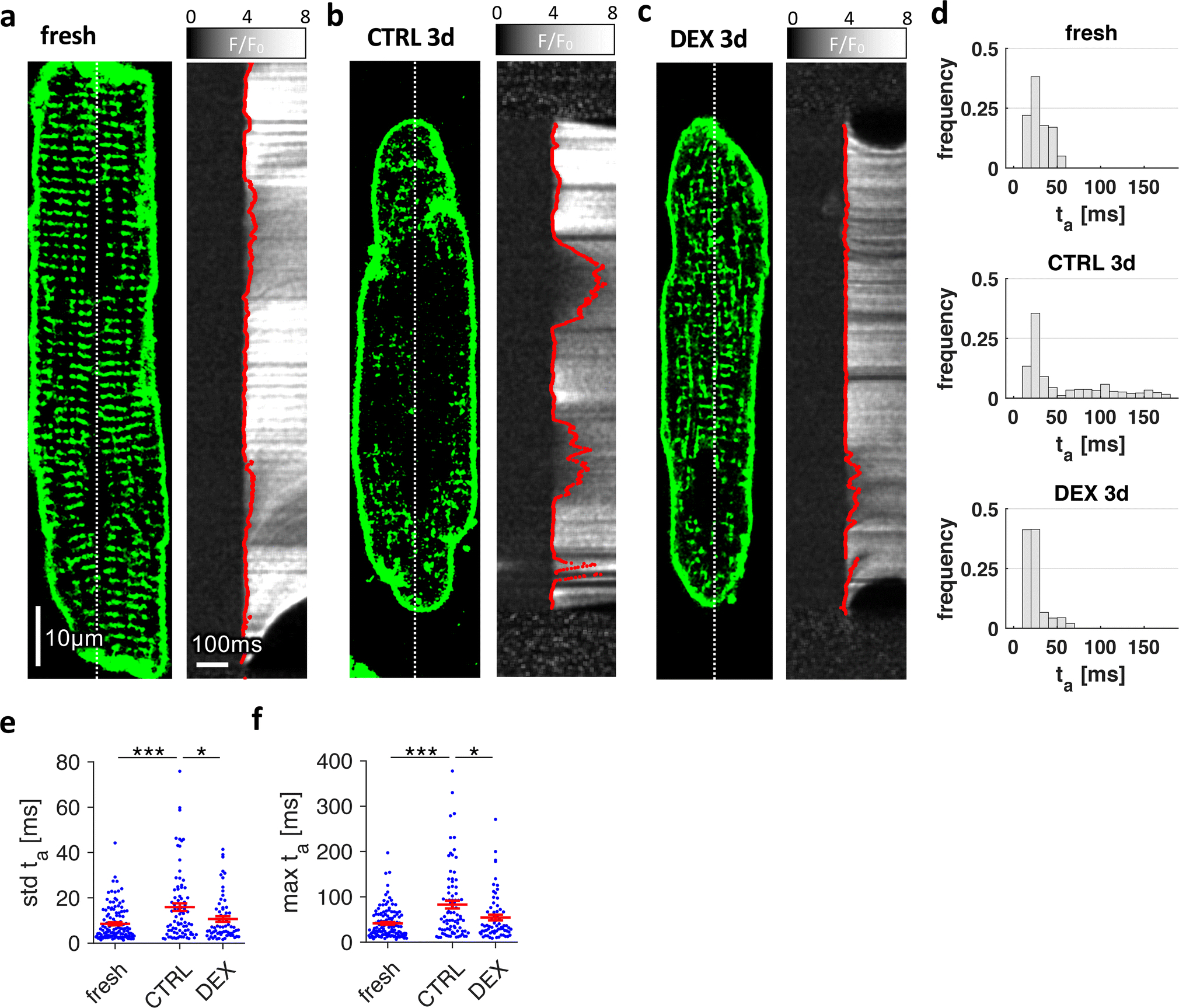
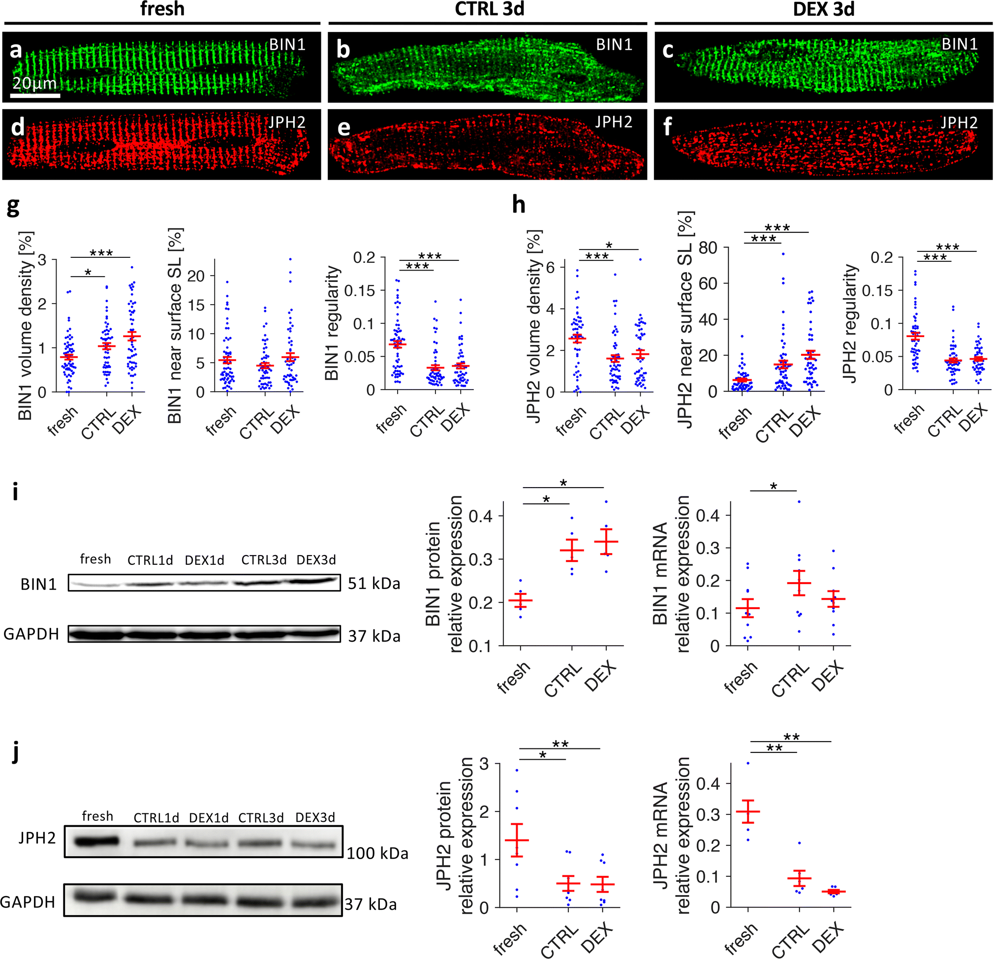
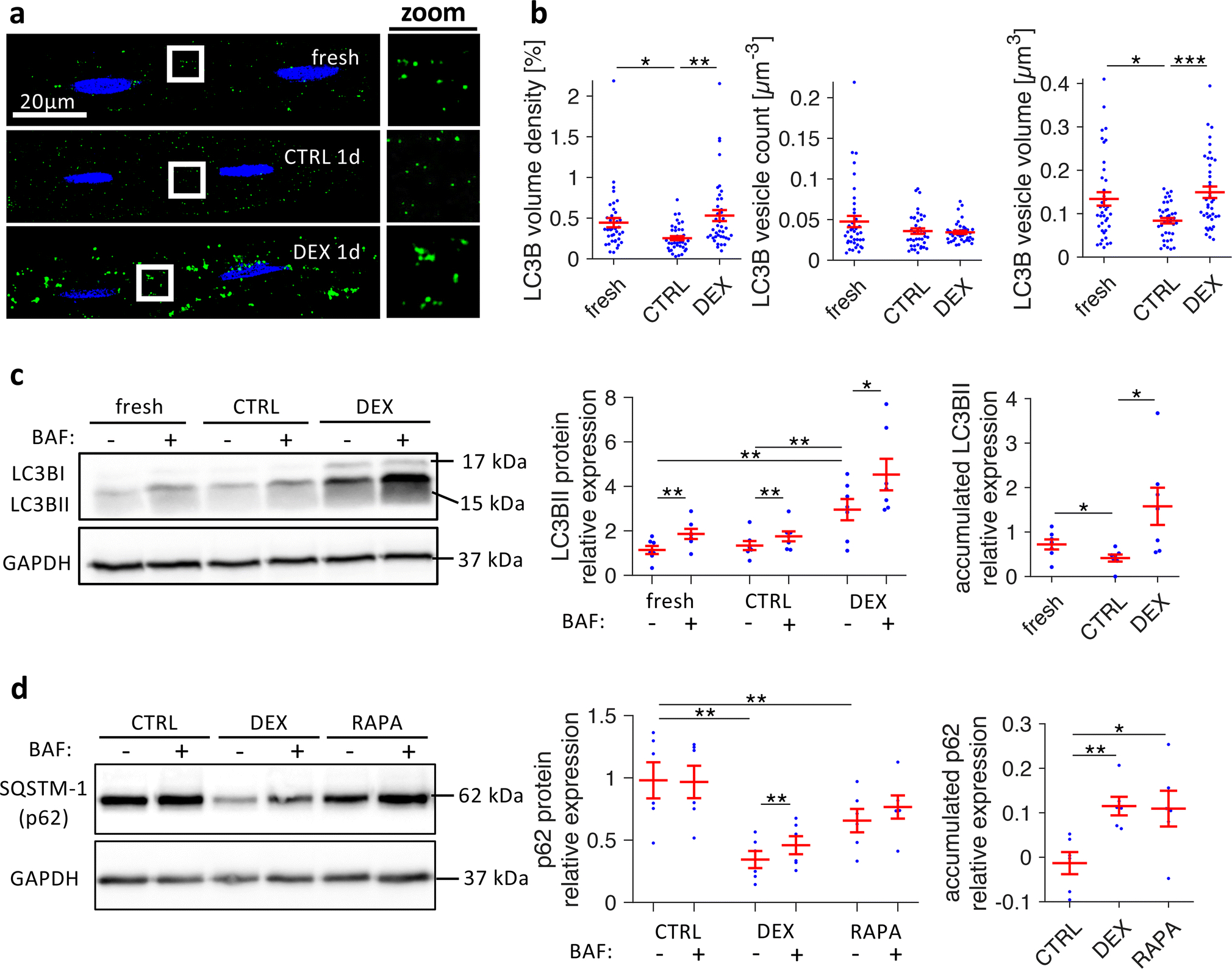
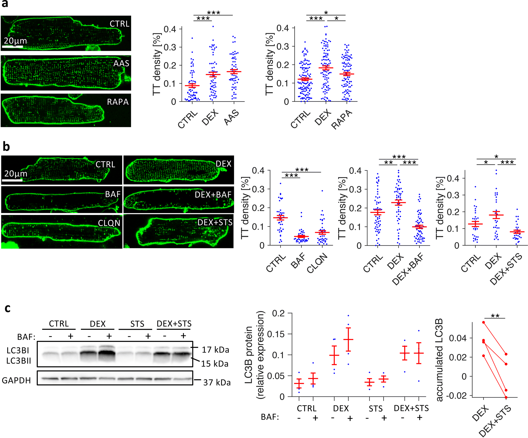
A major contributor to contractile dysfunction in heart failure is remodelling and loss of the cardiomyocyte transverse tubular system (t-system), but underlying mechanisms and signalling pathways remain elusive. It has been shown that dexamethasone promotes t-tubule development in stem cell-derived cardiomyocytes and that cardiomyocyte-specific glucocorticoid receptor (GR) knockout (GRKO) leads to heart failure. Here, we studied if the t-system is altered in GRKO hearts and if GR signalling is required for t-system preservation in adult cardiomyocytes. Confocal and 3D STED microscopy of myocardium from cardiomyocyte-specific GRKO mice revealed decreased t-system density and increased distances between ryanodine receptors (RyR) and L-type Ca2+ channels (LTCC). Because t-system remodelling and heart failure are intertwined, we investigated the underlying mechanisms in vitro. Ventricular cardiomyocytes from failing human and healthy adult rat hearts cultured in the absence of glucocorticoids (CTRL) showed distinctively lower t-system density than cells treated with dexamethasone (EC50 1.1 nM) or corticosterone. The GR antagonist mifepristone abrogated the effect of dexamethasone. Dexamethasone improved RyR-LTCC coupling and synchrony of intracellular Ca2+ release, but did not alter expression levels of t-system-associated proteins junctophilin-2 (JPH2), bridging integrator-1 (BIN1) or caveolin-3 (CAV3). Rather, dexamethasone upregulated LC3B and increased autophagic flux. The broad-spectrum protein kinase inhibitor staurosporine prevented dexamethasone-induced upregulation of autophagy and t-system preservation, and autophagy inhibitors bafilomycin A and chloroquine accelerated t-system loss. Conversely, induction of autophagy by rapamycin or amino acid starvation preserved the t-system. These findings suggest that GR signalling and autophagy are critically involved in t-system preservation and remodelling in the heart.

Keywords: Autophagy; Calcium signalling; Excitation–contraction coupling; Glucocorticoid receptor; Heart failure; Remodelling; Transverse tubular system.


Conflict of interest statement

Conflict of Interest

None declared.

Figures

Figure 1.
Figure 1.
Confocal images of cardiac tissue sections from three-month old mice, A-E, control (FLOX) and, F-J, cardiomyocyte-specific GR knockout (GRKO). Sections were stained for L-type Ca2+ channels (LTCC, red in A, C, F, H), ryanodine receptors (RyR, green in B, D, G, I), nuclei (DAPI, blue in A, C, F, H), and with wheat germ agglutinin (WGA) for the sarcolemma and extracellular matrix (magenta in in B, D, G, I). E and J, Color-coded distances from RyR clusters to their closest LTCC cluster (ΔRyR-LTCC). Scale bar in A also applies to B, F, G. Scale bar in C also applies to D, H, I. Scale bar in E also applies to J. K, Mean intracellular distance to closest sarcolemma (ΔSL) and, L, mean RyR cluster distance to closest sarcolemma (ΔRyR-SL) as measures of t-tubule density. M, Distribution of ΔRyR-LTCC in FLOX and GRKO tissue. The Kolmogorov-Smirnov (KS) test was applied to test for equal distributions. N=478,215/480,486 clusters. N, Mean ΔRyR-LTCC. Data were obtained from n=9/3 samples/animals in each group. *p<0.05 (two-tailed Welch’s t-test)
Figure 2.
Figure 2.
Three-dimensional STED microscopy of cardiac tissue sections from three-month old, A-G, control (FLOX) and, H-N, GRKO mice. Sections were stained for LTCCs (red in A, B, D, F, H, I, K, M) and RyRs (green in A, C, D, F, H, J, K, M). E, G and L, N, Color-coded distances from RyR clusters to their closest LTCC cluster (ΔRyR-LTCC). Scale bar in A also applies to H. Scale bar in B also applies to C-E and I-L. Scale bar in F also applies to G, M, N. O, Distribution of ΔRyR-LTCC in FLOX and GRKO tissue. KS test: Kolmogorov-Smirnov test for equal distribution. P, ΔRyR-LTCC obtained from n=359/494 analysed RyR clusters from 9/9 image stacks (6.6×6.6×3μm3) and 3/3 FLOX or GRKO animals, respectively. **p<0.01 (two-tailed Welchs’s t-test, clusters as statistical units)
Figure 3.
Figure 3.
Effect of corticosteroid agonists and antagonists on the t-system in cultured rat and human cardiomyocytes. A, Confocal images of Di8-ANEPPS-stained living rat myocytes of freshly isolated cells (fresh) and cells after 3 days of culture treated with vehicle (CTRL3d), 1μmol/L dexamethasone (DEX3d), 1μmol/L corticosterone (CORT3d), or 1μmol/L dexamethasone + 10μmol/L mifepristone (DEX+MIFE3d). B, Confocal images of freshly isolated and cultured cardiomyocytes from human failing hearts. Cells were cultured for 1 day and treated with vehicle (CTRL1d) or dexamethasone (DEX1d). C, T-tubule density (TT density), mean intracellular distance to nearest t-tubule (TT distance), and fraction of longitudinal t-tubule components (longitudinal TT) in fresh, CTRL3d and DEX3d cells from matched cell isolations. D, TT density and TT distance in CTRL and DEX human myocytes cultured for 1–3 days. E, Same parameters as shown in C in CTRL3d, DEX3d, CORT3d and DEX+MIFE3d cells from matched cell isolations. ***p<0.001, **p<0.01, *p<0.05, n (cells/animals) in C = 53/7, 158/20, 157/20 in fresh, CTRL3d, DEX3d, respectively, n (cells/patients) in D = 32/4, 34/4 in CTRL and DEX, respectively, n (cells/animals) in E = 79/10, 79/10, 80/10, 73/10 in CTRL3d, DEX3d, CORT3d, DEX+MIFE3d, respectively. Two-tailed Welch’s t-test was used for all tests, with multiple comparison correction where required.
Figure 4.
Figure 4.
Effect of dexamethasone on distributions and spatial relationship of L-type Ca2+ channels (LTCC) and ryanodine receptors (RyR) in cultured rat cardiomyocytes. Freshly isolated myocytes (fresh) and myocytes cultured for 3d in vehicle (CTRL3d) or 1μmol/L dexamethasone (DEX3d) were fixed, co-immunostained for LTCC and RyR and analysed by 3D confocal microscopy. A-C, LTCC, D-F, RyR, G-L, color-coded distances between RyR clusters and their closest LTCC cluster (ΔRyR-LTCC). M, ΔRyR-LTCC in the cytosol (≥ 0.5μm from the surface sarcolemma, SL) and ΔRyR-LTCC close (< 0.5μm) to the surface SL. N, RyR cluster density (clusters per μm3) and fraction of RyR signal detected close (< 0.5μm) to the surface SL. O, LTCC cluster density and fraction of LTCC signal detected close to the surface SL. Scale bar in A also applies to B-I. ***p<0.001, **p<0.01, *p<0.05, n (cells/animals) = 45/10, 43/7, 40/7 in fresh, CTRL3d, DEX3d, respectively. Two-tailed Welch’s t-test was used for all tests, with multiple comparison correction.
Figure 5.
Figure 5.
Effect of dexamethasone on stimulated cytosolic Ca2+ rise in cultured rat cardiomyocytes. Freshly isolated myocytes (fresh) and myocytes cultured for 3 days in vehicle (CTRL3d) or 1μmol/L dexamethasone (DEX3d) were stained with Di-8-ANEPPS, loaded with Fluo-4-AM as Ca2+ indicator and then field-stimulated. A-C, Examples of 2D confocal images with corresponding line scans recorded along the dotted lines with 1.89ms/line (530Hz). F/F0 shows the relative fluorescence intensity with respect to baseline intensity before stimulation. Red lines show the points of maximum increase in fluorescence intensity (dF/dtmax), determined by curve fitting algorithms. Local activation time of Ca2+ rise, ta, was defined as the time of dF/dtmax. D, Histograms of ta of the cells shown in A-C. E, Standard deviation (std) of ta, and, F, maximum local ta, obtained from fresh and cultured cells. Scale bar in A also applies to B and C. ***p<0.001, **p<0.01, *p<0.05, n (cells/animals) = 100/9, 82/8, 67/8 in fresh, CTRL3d, DEX3d, respectively. Two-tailed Welch’s t-test was used for all tests, with multiple comparison correction.
Figure 6.
Figure 6.
Effects of dexamethasone on distribution and expression of BIN1 and JPH2 in cultured rat cardiomyocytes. Freshly isolated myocytes (fresh) and myocytes cultured for 3 days in vehicle (CTRL3d) or 1μmol/L dexamethasone (DEX3d) were fixed, co-immunostained for BIN1 (green) and JPH2 (red), and analysed by three-dimensional confocal microscopy. A-C, BIN1, D-F, JPH2. Scale bar in A also applies to B-F. G, Quantitative analysis of BIN1 volume density, percentage of BIN1 near (< 0.5μm) the surface sarcolemma (SL), and BIN1 spectral density as a measure of spatial regularity. n (cells/animals) = 65/8, 56/8, 55/8 in fresh, CTRL3d, DEX3d, respectively, from matched cell isolations. H, Quantitative analyses of JPH2 immunostaining, as shown for BIN1 in G. n (cells/animals) = 53/7, 56/7, 50/7 in fresh, CTRL3d, DEX3d, respectively, from matched cell isolations (two-tailed Welch’s t-test with multiple comparison correction). I, BIN1 protein and mRNA expression levels. BIN1 protein expression was assessed by Western blot quantification (normalization by Ponceau staining) in fresh, CTRL3d and DEX3d cells, n=5 matched cell isolations. BIN1 mRNA expression assessed by quantitative RT-PCR in fresh, CTRL1d and DEX1d cells (normalized to reference genes, see Methods), n=10 matched cell isolations (paired t-test with multiple comparison correction). J, JPH2 protein and mRNA expression levels as described for BIN1 in I. Western blot: n=8 matched cell isolations, qPCR: n=6 matched cell isolations (paired t-test with multiple comparison correction). ***p<0.001, **p<0.01, *p<0.05
Figure 7.
Figure 7.
Effects of dexamethasone on autophagic flux in cultured rat cardiomyocytes. A, Freshly isolated myocytes (fresh) and myocytes cultured for 1 day in vehicle (CTRL1d) or 1μmol/L dexamethasone (DEX1d) were fixed, co-stained for LC3BII (green) and nuclei (blue), and imaged with confocal microscopy. Boxed regions are shown as magnified views next to each image (zoom). B, Quantitative analysis of LC3BII confocal image stacks, showing LC3BII volume density, LC3BII vesicle count (number of vesicles per μm3), and LC3BII vesicle size (volume of vesicles in μm3). n (cells/animals) = 39/5, 40/5, 40/5 in fresh, CTRL1d, DEX1d, respectively, from matched cell isolations. ***p<0.001, **p<0.01, *p<0.05 (two-tailed Welch’s t-test with multiple comparison correction). C, Example and quantitative analysis of LC3BII Western blots from fresh myocytes and myocytes cultured for 1d (CTRL, DEX) that were treated with either 100nmol/L bafilomycin A1 (BAF+) or with vehicle (BAF-) for 2.5 h before freezing, to assess LC3BII accumulation as a measure of autophagic flux. Accumulated LC3BII due to BAF treatment was calculated by pairwise subtraction of BAF− from BAF+ LC3BII protein levels from n = 7 matched cell isolations. D, Example and quantitative analysis of SQSTM-1 (p62) Western blots from myocytes cultured for 1d (CTRL, DEX, 1μmol/L rapamycin (RAPA)), treated with or without bafilomycin A1 for 2.5 h. Accumulated SQSTM-1 due to BAF treatment was calculated by pairwise subtraction of BAF− from BAF+ SQSTM-1 protein levels from n = 6 matched cell isolations. Protein expressions of LC3BII and SQSTM-1 were quantified by normalization to Ponceau staining and a reference sample. **p<0.01, *p<0.05 (paired t-tests)
Figure 8.
Figure 8.
Effects of autophagy enhancers and blockers on t-system density in isolated rat cardiomyocytes. A, Confocal images and quantification of t-tubule (TT) skeleton density of Di8-ANEPPS-stained myocytes after 3 days in culture, treated with vehicle (CTRL), amino-acid free medium (amino acid starvation, AAS), or 1μmol/L rapamycin (RAPA). n (cells/animals) = 38/5, 40/5, 38/5 in CTRL, DEX, AAS, respectively, and 126/14, 97/14, 97/14 in CTRL, DEX, RAPA, respectively. B, Confocal images and quantification of TT density of myocytes after 2 days in culture, treated with vehicle (CTRL), 100nmol/L bafilomycin A1 (BAF), 5μmol/L chloroquine (CLQN), 1μmol/L dexamethasone (DEX), DEX + 100nmol/L bafilomycin A1 (DEX+BAF) or DEX + 10nmol/L staurosporine (DEX+STS). n (cells/animals) = 40/5, 40/5, 40/5 in CTRL, BAF, CLQN, respectively, and 60/8, 61/8, 61/8 in CTRL, DEX, DEX+BAF, respectively, and 30/4, 31/4, 29/4 in CTRL, DEX, DEX+STS, respectively. Example images were chosen to be representative of mean TT densities. ***p<0.001, **p<0.05 (Welch’s t-test with multiple comparison correction) C, Example and quantitative analysis of LC3BII Western blots from myocytes cultured for 1d (CTRL, DEX, 10 nmol/L staurosporine (STS), DEX+STS) that were treated with either 100nmol/L bafilomycin A1 (BAF+) or with vehicle (BAF−) for 2.5 h before freezing, to assess LC3BII accumulation as a measure of autophagic flux. Accumulated LC3BII due to BAF treatment was calculated by pairwise subtraction of BAF− from BAF+ LC3BII protein levels from n = 4 matched cell isolations. **p<0.01 (paired t-test)

References

    1. Al-Qusairi L, Laporte J (2011) T-tubule biogenesis and triad formation in skeletal muscle and implication in human diseases. Skelet Muscle 1:26 doi:10.1186/2044-5040-1-26 - DOI - PMC - PubMed
    1. Banyasz T, Lozinskiy I, Payne CE, Edelmann S, Norton B, Chen B, Chen-Izu Y, Izu LT, Balke CW (2008) Transformation of adult rat cardiac myocytes in primary culture. Exp Physiol 93:370–382 doi:10.1113/expphysiol.2007.040659 - DOI - PubMed
    1. Beavers DL, Landstrom AP, Chiang DY, Wehrens XH (2014) Emerging roles of junctophilin-2 in the heart and implications for cardiac diseases. Cardiovasc Res 103:198–205 doi:10.1093/cvr/cvu151 - DOI - PMC - PubMed
    1. Bishu K, Ogut O, Kushwaha S, Mohammed SF, Ohtani T, Xu X, Brozovich FV, Redfield MM (2013) Anti-remodeling effects of rapamycin in experimental heart failure: dose response and interaction with angiotensin receptor blockade. PLoS One 8:e81325 doi:10.1371/journal.pone.0081325 - DOI - PMC - PubMed
    1. Bryant SM, Kong CHT, Watson JJ, Gadeberg HC, Roth DM, Patel HH, Cannell MB, James AF, Orchard CH (2018) Caveolin-3 KO disrupts t-tubule structure and decreases t-tubular ICa density in mouse ventricular myocytes. Am J Physiol Heart Circ Physiol 315:H1101–H1111 doi:10.1152/ajpheart.00209.2018 - DOI - PMC - PubMed

Publication types

LinkOut - more resources