. 2019 Dec 1;317(6):H1231-H1242.
doi: 10.1152/ajpheart.00237.2019. Epub 2019 Nov 1.

Cardiac ischemia-reperfusion injury induces ROS-dependent loss of PKA regulatory subunit RIα

Affiliations

Cardiac ischemia-reperfusion injury induces ROS-dependent loss of PKA regulatory subunit RIα

Kristofer J Haushalter et al. Am J Physiol Heart Circ Physiol. .

Abstract

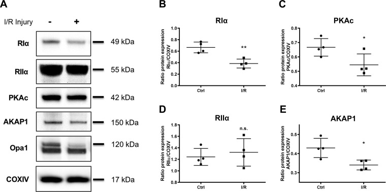
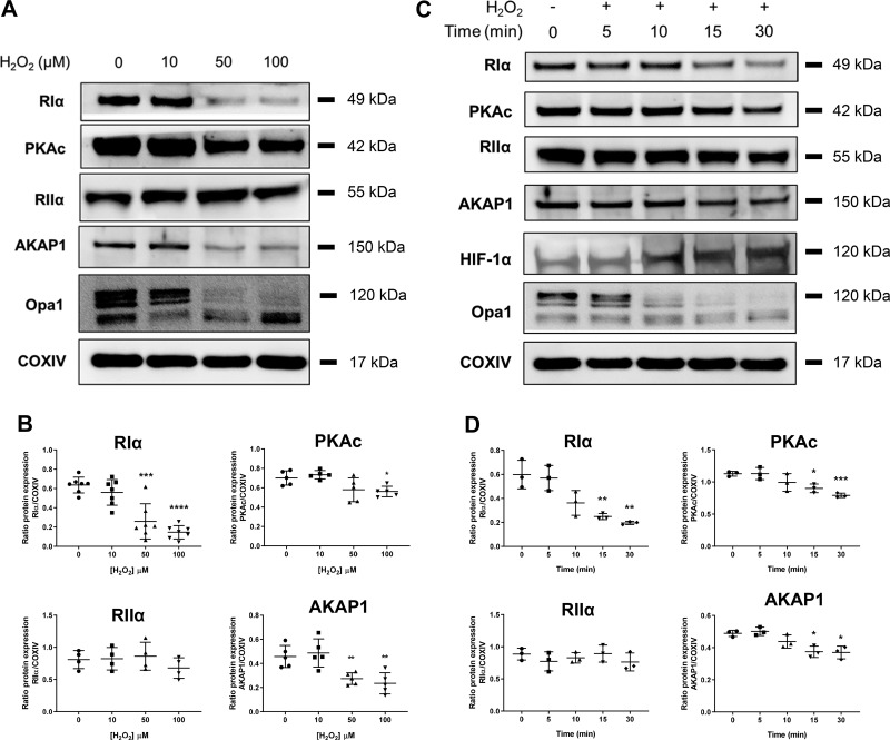
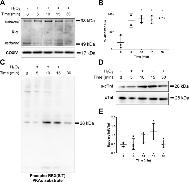
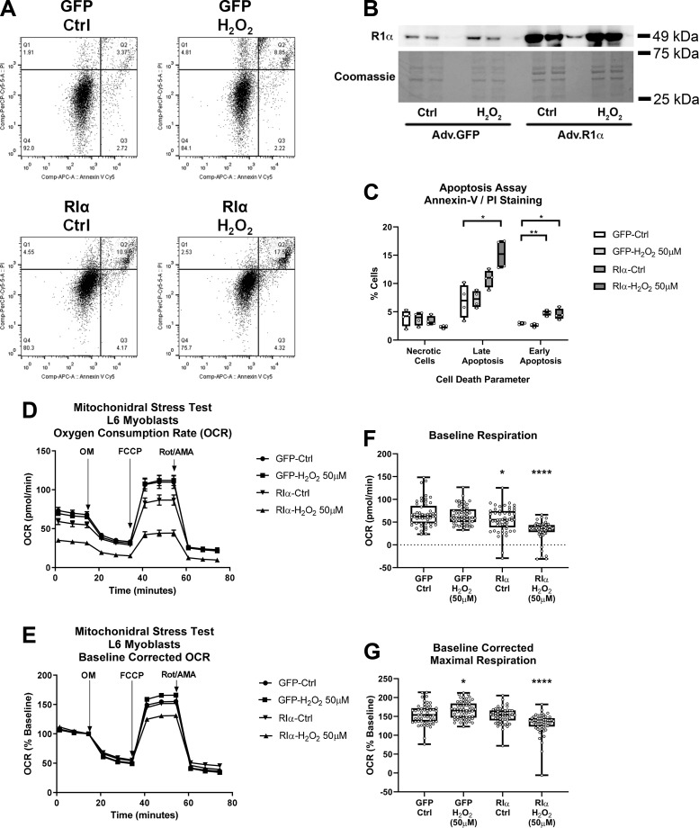
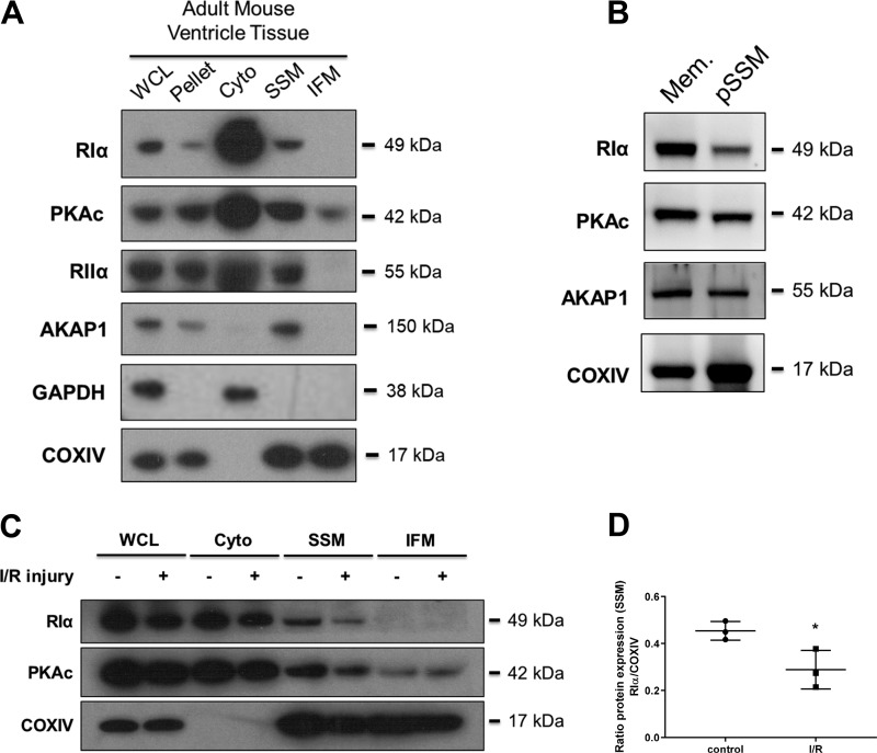
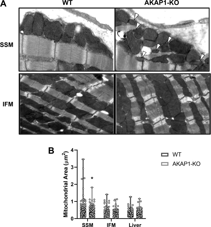
Type I PKA regulatory α-subunit (RIα; encoded by the Prkar1a gene) serves as the predominant inhibitor protein of the catalytic subunit of cAMP-dependent protein kinase (PKAc). However, recent evidence suggests that PKA signaling can be initiated by cAMP-independent events, especially within the context of cellular oxidative stress such as ischemia-reperfusion (I/R) injury. We determined whether RIα is actively involved in the regulation of PKA activity via reactive oxygen species (ROS)-dependent mechanisms during I/R stress in the heart. Induction of ex vivo global I/R injury in mouse hearts selectively downregulated RIα protein expression, whereas RII subunit expression appears to remain unaltered. Cardiac myocyte cell culture models were used to determine that oxidant stimulus (i.e., H2O2) alone is sufficient to induce RIα protein downregulation. Transient increase of RIα expression (via adenoviral overexpression) negatively affects cell survival and function upon oxidative stress as measured by increased induction of apoptosis and decreased mitochondrial respiration. Furthermore, analysis of mitochondrial subcellular fractions in heart tissue showed that PKA-associated proteins are enriched in subsarcolemmal mitochondria (SSM) fractions and that loss of RIα is most pronounced at SSM upon I/R injury. These data were supported via electron microscopy in A-kinase anchoring protein 1 (AKAP1)-knockout mice, where loss of AKAP1 expression leads to aberrant mitochondrial morphology manifested in SSM but not interfibrillar mitochondria. Thus, we conclude that modification of RIα via ROS-dependent mechanisms induced by I/R injury has the potential to sensitize PKA signaling in the cell without the direct use of the canonical cAMP-dependent activation pathway.NEW & NOTEWORTHY We uncovered a previously undescribed phenomenon involving oxidation-induced activation of PKA signaling in the progression of cardiac ischemia-reperfusion injury. Type I PKA regulatory subunit RIα, but not type II PKA regulatory subunits, is dynamically regulated by oxidative stress to trigger the activation of the catalytic subunit of PKA in cardiac myocytes. This effect may play a critical role in the regulation of subsarcolemmal mitochondria function upon the induction of ischemic injury in the heart.

Keywords: A-kinase anchoring protein 1; PKA regulatory subunit RIα; ischemia-reperfusion injury; mitochondria; oxidative stress.


Conflict of interest statement

H. H. Patel has equity as a founder in CavoGene LifeSciences Holdings, LLC.

Figures

Fig. 1.
Fig. 1.
Type I cAMP-dependent protein kinase (PKA) regulatory α-subunit (RIα) expression is reduced upon global ex vivo cardiac ischemia-reperfusion (I/R) injury. A: immunoblot analysis of tissue homogenate from adult mouse hearts exposed to ex vivo global I/R injury. Control perfusion heart samples were compared with hearts treated with 25 min ischemia followed by 30 min of reperfusion injury. Opa1, optic atrophy protein 1; COXIV, cytochrome-c oxidase. Corresponding scatter plots show protein expression quantification for RIα (B; P = 0.0039), catalytic subunit of PKA (PKAc) (C; P = 0.0487), RIIα (D; P = 0.5978), and A-kinase anchoring protein 1 (AKAP1) (E; P = 0.0297). Data are presented as means ± SD; n = 4 biological replicates. *P < 0.05; **P < 0.01; n.s., not significant.
Fig. 2.
Fig. 2.
Oxidative stress alone induces type I cAMP-dependent protein kinase (PKA) regulatory α-subunit (RIα) downregulation in adult mouse ventricular myocytes (AMVMs). A: immunoblot analysis of AMVM cell cultures treated for 30 min with the oxidant H2O2 at the indicated micromolar concentrations. Opa1, optic atrophy protein 1; COXIV, cytochrome-c oxidase. B: quantification of RIα, catalytic subunit of PKA (PKAc), RIIα, and A-kinase anchoring protein 1 (AKAP1) expression from H2O2 dose-response experiments in A. Data are presented as means ± SD; n = 4–7 biological replicates. *P < 0.05, **P < 0.01, ***P < 0.001, and ****P < 0.0001 compared with 0-µM treatment. C: immunoblot analysis of AMVM cell cultures treated with 100 μΜ H2O2 for the indicated time periods. D: quantification of RIα, PKAc, RIIα, and AKAP1 expression from 100 μM H2O2 time-course experiments from C. Data are presented as means ± SD; n = 3 biological replicates. *P < 0.05, **P < 0.01, and ***P < 0.001 compared with 0-min time point.
Fig. 3.
Fig. 3.
Oxidation of type I cAMP-dependent protein kinase (PKA) regulatory α-subunit (RIα) is correlated with increased catalytic subunit of PKA (PKAc) activity. A: immunoblot analysis comparing the extent of RIα protein oxidation upon time-course treatment with 100 μΜ Η2Ο2 in adult mouse ventricular myocytes cell cultures. COXIV, cytochrome-c oxidase. Dimer bands are considered oxidized due to the requirement of covalent disulfide bonds to maintain dimerization upon denaturing conditions of nonreducing SDS-PAGE. B: quantification of RIα protein oxidation, expressed as a percentage of oxidized RIα compared with total RIα protein (n = 2 to 3 biological replicates). Data are presented as means ± SD. *P < 0.05 compared with 0-min time point. C: immunoblot analysis comparing phospho-RRX(S/T) PKAc substrate phosphorylation within a time-course treatment with 100 μM H2O2 (n = 4 biological replicates). D: immunoblot analysis of cardiac troponin I (cTnI) phosphorylation within a time-course treatment with 100 µM H2O2. E: quantification of cTnI phosphorylation (p-cTnI) from D (n = 3–4 biological replicates). Data are presented as means ± SD. *P < 0.05 compared with 0-min time point.
Fig. 4.
Fig. 4.
Overexpression of type I cAMP-dependent protein kinase (PKA) regulatory α-subunit (RIα) in adult mouse ventricular myocytes (AMVMs) increases apoptosis upon oxidative stress. COXIV, cytochrome-c oxidase. A: immunoblot analysis of AMVM cell cultures first infected with adenovirus to overexpress regulatory α-subunit (RΙα) or green fluorescent protein (GFP) controls (Ad-RΙα and Ad-GFP, respectively) and then exposed to oxidant stress (10 µM H2O2) for 30 min. B: quantification of RIα expression from data in A (n = 4 biological replicates). Data are presented as means ± SD. *P < 0.05 compared with “Ad-GFP control (Ctrl)” condition. C: quantification of cleaved poly-ADP ribose polymerase (Cl PARP) expression from data in A (n = 4 biological replicates). Data are presented as means ± SD. *P < 0.05 compared with “Ad-GFP Ctrl” condition. D: immunoblot analysis of cardiac troponin I phosphorylation (p-TnI) from AMVM cells treated similarly as in A. E: quantification of p-cTnI data from D (n = 3 biological replicates). Data are presented as means ± SD. *P < 0.05; **P < 0.01.
Fig. 5.
Fig. 5.
Overexpression of type I cAMP-dependent protein kinase (PKA) regulatory α-subunit (RIα) in L6 myoblasts increases apoptosis and decreases mitochondrial function upon oxidative stress. A: representative scatter plot flow cytometry analysis of L6 myoblast cell cultures first infected with adenovirus to overexpress RΙα or green fluorescent protein (GFP) controls and then exposed to oxidant stress (50 µM H2O2) for 30 min, followed by staining with annexin-V and propidium iodide (PI) to detect apoptotic and necrotic cells. B: representative immunoblot and Coomassie stain analysis of RΙα expression from L6 myoblast cell cultures analyzed in A (n = 4 replicates). Adv, adenovirus. C: quantification of cell death parameters from data in A (necrotic cells, annexin-V/PI+; late apoptosis, annexin-V+/PI+; early apoptosis, annexin-V+/PI) (n = 4 replicates). Data are presented as means ± SD. *P < 0.05 compared with “GFP control (Ctrl)” condition, and **P < 0.01 compared with “GFP Ctrl” condition. D: Seahorse extracellular flux time-course analysis of mitochondrial “stress test” assays, assessing oxygen consumption rate (OCR) in GFP- or RIα-overexpressing L6 myoblasts treated with or without 50 µM H2O2. FCCP, carbonyl cyanide p-trifluoromethoxyphenylhydrazone; OM, oligomycin; Rot/AMA, rotenone/antimycin A. E: baseline corrected time-course analysis from data in D. F: quantification of baseline OCR data from D (n = 53–57 technical replicates summed over 3 independent experiments. Data are presented as means ± SE. *P < 0.05 and ****P < 0.0001 compared with “GFP Ctrl” condition. G: quantification of baseline corrected maximal respiration data from E (n = 53–57 technical replicates summed over 3 independent experiments). Data are presented as means ± SE. *P < 0.05 and ****P < 0.0001 compared with “GFP Ctrl” condition.
Fig. 6.
Fig. 6.
cAMP-dependent protein kinase (PKA) proteins are specifically enriched in subsarcolemmal mitochondria (SSM), and type I PKA regulatory α-subunit (RIα) expression is decreased in SSM with ischemia-reperfusion (I/R) injury. A: immunoblot analysis of PKA-associated proteins from untreated adult mouse heart ventricular tissue fractionated by differential centrifugation to separate mitochondrial subpopulations. AKAP1, A-kinase anchoring protein 1; COXVI, cytochrome-c oxidase; WCL, whole cell lysate; Pellet, nuclear/cytoskeleton pellet; Cyto, cytosol. B: immunoblot analysis of PKA-associated proteins within SSM subjected to density gradient ultracentrifugation. Mem, purified membrane; pSSM, purified SSM. C: immunoblot analysis of RΙα, catalytic subunit of PKA (PKAc), and COXIV from fractionated adult mouse heart ventricular tissue, comparing hearts that received either control perfusion or I/R injury. D: quantification of RIα expression from data in C, expressing changes of SSM-localized protein either with or without I/R injury stimulus (n = 3 biological replicates). Data are presented as means ± SD. *P < 0.05, compared on control condition.
Fig. 7.
Fig. 7.
Subsarcolemmal mitochondria (SSM) ultrastructure is perturbed in A-kinase anchoring protein 1 knockout (AKAP1-KO) mice. A: image analysis from transmission electron microscopy (TEM) performed on untreated mouse heart tissues from either wild-type (WT) or AKAP1-KO mice. Arrowheads indicate areas of decreased mitochondrial area. B: quantification of mitochondrial area (μm2) for heart SSM, heart interfibrillar mitochondria (IFM), and liver mitochondria, comparing WT and AKAP1-KO mice (n = 84–124 technical replicates summed over 10 biological replicates). Data are presented as means ± SE. *P < 0.05, compared with WT.

Similar articles

Cited by

References

    1. Aye TT, Soni S, van Veen TA, van der Heyden MA, Cappadona S, Varro A, de Weger RA, de Jonge N, Vos MA, Heck AJ, Scholten A. Reorganized PKA-AKAP associations in the failing human heart. J Mol Cell Cardiol 52: 511–518, 2012. doi:10.1016/j.yjmcc.2011.06.003. - DOI - PubMed
    1. Banky P, Huang LJ, Taylor SS. Dimerization/docking domain of the type Ialpha regulatory subunit of cAMP-dependent protein kinase. Requirements for dimerization and docking are distinct but overlapping. J Biol Chem 273: 35048–35055, 1998. doi:10.1074/jbc.273.52.35048. - DOI - PubMed
    1. Brennan JP, Bardswell SC, Burgoyne JR, Fuller W, Schröder E, Wait R, Begum S, Kentish JC, Eaton P. Oxidant-induced activation of type I protein kinase A is mediated by RI subunit interprotein disulfide bond formation. J Biol Chem 281: 21827–21836, 2006. doi:10.1074/jbc.M603952200. - DOI - PubMed
    1. Burgoyne JR, Eaton P. Transnitrosylating nitric oxide species directly activate type I protein kinase A, providing a novel adenylate cyclase-independent cross-talk to β-adrenergic-like signaling. J Biol Chem 284: 29260–29268, 2009. doi:10.1074/jbc.M109.046722. - DOI - PMC - PubMed
    1. Carlucci A, Adornetto A, Scorziello A, Viggiano D, Foca M, Cuomo O, Annunziato L, Gottesman M, Feliciello A. Proteolysis of AKAP121 regulates mitochondrial activity during cellular hypoxia and brain ischaemia. EMBO J 27: 1073–1084, 2008. doi:10.1038/emboj.2008.33. - DOI - PMC - PubMed

Publication types

MeSH terms

Substances

LinkOut - more resources