Review
. 2019 Nov;22(5):219-231.
doi: 10.14744/AnatolJCardiol.2019.91380.

Perivascular adipose tissue in cardiovascular diseases-an update

Affiliations
Review

Perivascular adipose tissue in cardiovascular diseases-an update

Adriana Grigoras et al. Anatol J Cardiol. 2019 Nov.

Abstract

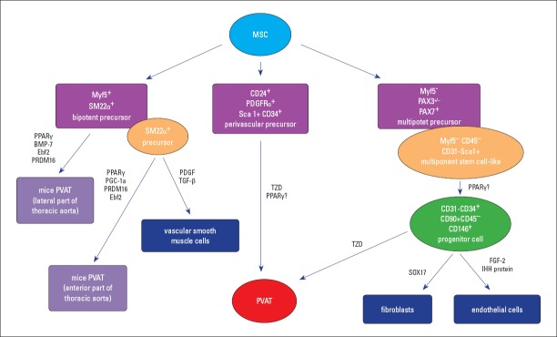
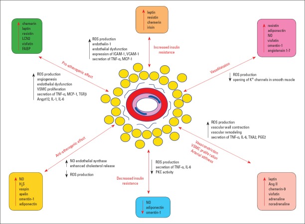
The perivascular adipose tissue (PVAT) has been recently recognized as an important factor in vascular biology, with implications in the pathogenesis of cardiovascular diseases. The cell types and the precursor cells of PVAT appear to be different according to their location, with the component cell type including white, brown, and beige adipocytes. PVAT releases a panel of adipokines and cytokines that maintain vascular homeostasis, but it also has the ability of intervention in the pathogenesis of the atherosclerotic plaques development and in the vascular tone modulation. In this review, we summarize the current knowledge and discuss the role of PVAT as a major contributing factor in the pathogenesis of ischemic coronary disease, hypertension, obesity, and diabetes mellitus. The new perspective of PVAT as an endocrine organ, along with the recent knowledge of the mechanisms involved in dysfunctional PVAT intervention in local vascular homeostasis perturbations, nowadays represent a new area of research in cardiovascular pathology, aiming to discover new therapeutic methods.


Conflict of interest statement

Conflict of interest: None declared.

Figures

Figure 1
Figure 1
Perivascular adipose tissue development Perivascular adipose tissue develops from a perivasculary PDGFRα+ precursor or from a Myf5-/Pax3+/− precursor cell for both fibroblasts and endothelial cells, a process regulated by SOX17, FGF-2, and IHH proteins. In experimental animals, PVAT from the lateral and anterior part of the thoracic aorta (tPVAT) originates from Myf5+ SM22α+, followed by preadipocytes differentiation into mature adipocytes, a process regulated by stimulatory factors, such as PPARγ, BMP-7, Ebf2, PRDM16, and PGC-1a. Another possibility of differentiation of the SM22α+ precursor is toward vascular smooth muscle cells, under the influence of PDGF and TGF-β. BMP-7 - bone morphogenetic protein 7; Ebf2 - early B-cell factor 2; FGF-2 - fibroblast growth factor 2; IHH protein - Indian hedgehog homolog; Myf5 - myogenic factor 5; MSC - mesenchymal stem cell; PDGF - platelet-derived growth factor; PGC-1a - peroxisome proliferator-activated receptor gamma coactivator 1 alpha; PPARγ - peroxisome proliferator-activated receptor gamma; PRDM16 - PR domain containing 16; PVAT - perivascular adipose tissue; SOX17 - transcription factor SOX-17; TGF-β - transforming growth factor beta; tPVAT - PVAT associated to thoracic aorta; TZD- thiazolidinedione
Figure 2
Figure 2
Perivascular adipose tissue implications in pathology Perivascular adipose tissue implications in pathology are mainly directed to the following pathways: increased insulin resistance (by an increased ROS and endothelin-1 production, endothelial dysfunction, the ICAM-1 and VCAM-1 expression, and the secretion of TNF-α and MCP-1) vs. decreased insulin resistance, vasoconstriction vs. dilatation, VSMC proliferation, arterial stiffness (by increased ROS production, strong vascular wall contraction, amplified remodeling, and enhanced secretion of TNF-α, IL-1, TXA2, and PGE2), and pro-atherogenic effects (by enhanced ROS genesis, angiogenesis stimulation, increased endothelial dysfunction, VSMC proliferation, and increased release of TNF-α, MCP-1, TGF-β, Angptl2, IL-1, and IL-6) vs. anti-atherogenic effects. Ang II - angiotensin II; Angptl2 - adipose tissue-specific angiopoietin-like 2; ICAM-1 - intercellular adhesion molecule 1; H2S - hydrogen sulfide; IL - interleukin; FABP - fatty acid binding protein 4; LCN2 - lipocalin-2; MCP-1 - monocyte chemoattractant protein-1; NO - nitric oxide; PGE2 - prostaglandin E2; PKC - protein kinase C; ROS - reactive oxygen species; TGF-β - transforming growth factor beta; TNF-α - tumor necrosis alpha; TXA2 - thromboxane A2; VCAM-1 - vascular cell adhesion protein 1; VSMC - vascular smooth muscle cells

Comment in

References

    1. Hildebrand S, Stümer J, Pfeifer A. PVAT and Its Relation to Brown, Beige, and White Adipose Tissue in Development and Function. Front Physiol. 2018;9:70. - PMC - PubMed
    1. Costa RM, Neves KB, Tostes RC, Lobato NS. Perivascular Adipose Tissue as a Relevant Fat Depot for Cardiovascular Risk in Obesity. Front Physiol. 2018;9:253. - PMC - PubMed
    1. Soltis EE, Cassis LA. Influence of perivascular adipose tissue on rat aortic smooth muscle responsiveness. Clin Exp Hypertens A. 1991;13:277–96. - PubMed
    1. Okamoto E, Couse T, De Leon H, Vinten-Johansen J, Goodman RB, Scott NA, et al. Perivascular inflammation after balloon angioplasty of porcine coronary arteries. Circulation. 2001;104:2228–35. - PubMed
    1. van Dam AD, Boon MR, Berbée JFP, Rensen PCN, van Harmelen V. Targeting white, brown and perivascular adipose tissue in atherosclerosis development. Eur J Pharmacol. 2017;816:82–92. - PubMed

MeSH terms

LinkOut - more resources