

Clinical and molecular characterization of primary sclerosing epithelioid fibrosarcoma of bone and review of the literature
- PMID: 31675134
- PMCID: PMC7082133
- DOI: 10.1002/gcc.22822
Clinical and molecular characterization of primary sclerosing epithelioid fibrosarcoma of bone and review of the literature
Abstract
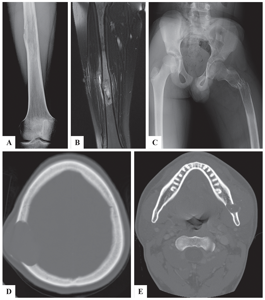
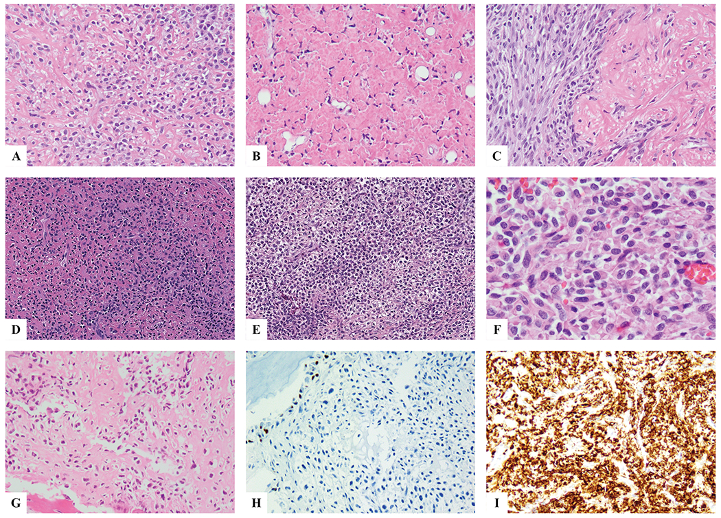
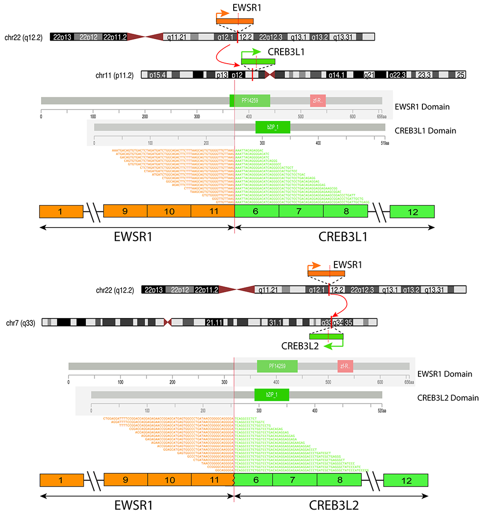
Sclerosing epithelioid fibrosarcoma (SEF) is a rare sarcoma subtype characterized by monomorphic epithelioid cells embedded in a densely sclerotic collagenous matrix. The overwhelming majority of tumors arise in soft tissues; however, rare cases have been documented to occur primarily in bone. The hallmarks of soft tissue SEF include MUC4 immunoreactivity and the presence of an EWSR1-CREB3L1 fusion. Rare cases with alternative fusions have also been reported such as EWSR1-CREB3L2 and FUS-CREB3L2 transcripts. The molecular alterations of skeletal SEF have not been well-defined, with only rare cases analyzed to date. In this study we investigated the clinicopathologic and molecular features of seven patients presenting with primary osseous SEF. There were 3 males and 4 females, with a mean age at diagnosis of 38 years. All cases had microscopic features within the histologic spectrum of SEF and showed strong and diffuse MUC4 positivity, while lacking SATB2 expression. However, due to its unusual presentation within bone, four cases were initially misinterpreted as either osteosarcoma, Ewing sarcoma or chondroblastoma. Half of the patients with follow-up data developed metastasis. The cases were tested by targeted RNA sequencing, MSK-IMPACT, and/or fluorescence in situ hybridization, showing EWSR1-CREB3L1 in six cases and EWSR1-CREB3L2 in one case. The fusion transcripts were composed of EWSR1 exon 11 to either exon 6 of CREB3L1 or CREB3L2. In summary, due to their rarity in the bone, skeletal SEF are often misdiagnosed, resulting in inadequate treatment modalities. Similar to their soft tissue counterpart, bone SEF follow an aggressive clinical behavior and show similar EWSR1-CREB3L1/CREB3L2 fusions.
Keywords: CREB3L1; CREB3L2; EWSR1; fusions; sclerosing epithelioid fibrosarcoma.
© 2019 Wiley Periodicals, Inc.
Figures
References
-
- Fletcher C, Bridge JA, Hogendoorn PC, et al. WHO Classification of Tumours of Soft Tissue and Bone. 4th Edition: IARC: Lyon; 2013.
-
- Guillou L, Benhattar J, Gengler C, et al. Translocation-positive low-grade fibromyxoid sarcoma: clinicopathologic and molecular analysis of a series expanding the morphologic spectrum and suggesting potential relationship to sclerosing epithelioid fibrosarcoma: a study from the French Sarcoma Group. Am J Surg Pathol. 2007;31:1387–1402. - PubMed
-
- Antonescu CR, Rosenblum MK, Pereira P, et al. Sclerosing epithelioid fibrosarcoma: a study of 16 cases and confirmation of a clinicopathologically distinct tumor. Am J Surg Pathol. 2001;25:699–709. - PubMed
-
- Wojcik JB, Bellizzi AM, Dal Cin P, et al. Primary sclerosing epithelioid fibrosarcoma of bone: analysis of a series. Am J Surg Pathol. 2014;38:1538–1544. - PubMed
