. 2019 Nov 27;62(22):10182-10203.
doi: 10.1021/acs.jmedchem.9b01065. Epub 2019 Nov 18.

Drug Design Targeting T-Cell Factor-Driven Epithelial-Mesenchymal Transition as a Therapeutic Strategy for Colorectal Cancer

Affiliations

Drug Design Targeting T-Cell Factor-Driven Epithelial-Mesenchymal Transition as a Therapeutic Strategy for Colorectal Cancer

Adedoyin D Abraham et al. J Med Chem. .

Abstract

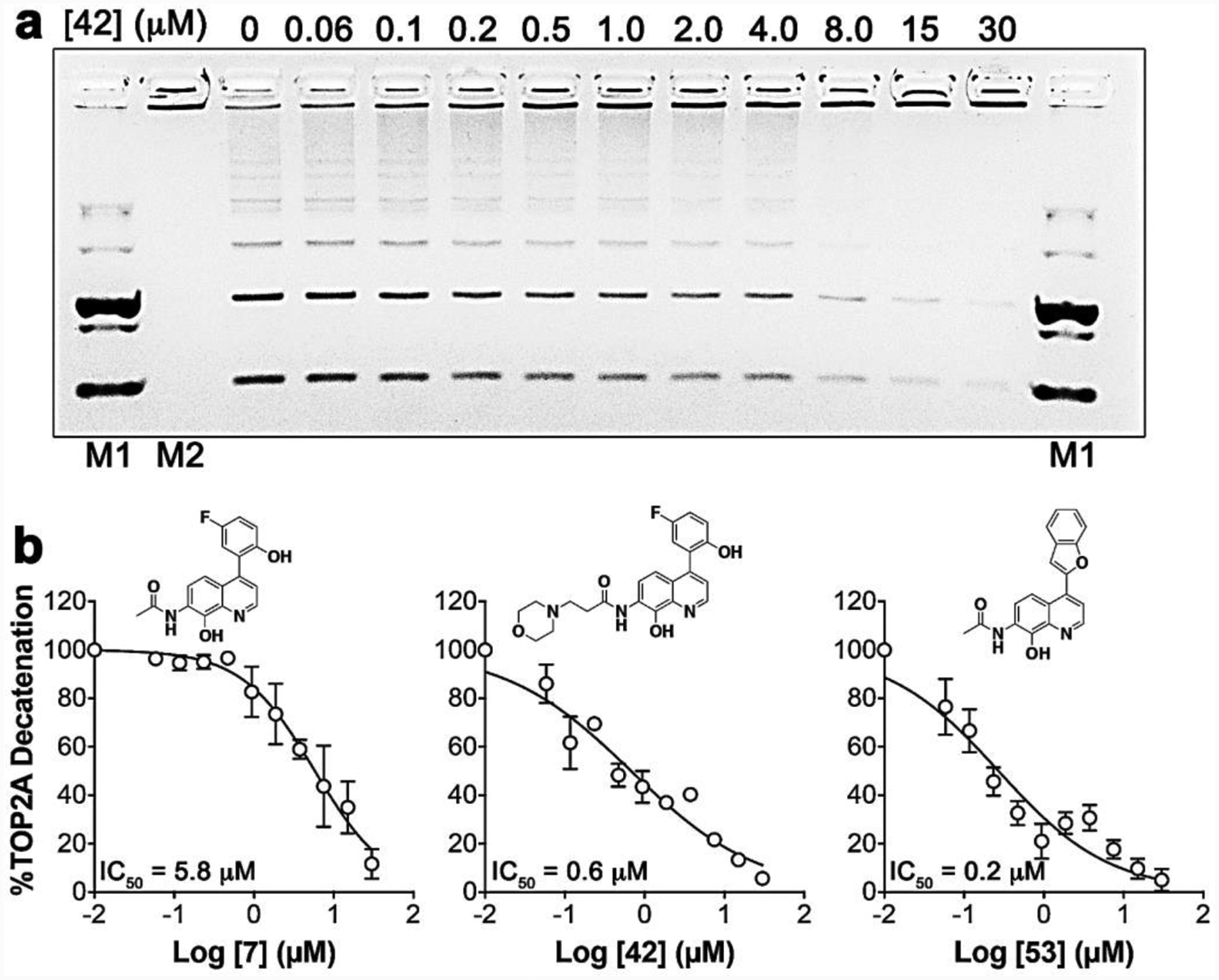
Metastasis is the cause of 90% of mortality in cancer patients. For metastatic colorectal cancer (mCRC), the standard-of-care drug therapies only palliate the symptoms but are ineffective, evidenced by a low survival rate of ∼11%. T-cell factor (TCF) transcription is a major driving force in CRC, and we have characterized it to be a master regulator of epithelial-mesenchymal transition (EMT). EMT transforms relatively benign epithelial tumor cells into quasi-mesenchymal or mesenchymal cells that possess cancer stem cell properties, promoting multidrug resistance and metastasis. We have identified topoisomerase IIα (TOP2A) as a DNA-binding factor required for TCF-transcription. Herein, we describe the design, synthesis, biological evaluation, and in vitro and in vivo pharmacokinetic analysis of TOP2A ATP-competitive inhibitors that prevent TCF-transcription and modulate or reverse EMT in mCRC. Unlike TOP2A poisons, ATP-competitive inhibitors do not damage DNA, potentially limiting adverse effects. This work demonstrates a new therapeutic strategy targeting TOP2A for the treatment of mCRC and potentially other types of cancers.


Figures

Figure 1.
Figure 1.. Structure based drug design inspired by neo.
(a) 3D rendering of the molecular model of neo docked into the ATP-binding site of the N-terminal domain of TOP2A (PDB:1ZXM, 1.87Å). The colored regions represent hydrophobic (blue), hydrophilic (green), Walker A motif (orange) residues, and Mg2+ (pink sphere). Based on this model, the pharmacophore of neo was deduced to be a substituted quinoline shown as red bonds in the chemical structure. Novel TOP2A ATP-competitive inhibitor prototypes 3 (b) and 7 (c) were designed using CADD and the pharmacophore of neo. TOP2A ATP-binding site interactions are shown with a 2D rendering as dashed lines: H-bonds (black), Mg2+ π-cation or chelation (orange), π-sigma (pink), π-loan pair (blue), and halogen hydrogen bond (red).
Figure 2.
Figure 2.
TOP2A enzyme Michaelis-Menten inhibition studies measuring ATP hydrolysis with (a) neo, (b) 3, and (c) 7. Like neo, the quinoline based pharmacophores of 3 and 7 display an ATP-competitive mode of inhibition against TOP2A, evidenced by shifts in KM with no changes in Vmax. (d) TOP2A ATP-competitive inhibitors Neo, 7, and BAP1 do not cause DNA DSBs, over a concentration range from 0–40 μM, measured by H2AX S139 phosphorylation immunofluorescence, while the TOP2A poison, etoposide, caused significant DNA damage over a concentration range from 5 to 100 μM. (e) Representative fluorescence images showing that neo and 7 do not cause induction of γ-HA2X, while etoposide caused significant induction of γ-HA2X.
Figure 3.
Figure 3.. Antitumor activity of prototypes 3 and 7.
Compounds 7 (a) and 3 (b) demonstrate the ability to significantly inhibit TOP2A-dependent TCF-transcription using SW620 tumor organoids transduced with the TOPflash luminescent reporter. Compounds 7 (c) and 3 (d) also demonstrated the ability to reverse EMT using SW620 tumor organoids transduced with EcadPro-RFP or VimPro-GFP EMT biomarker reporters over a 72 h treatment time course as indicated. Reversion of EMT is characterized by downregulation of vimentin and upregulation of E-cadherin promoter activity. Representative tumor organoid images are shown above each bar graph. (e) Compounds 7 and 3 also significantly inhibit colony formation over seven days, which is a measure of the loss of cancer stem cell stemness. (f) SW620 tumor organoids were treated with 7 and 3 for 72 h, tumor organoids were dissociated, and viable cell invasive potential was measured for additional 72h incubation. Compounds 3 and 7 both significantly inhibit the invasive potential of SW620 cells. Significance was determined using the Student’s t-test analysis.
Figure 4.
Figure 4.. Dose response studies measuring inhibition of TOP2A decatenation.
(a) Prototype 7 analog dose response curves were generated using DNA decatenation gels as demonstrated with compound 42. Control lanes with DNA markers, include: decatenated DNA (M1) and Catenated DNA (M2). Dose response studies were conducted using 2-fold dilutions as indicated. (b) Representative dose response curves with analogs 7, 42 and 53.
Figure 5.
Figure 5.. Structure activity relationship summary.
(a) Compound 7 docked into the TOP2A ATP binding site highlighting fluorine H-bond interaction (black dashed line). (b) Similarly, the trifluoromethyl group of 32 forms fluorine H-bonds with Arg98 (black dashed lines). (c) The SAR summary of TOP2A inhibitors, demonstrating water solubility with compound 42.
Figure 6.
Figure 6.
(a) dose responses for the reversion of EMT biomarker reporter activity in SW620 tumor organoids. (b) Representative dose response images for both vimentin and E-cadherin reporter activity. At higher doses we observed potential early stages of cell death indicated by loss of RFP reporter activity. (c) representative brightfield images indicating TOP2A inhibitors prevent SW620 tumor organoid growth compared to DMSO (scale bars = 250 μm). (d) SW620 tumor organoid volumes were measured at 72 h quantifying compound effects on tumor growth. (e) At the highest doses, all compounds display an induction of apoptosis based on cleaved caspase 3 and 7 activity with 42 and 60 at ~9-fold and ~3-, respectively.
Figure 7.
Figure 7.. TOP2A inhibitor effects on malignant properties of SW620 tumor organoids.
(a) The clonogenic assay results with TOP2A inhibitors demonstrate significant inhibition of CSC colony formation. The inset is a representative image of a DMSO control well of a 6-well plate. The expansion view shows detail of colonies formed, which are identified using area (μm2) min: 1×10⁴ and max: 4×10⁵, and masked in orange color for contrast. (b) The invasion assay results with TOP2A inhibitors demonstrate significant loss of invasive potential over days 1 through 4. The results in panel a and b were determined to be statistically significant using one-way ANOVA analysis, where P ≤ 0.0008 for each analog tested.
Scheme 1.
Scheme 1.
Syntheses of quinolines 3 and 7 prototype TOP2A ATP-competitive inhibitors.
Scheme 2.
Scheme 2.
Syntheses of prototype 7 designed analogs.
Scheme 3.
Scheme 3.
Syntheses of prototype 7 analogs with position 4 aromatic ring variations.

References

    1. Howlader N; Noone A; Krapcho M; Garshell J; Miller D; Altekruse S; Kosary C; Yu M; Ruhl J; Tatalovich Z; Mariotto A; Lewis D; Chen H; Feuer E; Cronin K. e. SEER Cancer Statistics Review, 1975–2013, National Cancer Institute; Bethesda, MD, http://seer.cancer.gov/csr/1975_2013/, based on November 2015 SEER data submission, posted to the SEER web site, April 2016. ((accessed Nov 7, 2004)).
    1. Clevers H Wnt/β-catenin signaling in development and disease. Cell 2006, 127, 469–480. - PubMed
    1. Cancer Genome Atlas N Comprehensive molecular characterization of human colon and rectal cancer. Nature 2012, 487, 330–337. - PMC - PubMed
    1. Sánchez-Tilló E; de Barrios O; Siles L; Cuatrecasas M; Castells A; Postigo A β-catenin/TCF4 complex induces the epithelial-to-mesenchymal transition (EMT)-activator ZEB1 to regulate tumor invasiveness. Proc. Natl. Acad. Sci. U.S.A 2011, 108, 19204–19209. - PMC - PubMed
    1. Chaffer CL; San Juan BP; Lim E; Weinberg RA EMT, cell plasticity and metastasis. Cancer Metast. Rev 2016. - PubMed

Publication types

MeSH terms

Substances