. 2019 Nov 1;17(11):e3000499.
doi: 10.1371/journal.pbio.3000499. eCollection 2019 Nov.

CEH-60/PBX regulates vitellogenesis and cuticle permeability through intestinal interaction with UNC-62/MEIS in Caenorhabditis elegans

Affiliations

CEH-60/PBX regulates vitellogenesis and cuticle permeability through intestinal interaction with UNC-62/MEIS in Caenorhabditis elegans

Pieter Van de Walle et al. PLoS Biol. .

Abstract

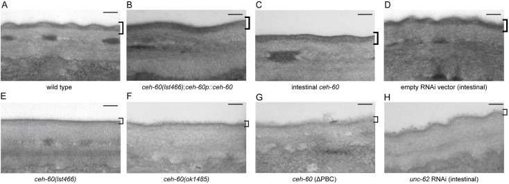
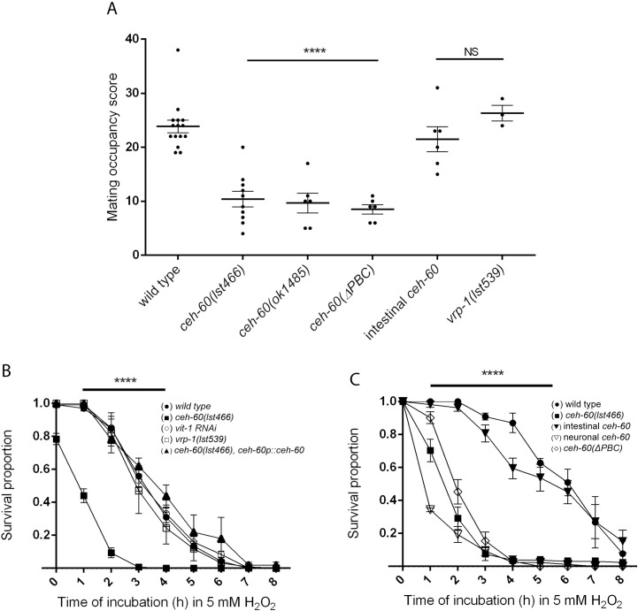
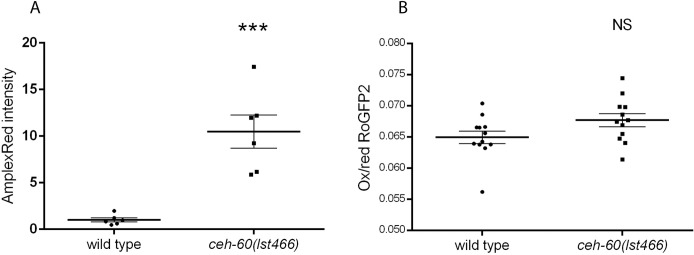
The onset of sexual maturity involves dramatic changes in physiology and gene expression in many animals. These include abundant yolk protein production in egg-laying species, an energetically costly process under extensive transcriptional control. Here, we used the model organism Caenorhabditis elegans to provide evidence for the spatiotemporally defined interaction of two evolutionarily conserved transcription factors, CEH-60/PBX and UNC-62/MEIS, acting as a gateway to yolk protein production. Via proteomics, bimolecular fluorescence complementation (BiFC), and biochemical and functional readouts, we show that this interaction occurs in the intestine of animals at the onset of sexual maturity and suffices to support the reproductive program. Our electron micrographs and functional assays provide evidence that intestinal PBX/MEIS cooperation drives another process that depends on lipid mobilization: the formation of an impermeable epicuticle. Without this lipid-rich protective layer, mutant animals are hypersensitive to exogenous oxidative stress and are poor partners for mating. Dedicated communication between the hypodermis and intestine in C. elegans likely supports these physiological outcomes, and we propose a fundamental role for the conserved PBX/MEIS interaction in multicellular signaling networks that rely on lipid homeostasis.


Conflict of interest statement

The authors have declared that no competing interests exist.

Figures

Fig 1
Fig 1. ceh-60 is expressed in the intestine from the L4 larval stage onwards.
Bright-field and GFP images of a ceh-60p::ceh-60::SL2::gfp reporter strain showing expression in the intestine (*) and pharynx (arrow) of (A) adult and (B) L4 animals. Intestinal expression is weaker in L4 larvae than in adults. (C) Expression in the intestine is not clearly observed in the L3 larval stage or before. Neuronal expression, while present (S2 Fig), is not clearly visible at this magnification. White dotted lines mark the outline of the L3 animal. Scale bar, 200 μm. GFP, green fluorescent protein.
Fig 2
Fig 2. Intestinal expression and an intact PBC domain are essential for CEH-60’s vitellogenesis-regulating function.
(A) Upon electrophoresis of total protein extracts, yolk proteins (YP170, YP115, and YP88) are present as abundant bands in wild-type animals but not in ceh-60(lst466) or in ceh-60(ok1485). Restoring ceh-60 expression under its own promoter (ceh-60p::ceh-60) or in the intestine (elt-2p::ceh-60) rescues the presence of yolk proteins in ceh-60(lst466) mutants, but expressing ceh-60 in the AWC neurons (odr-1p::ceh-60) or the pharyngeal muscles (myo-2p::ceh-60) does not. Truncating the PBC domain of resupplied CEH-60 (ceh-60p::ceh-60(ΔPBC)) also does not rescue yolk protein production in ceh-60 mutants. Yolk protein band identity is based on [7,27]. (B) Quantification of yolk proteins YP170, YP115, and YP88, normalized against total protein present in a lane and rescaled so that each YP has a mean abundance of 1 in wild type. **p < 0.01, ***p < 0.001, ****p < 0.0001. Error bars = SEM. N ≥ 3. Underlying data are available in S1 Data. NS, not significant; YP, yolk protein.
Fig 3
Fig 3. Bimolecular fluorescence shows in vivo interaction between UNC-62 and CEH-60 in the adult intestine.
(A) Bright-field and YFP images of animals carrying hsp-16.41p::ceh-20::VC155 and hsp-16.41p::unc-62::VN173. In vivo interaction between CEH-20 and UNC-62 reconstitutes fluorescence ubiquitously after heat shock (33°C, 2 hours), most clearly visible in the intestinal nuclei. (B) Interaction between CEH-60 and UNC-62 reconstitutes fluorescence in animals carrying hsp-16.41p::ceh-60::VC55 and hsp-16.41p::unc-62::VN173 after heat shock (33°C, 2 hours). (C) Interaction between CEH-60 and UNC-62 is not observed upon expression of a version of CEH-60 in which the PBC domain is not present, CEH-60(ΔPBC), indicating that this domain is needed for the interaction. (D) Quantification of Venus fluorescence for interaction between UNC-62 and CEH-20, CEH-60, or CEH-60(ΔPBC). One dot represents average fluorescence intensity in 6 intestinal nuclei per animal. ****p < 0.0001. N ≥ 6. Underlying data are available in S1 Data. (E) Bright-field and YFP images of animals carrying ceh-60p::ceh-60::VC155 and unc-62p::unc-62::VN173 transgenes, providing spatiotemporal specificity to the endogenous in vivo interaction of CEH-60 and UNC-62 in the adult intestine (solid arrows). No YFP signal was observed in other tissues besides the intestine. Reflecting the extrachromosomal nature of the transgenes, not all transgenic adult animals showed clear YFP in the intestine. Higher magnifications were used for endogenous BiFC because the YFP signal is much weaker compared to that of induced heat-shock promoters (A-C). Dotted arrow = signal of co-injection marker unc-122p::DsRed, * = fluorescent gut granule. Scale bar for A, B, and C = 100 μm. Scale bar for D = 50 μm. (F) Western blots detecting HA-tagged CEH-20, HA-tagged CEH-60, HA-tagged CEH-60(ΔPBC), or MYC-tagged UNC-62 in anti-HA immunoprecipitations using anti-HA and anti-MYC antibodies. CEH-20 and CEH-60, but not CEH-60(ΔPBC), co-immunoprecipitate UNC-62. A.U., arbitrary unit; BiFC, bimolecular fluorescence complementation; NS, not significant; YFP, yellow fluorescent protein.
Fig 4
Fig 4. ceh-60 mutants have a more permeable cuticle.
(A) Representative images of acridine orange staining in wild-type and ceh-60(lst466) animals. Dotted white lines in wild-type acridine orange images show worm outline. Scale bar = 200 μm. (B) Acridine orange stains ceh-60 mutants but not wild-type animals. Expressing wild-type ceh-60 under the control of its own promoter (ceh-60p::ceh-60) or under an intestinal promoter (elt-2p::ceh-60) rescues the defect in ceh-60(lst466) mutants, but neuronal (odr-1p::ceh-60), pharyngeal (myo-2p::ceh-60), or PBC-truncated (ceh-60p::ceh-60(ΔPBC)) expression does not. Fluorescence intensity is shown on a logarithmic scale for clarity. ****p < 0.0001. N ≥ 20. Underlying data are available in S1 Data. A.U., arbitrary unit; NS, not significant.
Fig 5
Fig 5. TEM unveils epicuticle problems in ceh-60 mutants.
The epicuticle is visible as a thick electron-dense layer at the outer side of the cuticle in wild-type animals (A), or when ceh-60(lst466) mutants are rescued with endogenous (ceh-60p::ceh-60 [B]) or intestinal (elt-2::ceh-60 [C]) ceh-60 expression. In contrast, the epicuticle is a thin and underdeveloped layer in ceh-60 mutants (E,F), or when a variant of CEH-60 with truncated PBC-interaction domain is expressed (ceh-60p::ceh-60(ΔPBC) [G]). Treatment with intestinal unc-62 RNAi (H) results in an epicuticle that is intermediate between those of empty vector–treated animals (D) and ceh-60 mutants. Square brackets indicate the epicuticle region in all panels. Scale bar = 100 nm. RNAi, RNA interference; TEM, transmission electron microscopy.
Fig 6
Fig 6. CEH-60 is essential for normal mating contact and survival of oxidative stress.
(A) The mating occupancy score of ceh-60 but not vrp-1 mutants is lower than wild-type animals, indicating a defect in mating contact that is not caused by lowered yolk protein production. Mating contact deficiency is rescued by expressing ceh-60 under the control of its own promoter (ceh-60p::ceh-60) or in the intestine (elt-2p::ceh-60) of ceh-60(lst466) mutant animals, but not when expressing PBC-truncated ceh-60 (ceh-60p::ceh-60(ΔPBC)). N = 3 for vrp-1(lst539). N ≥ 6 for other conditions. (B) Oxidative stress survival as measured by fraction of worms alive during incubation in 5 mM H2O2 is lower in ceh-60(lst466) mutants (■) than in controls (●). The down-regulation of VITs in vit-1 RNAi treated animals (○) or vrp-1(lst539) animals (□) does not cause increased susceptibility to oxidative stress. Expression of ceh-60 under its own promoter (ceh-60p::ceh-60) in ceh-60(lst466) mutants (▲) is able to rescue stress survival. (C) Oxidative stress survival in ceh-60(lst466) animals (■) is rescued by intestinal expression of ceh-60 (elt-2p::ceh-60, ) but not by expression of ceh-60 in the AWC neurons (odr-1p::ceh-60, ▽) or expression of ceh-60 with a truncated PBC-interaction domain (ceh-60p::ceh-60(ΔPBC), ◇). N ≥ 3. Error bars indicate SEM. ***p < 0.001, ****p < 0.0001. Underlying data are available in S1 Data. RNAi, RNA interference; VIT, vitellogenin.
Fig 7
Fig 7. Dysfunctional CEH-60 causes a cuticle that is hyperpermeable to endogenous ROS.
(A) In supernatants collected from ceh-60(lst466) animals, the intensity of Amplex Red absorbance is much higher than in that of wild-type animals. Values are normalized to control. N = 6. (B) Endogenous ROS production measured with the in vivo genetic ROS sensor RoGFP2, indicated as the ratio of oxidized over reduced GFP, is the same in ceh-60 and wild-type animals. Error bars, SEM; ***p < 0.001. N = 12. Underlying data are available in S1 Data. GFP, green fluorescent protein; NS, not significant; Ox/red, ratio of oxidized over reduced GFP; RoGFP2, reduction-oxidation sensitive green fluorescent protein; ROS, reactive oxygen species.

References

    1. Bownes M, Lineruth M, Mauchline D. Egg production and fertility in Drosophila depend upon the number of yolk-protein gene copies. Mol Genet genomics. 1991;228: 324–327. 10.1007/BF00282485 - DOI - PubMed
    1. Yilmaz O, Patinote A, Nguyen T, Com E, Pineau C, Bobe J. Genome editing reveals reproductive and developmental dependencies on specific types of vitellogenin in zebrafish (Danio rerio). Molecular Reproduction and Development. 2019; 86: 1168–1188. 10.1002/mrd.23231 - DOI - PubMed
    1. Van Rompay L, Borghgraef C, Beets I, Caers J, Temmerman L. New genetic regulators question relevance of abundant yolk protein production in C. elegans. Sci Rep. 2015; 1–16. 10.1038/srep16381 - DOI - PMC - PubMed
    1. Grant B, Hirsh D. Receptor-mediated endocytosis in the Caenorhabditis elegans oocyte. Mol Biol Cell. 1999;10: 4311–4326. 10.1091/mbc.10.12.4311 - DOI - PMC - PubMed
    1. Kimble J, Sharrock J. Tissue-specific synthesis of yolk proteins in Caenorhabditis elegans. Dev Biol. 1982; 10.1016/0012-1606(83)90322-6 - DOI - PubMed

Publication types

MeSH terms