

Elevating pancreatic cystic lesion stratification: Current and future pancreatic cancer biomarker(s)
- PMID: 31676330
- PMCID: PMC6980327
- DOI: 10.1016/j.bbcan.2019.188318
Elevating pancreatic cystic lesion stratification: Current and future pancreatic cancer biomarker(s)
Abstract
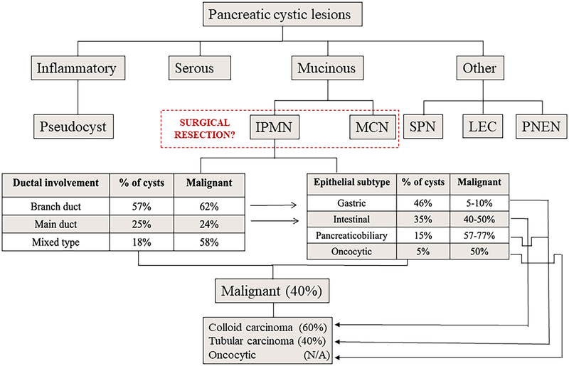
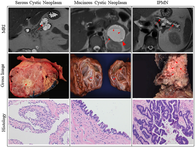
Pancreatic ductal adenocarcinoma (PDAC) is an incredibly deadly disease with a 5-year survival rate of 9%. The presence of pancreatic cystic lesions (PCLs) confers an increased likelihood of future pancreatic cancer in patients placing them in a high-risk category. Discerning concurrent malignancy and risk of future PCL progression to cancer must be carefully and accurately determined to improve survival outcomes and avoid unnecessary morbidity of pancreatic resection. Unfortunately, current image-based guidelines are inadequate to distinguish benign from malignant lesions. There continues to be a need for accurate molecular and imaging biomarker(s) capable of identifying malignant PCLs and predicting the malignant potential of PCLs to enable risk stratification and effective intervention management. This review provides an update on the current status of biomarkers from pancreatic cystic fluid, pancreatic juice, and seromic molecular analyses and discusses the potential of radiomics for differentiating PCLs harboring cancer from those that do not.
Keywords: Biomarker; Early detection; IPMN; Pancreatic cancer; Pancreatic cystic lesions; Pancreatic ductal adenocarcinoma; Radiomics.
Copyright © 2019. Published by Elsevier B.V.
Conflict of interest statement
Declaration of competing interest
Dr. Surinder Batra is a co-founder of Sanguine Diagnostics and Therapeutics.
Figures
References
-
- Siegel RL, Miller KD, Jemal A, Cancer statistics 2019, Cancer statistics, 2019, Cancer J. Clin 69 (1) (2019) 7–34. - PubMed
-
- Neoptolemos JP, Kleeff J, Michl P, Costello E, Greenhalf W, Palmer DH, Therapeutic developments in pancreatic cancer: current and future perspectives, Nat Rev Gastroenterol Hepatol 15 (6) (2018) 333–348. - PubMed
-
- Khorana AA, Mangu PB, Berlin J, Engebretson A, Hong TS, Maitra A, et al., Potentially curable pancreatic cancer: American Society of Clinical Oncology Clinical Practice Guideline Update, J. Clin. Oncol 35 (20) (2017) 2324–2328. - PubMed
-
- Mukewar SS, Sharma A, Phillip N, Gupta R, Aryal-Khanal A, de Pretis N, et al., Risk of pancreatic cancer in patients with pancreatic cysts and family history of pancreatic cancer, Clin. Gastroenterol. Hepatol 16 (7) (2018) 1123–1130. - PubMed
Publication types
MeSH terms
Substances
Grants and funding
LinkOut - more resources
Full Text Sources
Medical
