

Mesothelin Enhances Tumor Vascularity in Newly Forming Pancreatic Peritoneal Metastases
- PMID: 31676721
- PMCID: PMC8139242
- DOI: 10.1158/1541-7786.MCR-19-0688
Mesothelin Enhances Tumor Vascularity in Newly Forming Pancreatic Peritoneal Metastases
Abstract
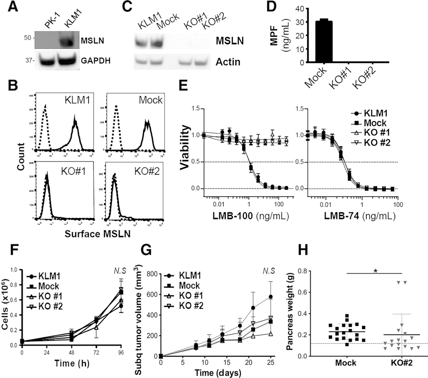
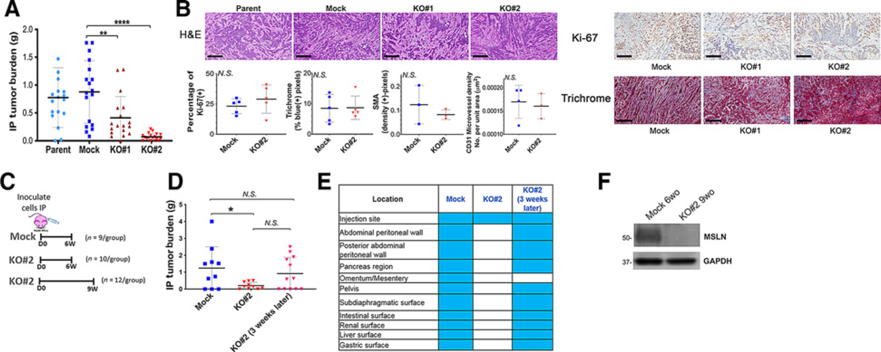
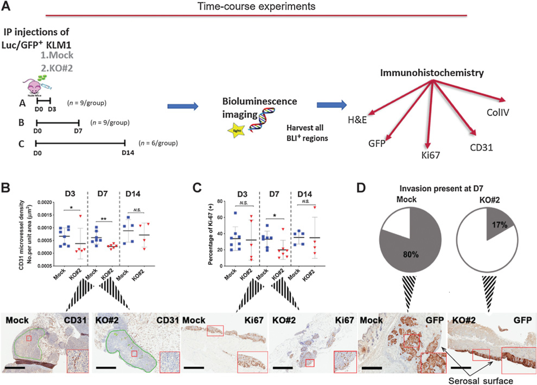
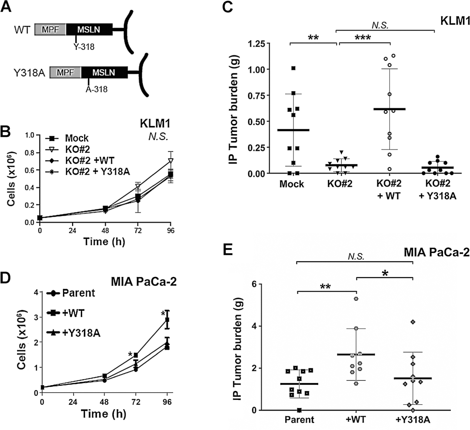
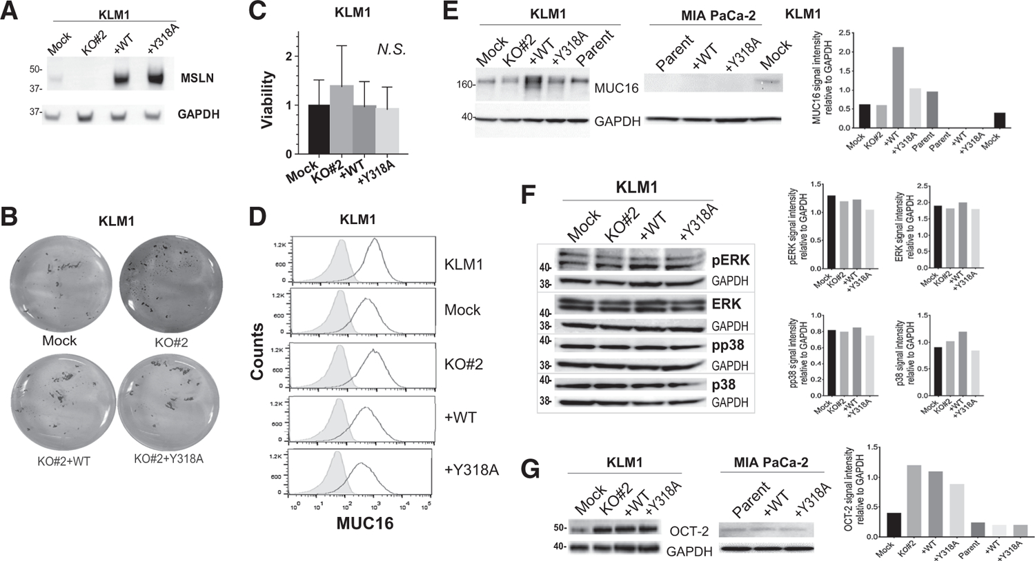
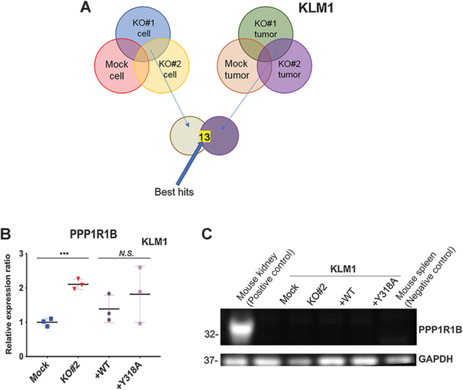
Over 90% of pancreatic ductal adenocarcinomas (PDAC) express mesothelin (MSLN). Overexpression or knockdown of MSLN has been implicated in PDAC aggressiveness. This activity has been ascribed to MSLN-induced activation of MAPK or NF-κB signaling pathways and to interaction of MSLN with its only known binding partner, MUC16. Here, we used CRISPR/Cas9 gene editing to delete MSLN from PDAC, then restored expression of wild-type (WT) or Y318A mutant MSLN by viral transduction. We found that MSLN KO cells grew in culture and as subcutaneous tumors in mouse xenografts at the same rate as WT cells but formed intraperitoneal metastases poorly. Complementation with WT MSLN restored intraperitoneal growth, whereas complementation with Y318A mutant MSLN, which does not bind MUC16, was ineffective at enhancing growth in both MUC16(+) and MUC16(-) models. Restoration of WT MSLN did enhance growth but did not affect cell-to-cell binding, cell viability in suspension or signaling pathways previously identified as contributing to the protumorigenic effect of MSLN. RNA deep sequencing of tumor cells identified no changes in transcriptional profile that could explain the observed phenotype. Furthermore, no histologic changes in tumor cell proliferation or morphology were observed in mature tumors. Examination of nascent MSLN KO tumors revealed decreased microvascular density as intraperitoneal tumors were forming, followed by decreased proliferation, which resolved by 2 weeks postimplantation. These data support a model whereby MSLN expression by tumor cells contributes to metastatic colonization. IMPLICATIONS: MSLN confers a growth advantage to tumor cells during colonization of peritoneal metastasis. Therapeutic blockade of MSLN might limit peritoneal spread.
©2019 American Association for Cancer Research.
Conflict of interest statement
Disclosure of Potential Conflicts of Interest
No potential conflicts of interest were disclosed.
Figures
Similar articles
-
Excess shed mesothelin disrupts pancreatic cancer cell clustering to impair peritoneal colonization.FASEB J. 2024 Dec 13;38(24):e70247. doi: 10.1096/fj.202400446R. FASEB J. 2024. PMID: 39673668 Free PMC article.
-
Host Mesothelin Expression Increases Ovarian Cancer Metastasis in the Peritoneal Microenvironment.Int J Mol Sci. 2021 Nov 18;22(22):12443. doi: 10.3390/ijms222212443. Int J Mol Sci. 2021. PMID: 34830322 Free PMC article.
-
Mesothelin-based CAR-T cells exhibit potent antitumor activity against ovarian cancer.J Transl Med. 2024 Apr 18;22(1):367. doi: 10.1186/s12967-024-05174-y. J Transl Med. 2024. PMID: 38637885 Free PMC article.
-
Peritoneal dissemination of ovarian cancer: role of MUC16-mesothelin interaction and implications for treatment.Expert Rev Anticancer Ther. 2018 Feb;18(2):177-186. doi: 10.1080/14737140.2018.1418326. Epub 2017 Dec 21. Expert Rev Anticancer Ther. 2018. PMID: 29241375 Review.
-
From Malignant Progression to Therapeutic Targeting: Current Insights of Mesothelin in Pancreatic Ductal Adenocarcinoma.Int J Mol Sci. 2020 Jun 6;21(11):4067. doi: 10.3390/ijms21114067. Int J Mol Sci. 2020. PMID: 32517181 Free PMC article. Review.
Cited by
-
Tumor resistance to anti-mesothelin CAR-T cells caused by binding to shed mesothelin is overcome by targeting a juxtamembrane epitope.Proc Natl Acad Sci U S A. 2024 Jan 23;121(4):e2317283121. doi: 10.1073/pnas.2317283121. Epub 2024 Jan 16. Proc Natl Acad Sci U S A. 2024. PMID: 38227666 Free PMC article.
-
Current advances and outlooks in immunotherapy for pancreatic ductal adenocarcinoma.Mol Cancer. 2020 Feb 15;19(1):32. doi: 10.1186/s12943-020-01151-3. Mol Cancer. 2020. PMID: 32061257 Free PMC article. Review.
-
High mesothelin expression by immunohistochemistry predicts improved survival in pleural mesothelioma.Histopathology. 2023 Aug;83(2):202-210. doi: 10.1111/his.14916. Epub 2023 Apr 11. Histopathology. 2023. PMID: 37040900 Free PMC article.
-
Anti-Tumor Efficacy of a Mesothelin-Based Nanovaccine in a KPC Orthotopic Mouse Model of Pancreatic Cancer.Vaccines (Basel). 2025 Mar 14;13(3):314. doi: 10.3390/vaccines13030314. Vaccines (Basel). 2025. PMID: 40266223 Free PMC article.
-
Structures of Cancer Antigen Mesothelin and Its Complexes with Therapeutic Antibodies.Cancer Res Commun. 2023 Feb 1;3(2):175-191. doi: 10.1158/2767-9764.CRC-22-0306. eCollection 2023 Feb. Cancer Res Commun. 2023. PMID: 36968141 Free PMC article.
References
-
- Argani P, Iacobuzio-Donahue C, Ryu B, Rosty C, Goggins M, Wilentz RE, et al. Mesothelin is overexpressed in the vast majority of ductal adenocarcinomas of the pancreas: identification of a new pancreatic cancer marker by serial analysis of gene expression (SAGE). Clin Cancer Res 2001;7:3862–8. - PubMed
-
- Hassan R, Laszik ZG, Lerner M, Raffeld M, Postier R, Brackett D. Mesothelin is overexpressed in pancreaticobiliary adenocarcinomas but not in normal pancreas and chronic pancreatitis. Am J Clin Pathol 2005;124:838–45. - PubMed
-
- Scales SJ, Gupta N, Pacheco G, Firestein R, French DM, Koeppen H, et al. An antimesothelin-monomethyl auristatin e conjugate with potent antitumor activity in ovarian, pancreatic, and mesothelioma models. Mol Cancer Ther 2014;13: 2630–40. - PubMed
Publication types
MeSH terms
Substances
Grants and funding
LinkOut - more resources
Full Text Sources
Medical
Research Materials
Miscellaneous
