Review
. 2020 Mar;20(3):173-185.
doi: 10.1038/s41577-019-0224-6. Epub 2019 Nov 1.

TIM3 comes of age as an inhibitory receptor

Affiliations
Review

TIM3 comes of age as an inhibitory receptor

Yochai Wolf et al. Nat Rev Immunol. 2020 Mar.

Abstract

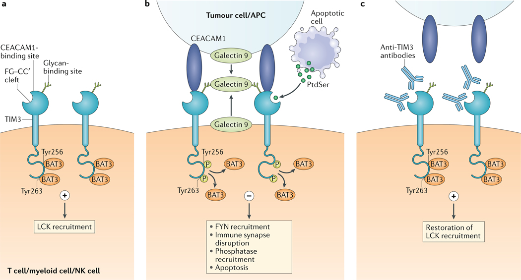
T cell immunoglobulin and mucin domain-containing protein 3 (TIM3), a member of the TIM family, was originally identified as a receptor expressed on interferon-γ-producing CD4+ and CD8+ T cells. Initial data indicated that TIM3 functioned as a 'co-inhibitory' or 'checkpoint' receptor, but due to the lack of a definable inhibitory signalling motif, it was also suggested that TIM3 might act as a co-stimulatory receptor. Recent studies have shown that TIM3 is part of a module that contains multiple co-inhibitory receptors (checkpoint receptors), which are co-expressed and co-regulated on dysfunctional or 'exhausted' T cells in chronic viral infections and cancer. Furthermore, co-blockade of TIM3 and programmed cell death 1 (PD1) can result in tumour regression in preclinical models and can improve anticancer T cell responses in patients with advanced cancers. Here, we highlight the developments in understanding TIM3 biology, including novel ligand identification and the discovery of loss-of-function mutations associated with human disease. In addition, we summarize emerging data from human clinical trials showing that TIM3 indeed acts as a 'checkpoint' receptor and that inhibition of TIM3 enhances the antitumour effect of PD1 blockade.


Figures

Fig. 1 |
Fig. 1 |. models for TIm3–ligand interactions.
a | In its ligand-unbound form, T cell immunoglobulin and mucin domain-containing protein 3 (TIM3) interacts with HLA-B-associated transcript 3 (BAT3) and maintains T cell activation by LCK recruitment. b | Galectin 9 can be bound to the surface of tumour cells and can also be secreted by tumour cells, antigen-presenting cells (APCs) and other cells in the parenchyma, or can be secreted in autocrine fashion by TIM3-expressing cells. Its carbohydrate recognition domains bind to the glycan-binding site on TIM3. Galectin 9 has two carbohydrate recognition domains and can promote the oligomerization of TIM3, thus potentially facilitating the formation of other TIM3–ligand complexes such as carcinoembyronic antigen-related cell adhesion molecule 1 (CEACAM1)–TIM3. In addition, phosphatidylserine (PtdSer) released from apoptotic cells can bind the FG–CC′ cleft-binding site on TIM3. On binding to galectin 9 or CEACAM1, Tyr256 and Tyr263 in the intracellular domain of TIM3 are phosphorylated; this releases BAT3 and allows recruitment of the tyrosine kinase FYN. This results in the disruption of immune synapse formation and in phosphatase recruitment. Ultimately, the cell becomes anergic or undergoes apoptosis, which is mediated by intracellular calcium release. c | Most of the TIM3-targeted antibodies that facilitate antitumour immunity interfere with either CEACAM1 or PtdSer binding to TIM3, thus maintaining the TIM3–BAT3 interaction. NK cell, natural killer cell.
Fig. 2 |
Fig. 2 |. TIm3 function in dendritic cells.
a | Endosomal Toll- like receptor 3 (TLR3), TLR7 and TLR9 sense viral double-stranded RNA, single-stranded RNA and bacterial or viral DNA, respectively, and activate several downstream transcription factors, such as nuclear factor-κB (NF-κB) and several interferon regulatory factors (IRFs), giving rise to the secretion of IL-12 and interferon-γ(IFNγ). In dendritic cells, the activation of TLR3, TLR7 and TLR9 can be suppressed by T cell immunoglobulin and mucin domain-containing protein 3 (TIM3)-mediated sequestration of high mobility group protein B1 (HMGB1), a protein that binds nucleic acids and facilitates their uptake, leading to TLR signalling. Thus, TIM3 serves as a molecular sink and prevents the entry of the nucleic acids into the cell, thereby dampening both antiviral and antitumour immune responses. Antibody-mediated blockade of TIM3 can relieve this suppression. b | TIM3 is highly expressed in type 1 conventional dendritic cells in tumours, where its blockade promotes the expression of CXC-chemokine ligand 9 (CXCL9) by type 1 conventional dendritic cells and the recruitment of CD8+ T cells that can respond to tumour antigens released by chemotherapy-induced apoptosis. More findings on TIM3 function in dendritic cells and other non-T cell leukocytes are described in TABLE 1. PtdSer, phosphatidylserine.
Fig. 3 |
Fig. 3 |. TIM3 gene single-point mutations give rise to loss of function in SPTCl.
a | Three mutations in the immunoglobulin V domain of T cell immunoglobulin and mucin domain-containing protein 3 (TIM3) have a different epidemiology in the general world population. The Tyr82Cys and Ile97Met mutations can cause disease either as homozygous or compound mutations, and the Thr101Ile mutation may cause disease as a compound mutation with the Tyr82Cys mutation, which suggests a similar function of these residues. b | The TIM3 mutations Tyr82Cys, Ile97Met and Thr101Ile lead to protein misfolding, resulting in TIM3 intracellular aggregates, which abrogate TIM3 signalling. c | Due to the broad expression and function of TIM3 in different immune cells, the absence of TIM3 signalling leads to systemic immune hyperactivation, resulting in myeloid cell dysregulation, a reduction in the number of regulatory T cells (Treg cells) in subcutaneous adipose tissue, excessive proliferation and malignancy of CD8+ T cells in the adipose tissue, which gives rise to subcutaneous fat lesions, and an indirect promotion of autoantibody production by B cells, resulting in autoimmune symptoms with lupus-l ike disease. dsDNA, double-s tranded DNA; HLH, haemophagocytic lymphohistiocytosis; SPTCL, subcutaneous panniculitis-like T cell lymphoma; TNF, tumour necrosis factor.

References

    1. Monney L et al. Th1-specific cell surface protein Tim-3 regulates macrophage activation and severity of an autoimmune disease. Nature 415, 536–541 (2002).

      This is the first report describing cloning of TIM3 and its definition as an immunoregulatory molecule.

    1. McIntire JJ et al. Identification of Tapr (an airway hyperreactivity regulatory locus) and the linked Tim gene family. Nat. Immunol. 2, 1109 (2001). - PubMed
    1. Meyers JH, Sabatos CA, Chakravarti S & Kuchroo VK The TIM gene family regulates autoimmune and allergic diseases. Trends Mol. Med. 11, 362–369 (2005). - PubMed
    1. Gao X et al. TIM-3 expression characterizes regulatory T cells in tumor tissues and is associated with lung cancer progression. PLOS ONE 7, e30676 (2012). - PMC - PubMed
    1. Anderson AC et al. Promotion of tissue inflammation by the immune receptor Tim-3 expressed on innate immune cells. Science 318, 1141–1143 (2007). - PubMed

Publication types

MeSH terms

Substances