. 2019 Nov 1;9(1):15812.
doi: 10.1038/s41598-019-52143-3.

Establishment of a novel human CIC-DUX4 sarcoma cell line, Kitra-SRS, with autocrine IGF-1R activation and metastatic potential to the lungs

Affiliations

Establishment of a novel human CIC-DUX4 sarcoma cell line, Kitra-SRS, with autocrine IGF-1R activation and metastatic potential to the lungs

Sho Nakai et al. Sci Rep. .

Erratum in

Abstract

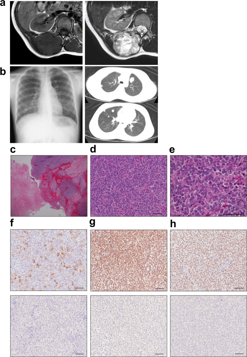
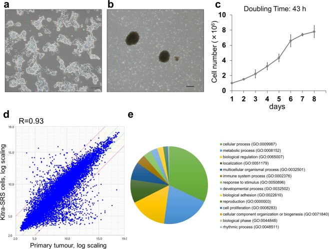
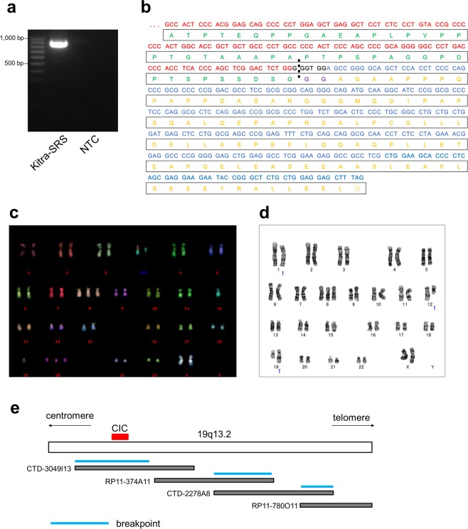
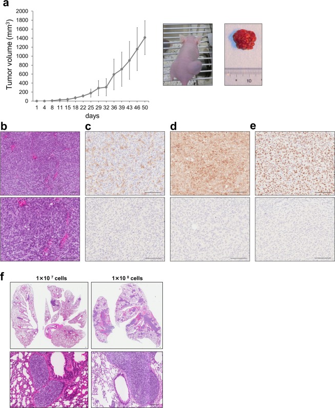
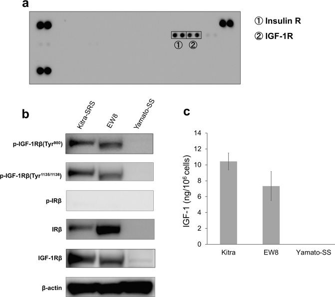
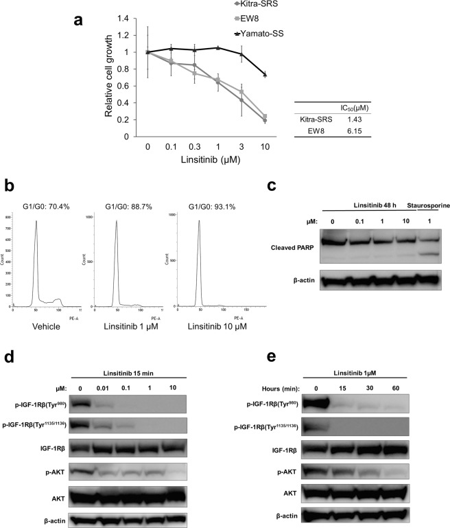
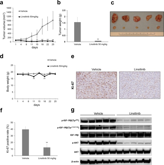
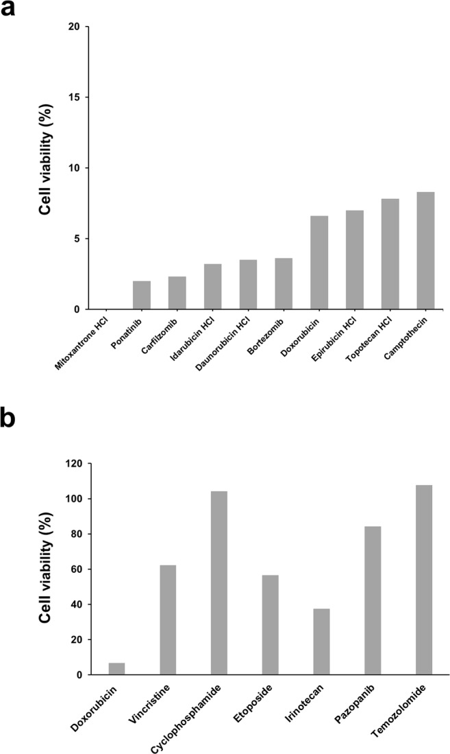
Approximately 60-70% of EWSR1-negative small blue round cell sarcomas harbour a rearrangement of CIC, most commonly CIC-DUX4. CIC-DUX4 sarcoma (CDS) is an aggressive and often fatal high-grade sarcoma appearing predominantly in children and young adults. Although cell lines and their xenograft models are essential tools for basic research and development of antitumour drugs, few cell lines currently exist for CDS. We successfully established a novel human CDS cell line designated Kitra-SRS and developed orthotopic tumour xenografts in nude mice. The CIC-DUX4 fusion gene in Kitra-SRS cells was generated by t(12;19) complex chromosomal rearrangements with an insertion of a chromosome segment including a DUX4 pseudogene component. Kitra-SRS xenografts were histologically similar to the original tumour and exhibited metastatic potential to the lungs. Kitra-SRS cells displayed autocrine activation of the insulin-like growth factor 1 (IGF-1)/IGF-1 receptor (IGF-1R) pathway. Accordingly, treatment with the IGF-1R inhibitor, linsitinib, attenuated Kitra-SRS cell growth and IGF-1-induced activation of IGF-1R/AKT signalling both in vitro and in vivo. Furthermore, upon screening 1134 FDA-approved drugs, the responses of Kitra-SRS cells to anticancer drugs appeared to reflect those of the primary tumour. Our model will be a useful modality for investigating the molecular pathology and therapy of CDS.


Conflict of interest statement

The authors declare no competing interests.

Figures

Figure 1
Figure 1
Appearance of the primary tumour in the back and lung metastatic lesions. (a) T1- and T2-weighted magnetic resonance images of the primary tumour. (b) X-rays and computed tomography of lung metastases. (ce) HE staining of the primary tumour tissue. (fh) Immunohistochemical analysis of CD99 (f), Bcl-2 (g), WT1 (h) and each negative control. Scale bars: 50 µm.
Figure 2
Figure 2
Phenotypic characteristics and gene expression signatures of Kitra-SRS cells. (a,b) Morphology of Kitra-SRS cells cultured in 2D standard plates (a) and in ultra-low attachment plates (b) assessed with phase-contrast microscopy. Scale bars: 100 µm. (c) Growth curve of Kitra-SRS cells in vitro. Each point represents the mean value ± SD (n = 3). (d) A scatter plot showing the correlation of gene expression between Kitra-SRS cells and the primary tumour. Expression values are derived from transcriptome analysis of two biological replicates. R represents the correlation coefficient. Green and red lines indicate absolute log-fold-change >1 and >3, respectively. (e) A pie chart depiction of PANTHER-GO slim biological processes that had more than 4-fold gene expression change.
Figure 3
Figure 3
Genetic analysis of Kitra-SRS cells. (a) RT-PCR with the CIC forward primer located in exon 16 and the DUX4 reverse primer in exon 1. No band is present for the negative control (NTC) of distilled water in lane 3. (b) Nucleotide and predicted amino acid sequences of the CIC-DUX4 fusions. Two additional amino acid residues that do not come from either CIC or DUX4 are present at the fusion point. Red indicates the CIC nucleotide sequence; blue, DUX4 nucleotide sequence; black, nucleotide sequence not belonging to CIC or DUX4; green, CIC amino acid sequence; yellow, DUX4 amino acid sequence; purple, amino acid sequence not belonging to CIC or DUX4. (c) A representative karyotype of Kitra-SRS cells at passage 20. M-FISH analysis showed four recurrent structural chromosomal rearrangements: 48, XX, del(1)(p32), +8, t(12;19) (q13;q13), +20. (d) A representative karyotype of Kitra-SRS cells at passage 100. G-banding showed four recurrent structural chromosomal rearrangements: 47, XX, del(1)(p?), +8, der(12)add(12)(p13)t(12;19) (q13;q13.1), der(19) t(12;19) (q13;q13.1). (e) A physical map of 19q13.2 and bacterial artificial chromosome clones used for identification of breakpoints.
Figure 4
Figure 4
Characteristics of Kitra-SRS tumours in vivo. (a) Growth curve of Kitra-SRS tumours in nude mice. Each point represents the mean value ± SD (n = 4). (b) HE staining of Kitra-SRS tumours. (ce) Immunohistochemical analysis of CD99 (c), Bcl-2 (d) and WT1 (e) and each negative control. (f) HE staining of lung sections in mice inoculated with 1 × 107 or 1 × 108 Kitra-SRS cells. Scale bars: 100 µm.
Figure 5
Figure 5
The IGF-1/IGF-1R pathway is activated in Kitra-SRS cells. (a) Phospho-RTK array analysis of Kitra-SRS cells. (b) Phosphorylation of IGF-1R and IR in Kitra-SRS, EW8, and Yamato-SS cells was assessed with western blotting. (c) Amount of IGF-1 secretion by these cells. Data in a bar graph represents the mean value ± SD, n = 3.
Figure 6
Figure 6
Linsitinib reduced the number of viable Kitra-SRS cells by inducing G1/G0 cell cycle arrest. (a) Kitra-SRS, EW8, and Yamato-SS were treated with 0–10 µM linsitinib for 72 h, and the number of viable cells was estimated with the WST-8 assay. The calculated IC50 values are shown in the table. Each point represents the mean value ± SD (n = 3). (b) Kitra-SRS cells were treated with 0–10 µM linsitinib for 48 h, stained with propidium iodide, and analysed for the cell cycle stage with flow cytometry. (c) Kitra-SRS cells were treated with 0–10 µM linsitinib for 48 h, and expression of cleaved PARP was evaluated with western blotting. Staurosporine was used as a positive control. (d,e) Kitra-SRS cells were treated with 0–10 µM linsitinib for 15 min (d) or 1 µM linsitinib for 0–60 min (e) and then subjected to western blotting with the indicated antibodies.
Figure 7
Figure 7
Linsitinib significantly abrogates Kitra-SRS tumour growth in xenograft mouse models. (ad) Mice bearing Kitra-SRS xenografts were treated with either 50 mg/kg linsitinib or vehicle (five mice/group). Tumour volume and body weight during treatment (a,d) and tumour weight and the appearance of resected tumours at the end point (b,c) are shown. (e) Immunohistochemical staining showing expression of Ki-67 in each treatment group. (f) Quantification of Ki-67-positive cells in Kitra-SRS tumours from each treatment group (five fields counted/group). (g) Tumour tissues from Kitra-SRS xenografts at the end point were harvested, and lysates were prepared for western blot analyses using the indicated antibodies. Data in a bar graph represent the mean value ± SD. **p < 0.01; significantly different from the vehicle group.
Figure 8
Figure 8
Screening of 1134 FDA-approved drugs in Kitra-SRS cells. Ten micromolar of 1134 FDA-approved drugs in our library were added to Kitra-SRS cells for 48 h. (a) Ten anticancer drugs that inhibited more than 80% cell viability in Kitra-SRS cells. (b) The antiproliferative efficacy of the drugs used in the donor patient.

References

    1. Italiano A, et al. High prevalence of CIC fusion with double-homeobox (DUX4) transcription factors in EWSR1-negative undifferentiated small blue round cell sarcomas. Genes Chromosomes Cancer. 2012;51:207–218. - PMC - PubMed
    1. Yoshida A, et al. CIC-rearranged Sarcomas: A Study of 20 Cases and Comparisons With Ewing Sarcomas. Am. J. Surg. Pathol. 2016;40:313–323. - PubMed
    1. Sturm D, et al. New Brain Tumor Entities Emerge from Molecular Classification of CNS-PNETs. Cell. 2016;164:1060–1072. - PMC - PubMed
    1. Huang SC, et al. Recurrent CIC Gene Abnormalities in Angiosarcomas: A Molecular Study of 120 Cases With Concurrent Investigation of PLCG1, KDR, MYC, and FLT4 Gene Alterations. Am. J. Surg. Pathol. 2016;40:645–655. - PMC - PubMed
    1. Sugita S, et al. A novel CIC-FOXO4 gene fusion in undifferentiated small round cell sarcoma: a genetically distinct variant of Ewing-like sarcoma. Am. J. Surg. Pathol. 2014;38:1571–1576. - PubMed

Publication types

Substances