. 2020 Feb;42(1):19-38.
doi: 10.1007/s11357-019-00103-0. Epub 2019 Nov 1.

The transfer of specific mitochondrial lipids and proteins to lipid droplets contributes to proteostasis upon stress and aging in the eukaryotic model system Saccharomyces cerevisiae

Affiliations

The transfer of specific mitochondrial lipids and proteins to lipid droplets contributes to proteostasis upon stress and aging in the eukaryotic model system Saccharomyces cerevisiae

Florian Geltinger et al. Geroscience. 2020 Feb.

Abstract

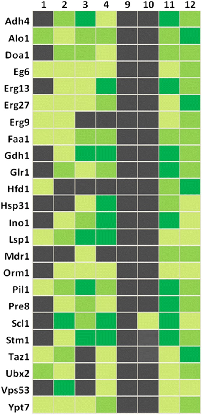
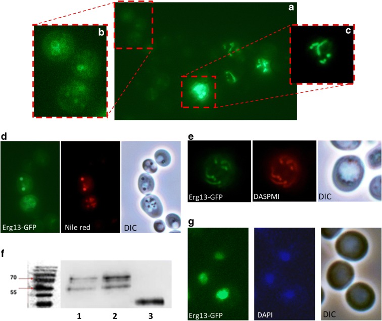
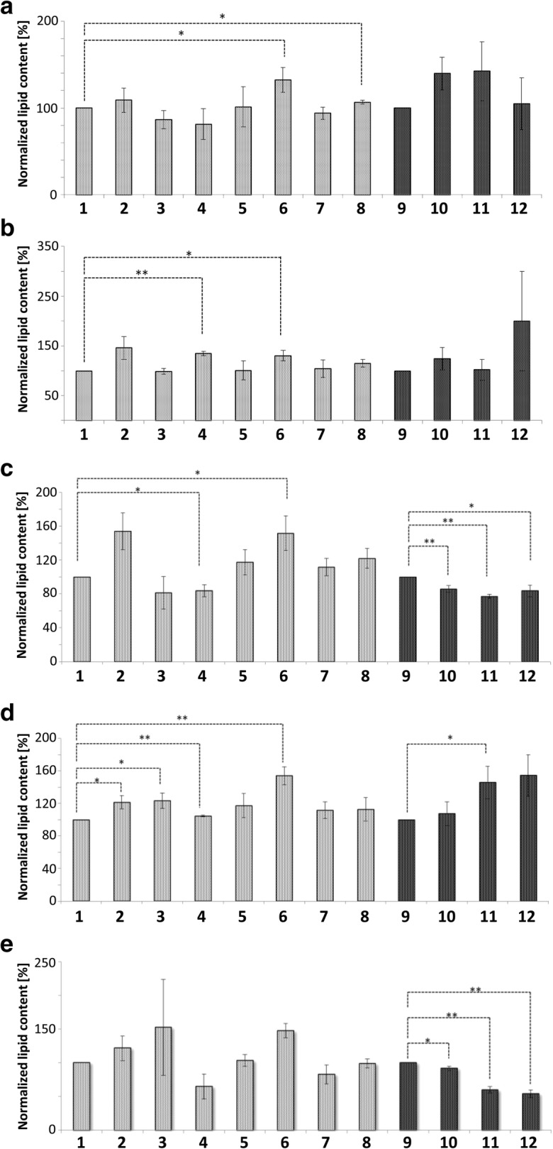
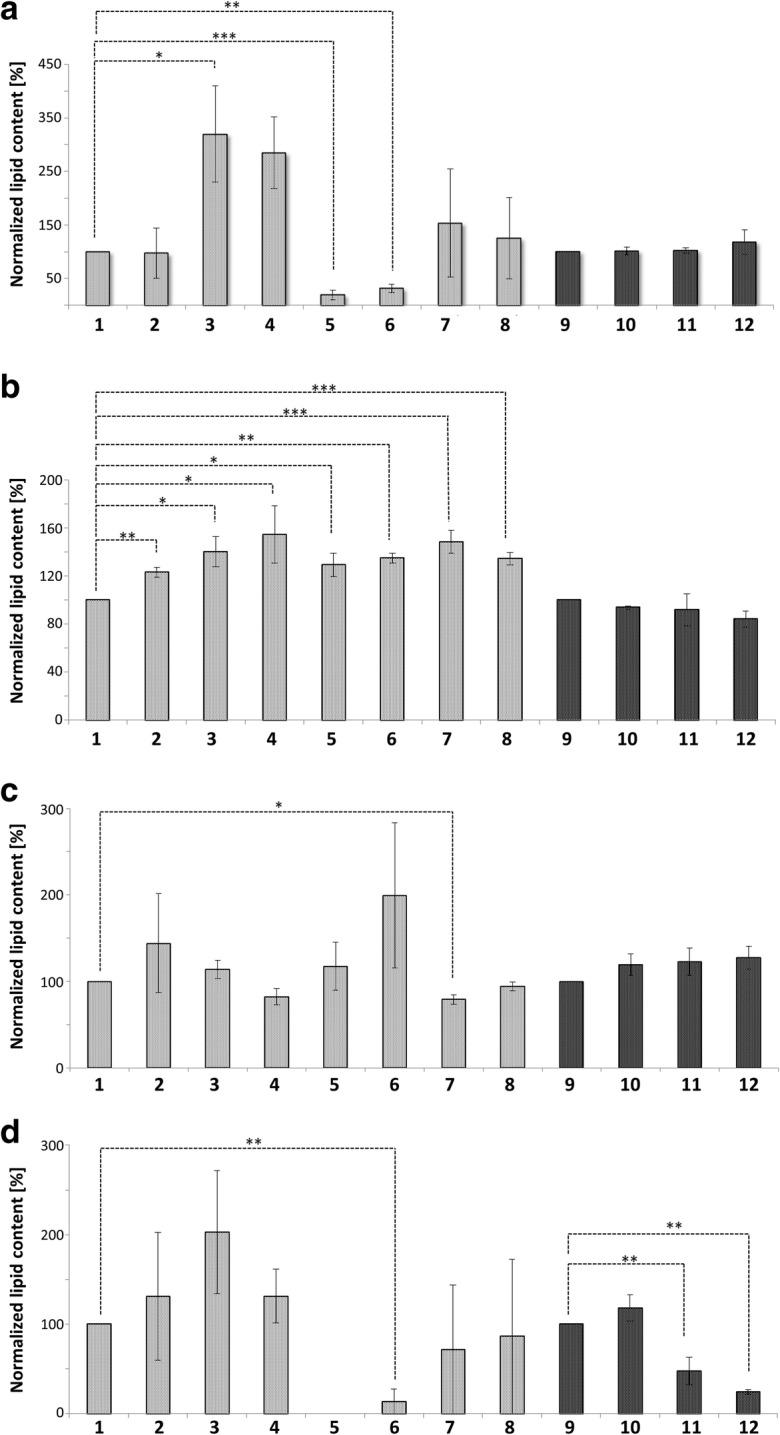
Originally Lipid droplets (LDs) were considered as being droplets for lipid storage only. Increasing evidence, however, demonstrates that LDs fulfill a pleiotropy of additional functions. Among them is the modulation of protein as well as lipid homeostasis. Under unfavorable pro-oxidative conditions, proteins can form aggregates which may exceed the overall proteolytic capacity of the proteasome. After stress termination LDs can adjust and support the removal of these aggregates. Additionally, LDs interact with mitochondria, specifically take over certain proteins and thus prevent apoptosis. LDs, which are loaded with these harmful proteins, are subsequently eliminated via lipophagy. Recently it was demonstrated that this autophagic process is a modulator of longevity. LDs do not only eliminate potentially dangerous proteins, but they are also able to prevent lipotoxicity by storing specific lipids. In the present study we used the model organism Saccharomyces cerevisiae to compare the proteome as well as lipidome of mitochondria and LDs under different conditions: replicative aging, stress and apoptosis. In this context we found an accumulation of proteins at LDs, supporting the role of LDs in proteostasis. Additionally, the composition of main lipid classes such as phosphatidylcholines, phosphatidylethanolamines, phosphatidylinositols, phosphatidylglycerols, triacylglycerols, ceramides, phosphatidic acids and ergosterol of LDs and mitochondria changed during stress conditions and aging.

Keywords: Lipid droplets; Lipidome; Mitochondria; Protein sink; Proteome; Replicative aging.


Figures

Fig. 1
Fig. 1
Survival upon BAX expression. The strains BY4741 pCM666 and BY4741 pCM666-mBAX were analyzed. BAX expression as induced by the addition of 200 mg/l doxycycline. Expression of BAX clearly reduces cell viability. The values represent the mean ± S.E.M. of 3 biological replicates, the significance of difference was analyzed by Student’s t test (*: p < 0.1; **: p < 0.05; ***: p < 0.01)
Fig. 2
Fig. 2
Venn diagram comparing the LD proteomes isolated from unstressed and mildly stressed cells (200 mg/L doxycycline). Numbers indicate identified proteins
Fig. 3
Fig. 3
Venn diagram comparing the proteome of LDs obtained from unstressed cells (BY4741), stressed cells (BY4741 + 200 mg/L doxycycline), apoptotic cells (BY4741 pCM666-mBAX +200 mg/l doxycycline) and aged cells. Numbers indicate identified proteins. Proteins are listed in supplementary Table 2
Fig. 4
Fig. 4
Venn diagram comparing the proteome of mitochondria obtained from unstressed cells (BY4741 as well as BY4741 Δare1 Δare2 Δlro1 Δdga1), stressed cells (BY4741 + 200 mg/L doxycycline), apoptotic cells (BY4741 pCM666-mBAX +200 mg/l doxycycline) and aged cells (BY4741 as well as LD deficient BY4741 Δare1 Δare2 Δlro1 Δdga1). Numbers indicate identified proteins. Proteins are listed in supplementary Table 4
Fig. 5
Fig. 5
Heatmap of selected protein localizations. Black indicates the absence or near absence of proteins. Different shades of green represent various amounts of proteins, from light green (low abundance) to dark green (high abundance). 1: Mitochondria from BY4741; 2: Mitochondria from replicatively aged BY4741 cells; 3: Mitochondria from stressed cells (BY4741 pCM666 + 200 mg/l doxycycline); 4: Mitochondria from apoptotic cells (BY4741 pCM666-mBAX +200 mg/l doxycycline); 9: LDs from BY4741; 10: LDs from replicatively aged BY4741 cells; 11: LDs from stressed cells (BY4741 pCM666 + 200 mg/l doxycycline); 12: LDs from apoptotic cells (BY4741 pCM666-mBAX +200 mg/l doxycycline)
Fig. 6
Fig. 6
LD and mitochondrial localization of Erg13. A) mBAX expressionErg13-GFP localizes either to LDs or mitochondria. A heterogeneous expression level of Erg13-GFP is evident. (B) is a magnification of (A) and shows Erg13-GFP stained LDs. (C) is a magnification of (A) with a reduced exposure time. Erg13-GFP resides at mitochondria. (D) Erg13-GFP co-localizes with LD specific Nile red staining and LDs in the DIC image. (E) The GFP signal of Erg13 completely overlaps with DASPMI (a mitochondrial specific dye). (F) Immunoblot of purified LDs detecting the Erg13-GFP fusion using an anti-GFP antibody. 1: BY4741 ERG13::GFP::HIS3MX6 YEp51 (unstressed cells); 2: BY4741 ERG13::GFP::HIS3MX6 YEp51-mBAX (apoptotic cells); 3: BY4741 pUG35-vBAX (control cells). Erg13-GFP (83 kDa) shows a low abundance at isolated LDs obtained from unstressed cells and shows a high abundance at LDs isolated from apoptotic cells. The positive control vBAX-GFP (33 kDA) confirms the isolation of LDs. (G) Erg13-GFP shows a perfect co-localization with the DNA specific dye DAPI. In the images (A), (B), (C), (D) and (E) the BY4741 ERG13::GFP::HIS3MX6 YEp51-mBAX background is used (apoptotic cells), whereas in (G) the BY4741 ERG13::GFP::HIS3MX6 background (unstressed cells) is shown
Fig. 7
Fig. 7
Glycerophospholipid content of Mitochondria and LDs. Mitochondria represented in gray, LDs in black. 1: Mitochondria from BY4741; 2: Mitochondria from replicatively aged BY4741 cells; 3: Mitochondria from stressed cells (BY4741 pCM666 + 200 mg/l doxycycline); 4: Mitochondria from apoptotic cells (BY4741 pCM666-mBAX +200 mg/l doxycycline); 5: Mitochondria from the strain BY4741 Δare1 Δ are2 Δlro1 Δdga1; 6: Mitochondria from the replicatively aged strain BY4741 Δare1 Δ are2 Δlro1 Δdga1; 7: Mitochondria from the stressed strain BY4741 Δare1 Δ are2 Δlro1 Δdga1 pCM666 + 200 mg/l doxycycline; 8: Mitochondria from the apoptotic strain BY4741 Δare1 Δ are2 Δlro1 Δdga1 pCM666-mBAX +200 mg/l doxycycline; 9: LDs from BY4741; 10: LDs from replicatively aged BY4741 cells; 11: LDs from stressed cells (BY4741 pCM666 + 200 mg/l doxycycline); 12: LDs from apoptotic cells (BY4741 pCM666-mBAX +200 mg/l doxycycline). In (A) phosphatidylcholines, in (B) phosphatidylethanolamines, in (C) phosphatidylinositols and in (D) phosphatidylglycerols
Fig. 8
Fig. 8
Lipid content of Mitochondria and LDs. Mitochondria represented in gray, LDs in black. 1: Mitochondria from BY4741; 2: Mitochondria from replicatively aged BY4741 cells; 3: Mitochondria from stressed cells (BY4741 pCM666 + 200 mg/l doxycycline); 4: Mitochondria from apoptotic cells (BY4741 pCM666-mBAX +200 mg/l doxycycline); 5: Mitochondria from the strain BY4741 Δare1 Δ are2 Δlro1 Δdga1; 6: Mitochondria from the replicatively aged strain BY4741 Δare1 Δ are2 Δlro1 Δdga1; 7: Mitochondria from the stressed strain BY4741 Δare1 Δ are2 Δlro1 Δdga1 pCM666 + 200 mg/l doxycycline; 8: Mitochondria from the apoptotic strain BY4741 Δare1 Δ are2 Δlro1 Δdga1 pCM666-mBAX +200 mg/l doxycycline; 9: LDs from BY4741; 10: LDs from replicatively aged BY4741 cells; 11: LDs from stressed cells (BY4741 pCM666 + 200 mg/l doxycycline); 12: Mitochondria from apoptotic cells (BY4741 pCM666-mBAX +200 mg/l doxycycline). In (A) triacylglycerols, in (B) ceramides, in (C) phosphatidic acids and in (D) ergosterols are presented

References

    1. Altmann K, Westermann B. Role of essential genes in mitochondrial morphogenesis in Saccharomyces cerevisiae. Mol Biol Cell. 2005;16:5410–5417. doi: 10.1091/mbc.e05-07-0678. - DOI - PMC - PubMed
    1. Bhattacharya S, Esquivel BD, White TC (2018) Overexpression or deletion of Ergosterol biosynthesis genes alters doubling time, response to stress agents, and drug susceptibility in Saccharomyces cerevisiae. Mbio 9. 10.1128/mBio.01291-18 - PMC - PubMed
    1. Bischof J, Salzmann M, Streubel MK, Hasek J, Geltinger F, Duschl J, Bresgen N, Briza P, Haskova D, Lejskova R, Sopjani M, Richter K, Rinnerthaler M. Clearing the outer mitochondrial membrane from harmful proteins via lipid droplets. Cell Death Discov. 2017;3:17016. doi: 10.1038/cddiscovery.2017.16. - DOI - PMC - PubMed
    1. Bitto A, Lerner CA, Nacarelli T, Crowe E, Torres C, Sell C. p62/SQSTM1 at the interface of aging, autophagy, and disease. Age. 2014;36:1123–1137. doi: 10.1007/s11357-014-9626-3. - DOI - PMC - PubMed
    1. Bohnert M. Wrapping up the fats-a structure of the lipid droplet biogenesis protein seipin. J Cell Biol. 2018;217:4053–4054. doi: 10.1083/jcb.201811021. - DOI - PMC - PubMed

Publication types

LinkOut - more resources