. 2020 Feb;139(2):287-303.
doi: 10.1007/s00401-019-02088-8. Epub 2019 Nov 1.

The histomolecular criteria established for adult anaplastic pilocytic astrocytoma are not applicable to the pediatric population

Affiliations

The histomolecular criteria established for adult anaplastic pilocytic astrocytoma are not applicable to the pediatric population

Albane Gareton et al. Acta Neuropathol. 2020 Feb.

Abstract

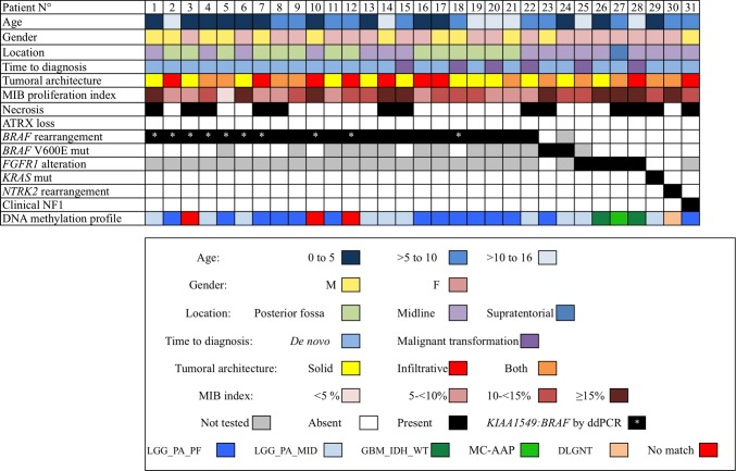
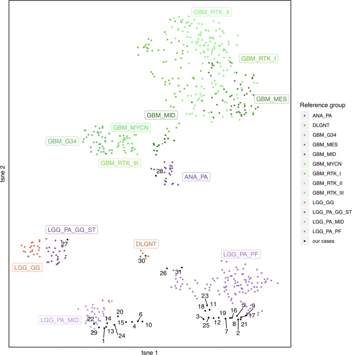
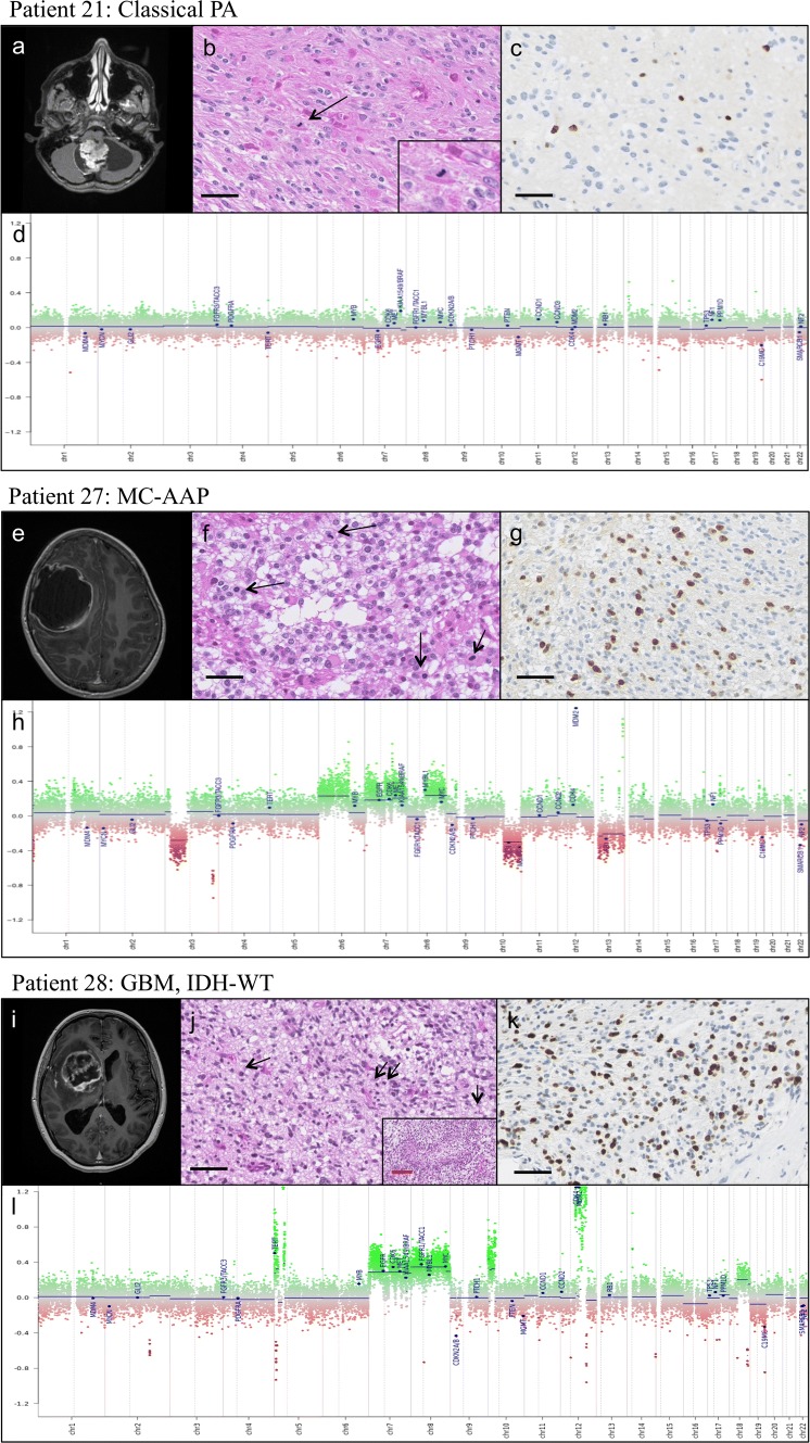
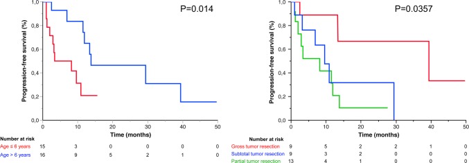
Pilocytic astrocytoma (PA) is the most common pediatric glioma, arising from a single driver MAPK pathway alteration. Classified as a grade I tumor according to the 2016 WHO classification, prognosis is excellent with a 10-year survival rate > 95% after surgery. However, rare cases present with anaplastic features, including an unexpected high mitotic/proliferative index, thus posing a diagnostic and therapeutic challenge. Based on small histomolecular series and case reports, such tumors arising at the time of diagnosis or recurrence have been designated by many names including pilocytic astrocytoma with anaplastic features (PAAF). Recent DNA methylation-profiling studies performed mainly on adult cases have revealed that PAAF exhibit a specific methylation signature, thus constituting a distinct methylation class from typical PA [methylation class anaplastic astrocytoma with piloid features-(MC-AAP)]. However, the diagnostic and prognostic significance of MC-AAP remains to be determined in children. We performed an integrative work on the largest pediatric cohort of PAAF, defined according to strict criteria: morphology compatible with the diagnosis of PA, with or without necrosis, ≥ 4 mitoses for 2.3 mm2, and MAPK pathway alteration. We subjected 31 tumors to clinical, imaging, morphological and molecular analyses, including DNA methylation profiling. We identified only one tumor belonging to the MC-AAP (3%), the others exhibiting a methylation profile typical for PA (77%), IDH-wild-type glioblastoma (7%), and diffuse leptomeningeal glioneuronal tumor (3%), while three cases (10%) did not match to a known DNA methylation class. No significant outcome differences were observed between PAAF with necrosis versus no necrosis (p = 0.07), or with 4-6 mitoses versus 7 or more mitoses (p = 0.857). Our findings argue that the diagnostic histomolecular criteria established for anaplasia in adult PA are not of diagnostic or prognostic value in a pediatric setting. Further extensive and comprehensive integrative studies are necessary to accurately define this exceptional entity in children.

Keywords: DNA methylation profiling; FGFR1; MAPK pathway; MC-AAP; Pediatric; Pilocytic astrocytoma with anaplastic features.


Conflict of interest statement

The authors declare that they have no conflicts of interest.

Figures

Fig. 1
Fig. 1
Clinical, Radiological, Histopathological, and Molecular Characterization of 31 pediatric PAAF
Fig. 2
Fig. 2
t-SNE plot of the 31 PAAF cases. Reference classes: ANA_PA anaplastic pilocytic astrocytoma, DLGNT diffuse leptomeningeal glioneuronal tumor, GBM_G34 glioblastoma with G34 mutation, GBM_MES glioblastoma, mesenchymal subtype, GBM_MID glioblastoma of the midline, GBM_MYCN glioblastoma, MYCN mutant GBM_RTK_1: glioblastoma, RTK I subtype, GBM_RTK_II glioblastoma, RTK II subtype, GBM_RTK_III glioblastoma, RTK III subtype, LGG_GG ganglioglioma, LGG_PA_GG_ST PA or ganglioglioma of the supratentorial hemispheres, LGG_PA_MID PA of the midline, LGG_PA_FP PA of the posterior fossa
Fig. 3
Fig. 3
Comparative radiological, histological, molecular and CNP characteristics of PA (patient 21), MC-AAP (patient 27), and GBM (patient 28). Patient 21 Classical PA: a MRI: 3D T1 with gadolinium injection, showing a classical cerebellar PA features with both cystic and nodular portions and strong contrast enhancement. b Histological features: biphasic architecture, Rosenthal fibers, eosinophilic granular bodies, and mitosis (see insert and arrowhead), c MIB1 labeling index estimated at 10%, d flat CNP characteristic of classical PA. Patient 27 MC-AAP: e MRI: 3D T1 with gadolinium injection showing a right parietal mostly cystic lesion with a thick, irregular Contrast enhancing border. f Histological features: oligodendroglial-like morphology, numerous mitoses (arrowheads). No Rosenthal fiber or eosinophilic granular bodies were observed, g MIB1 labeling index estimated at 30%, h CNP with many chromosomal gains and losses, as well as focal amplifications, including MDM2. Patient 28 GBM, IDH-WT:i MRI: 3D T1 with gadolinium injection showing a right mass growing from the optic chiasm into the basal ganglia. j Histological features: biphasic morphology with numerous mitoses (arrowheads) and palisading necrosis (see insert), k MIB1 labeling index estimated at 40%, l CNP showing multiple chromosomal gains and losses, including a loss of CDKN2A/B, and focal amplifications of MDM2 and CDK4. Black scale bar corresponds to 50 μm, red scale bar corresponds to 200 μm
Fig. 4
Fig. 4
OS and PFS of 31 pediatric PAAF, in months (Kaplan Meier curves)
Fig. 5
Fig. 5
PFS according to age and extent of resection

References

    1. Appay R, Fina F, Macagno N, Padovani L, Colin C, Barets D, et al. Duplications of KIAA1549 and BRAF screening by droplet digital PCR from formalin-fixed paraffin-embedded DNA is an accurate alternative for KIAA1549—BRAF fusion detection in pilocytic astrocytomas. Mod Pathol. 2018;31:1490. doi: 10.1038/s41379-018-0050-6. - DOI - PubMed
    1. Cancer Genome Atlas Research Network Comprehensive genomic characterization defines human glioblastoma genes and core pathways. Nature. 2008;455:1061–1068. doi: 10.1038/nature07385. - DOI - PMC - PubMed
    1. Capper D, Jones DTW, Sill M, Hovestadt V, Schrimpf D, Sturm D, et al. DNA methylation-based classification of central nervous system tumours. Nature. 2018;555:469–474. doi: 10.1038/nature26000. - DOI - PMC - PubMed
    1. Chiang JCH, Harreld JH, Orr BA, Sharma S, Ismail A, Segura AD, et al. Low-grade spinal glioneuronal tumors with BRAF gene fusion and 1p deletion but without leptomeningeal dissemination. Acta Neuropathol (Berl) 2017;134:159–162. doi: 10.1007/s00401-017-1728-4. - DOI - PMC - PubMed
    1. D’Angelo F, Ceccarelli M, Tala GL, Zhang J, Frattini V, Caruso FP, et al. The molecular landscape of glioma in patients with Neurofibromatosis 1. Nat Med. 2019;25:176. doi: 10.1038/s41591-018-0263-8. - DOI - PMC - PubMed