. 2020 Jan 30;26(1):51-60.
doi: 10.5056/jnm18208.

Morphology of the Esophageal Hiatus: Is It Different in 3 Types of Hiatus Hernias?

Affiliations

Morphology of the Esophageal Hiatus: Is It Different in 3 Types of Hiatus Hernias?

Dushyant Kumar et al. J Neurogastroenterol Motil. .

Abstract

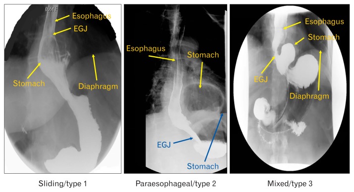
Background/aims: The esophageal hiatus is formed by the right crus of the diaphragm in the majority of subjects. Contraction of the hiatus exerts a sphincter-like action on the lower esophageal sphincter (LES). The aim is to study the hiatal anatomy (using CT scan imaging) and function (using high-resolution manometry [HRM]), and esophageal motor function in patients with sliding and paraesophageal hiatal hernia.

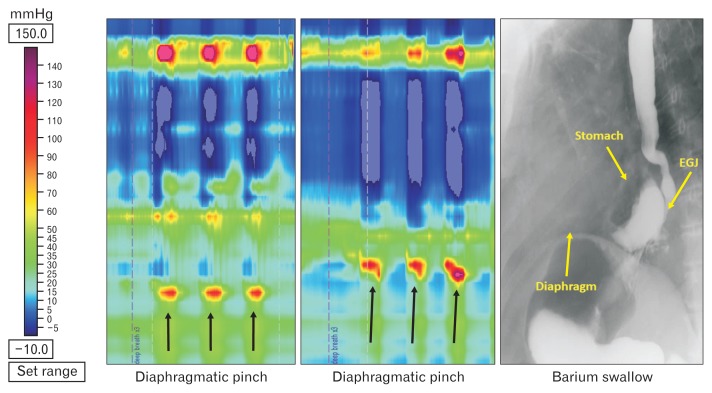
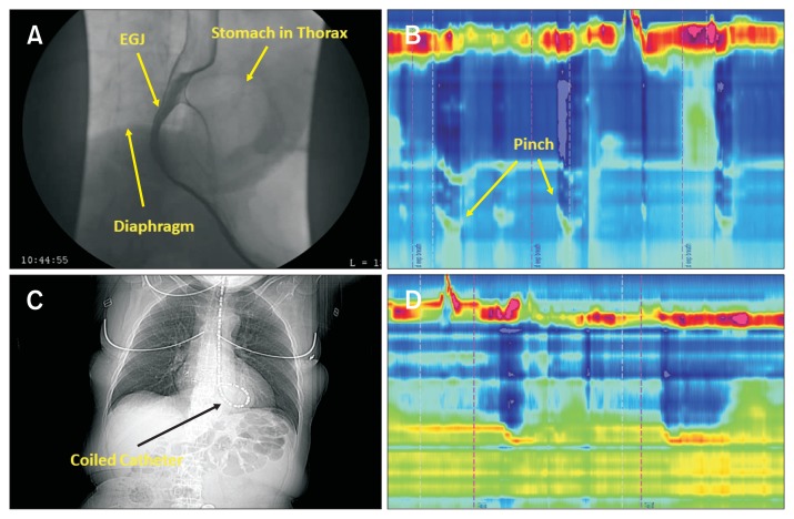
Methods: We assessed normal subjects (n = 20), patients with sliding type 1 hernia (n = 18), paraesophageal type 2 hernia (n = 19), and mixed type 3 hernia (n = 19). Hernia diagnosis was confirmed on the upper gastrointestinal series. The hiatal morphology was constructed from the CT scan images. The LES pressure and relaxation, percent peristalsis, bolus pressure, and hiatal squeeze pressure were assessed by HRM.

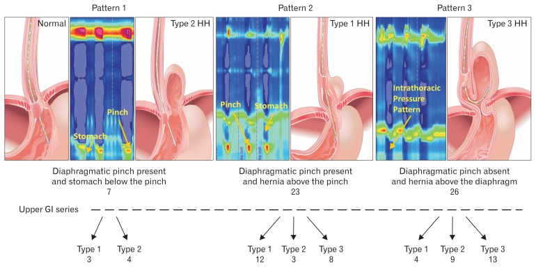
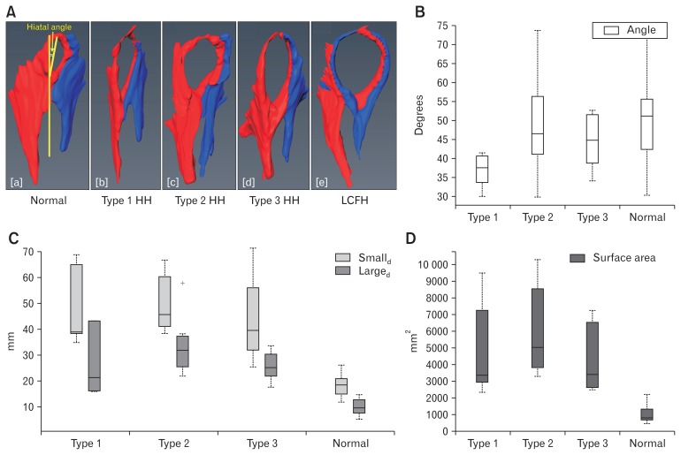
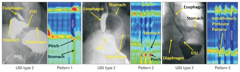
Results: The CT images revealed that the esophageal hiatus is formed by the right crus of the diaphragm in all normal subjects and 86% of hernia patients. The hiatus is elliptical in shape with a surface area of 1037 mm2 in normal subjects. The hiatal dimensions were larger in patients compared to normal subjects. The HRM revealed impaired LES relaxation and higher bolus pressure in patients with paraesophageal compared to the sliding hernia. The hiatal pinch on HRM was recognized in significantly higher number of patients with sliding as compared to paraesophageal hernia.

Conclusions: Using a novel approach, we provide details of the esophageal hiatus in patients with various kinds of hiatal hernia. Impaired LES relaxation in paraesophageal hernia may play a role in its pathophysiology and genesis of symptoms.

Keywords: Esophageal peristalsis; Hiatal; Lower esophageal sphincter; Manometry; Tomography X-ray computed; hernia.


Conflict of interest statement

Conflicts of interest: None.

Figures

Figure 1
Figure 1
Radiographic patterns in 3 types of hiatal hernia (HH) patients. EGJ, esophagogastric junction.
Figure 2
Figure 2
Esophagogastric junction (EGJ) patterns of hiatal hernia (HH) on manometry and relationship with the radiological diagnosis in 3 types of hernia (see text for explanation). EGJ pattern 1 expected in patients with normal subjects and patients with paraesophageal hernia type 2. High-resolution manometry (HRM) EGJ pattern 2 expected in patient with sliding HH (type 1) and pattern 3 expected in patient with type 3 HH. GI, gastrointestinal.
Figure 3
Figure 3
Hiatal anatomy and hiatal dimension. (A) Anatomy of esophageal diaphragmatic hiatus in normal subjects and patients with various types of hiatal hernia (HH). The hiatus is larger in dimension in 3 types of HH compared to normal subjects. (B–D) Hiatal dimensions (angle, large diameter [Larged] and small diameter [Smalld], and surface area) in normal subjects, patients with type 1, type 2, and type 3 HH. (B) Angle of the hiatus in relationship to spine, (C) long and short dimensions of hiatus, and (D) cross sectional area of the hiatus. Normal subjects have significantly smaller surface area, large and small diameters compared to 3 types of hernia but there is no difference among 3 type of HH. Data showed in median and interquartile range. LCFH, left crus forming right hiatal margin. +Outlier value in the group.
Figure 4
Figure 4
Manometry data (median and interquartile range) in 3 types of hiatal hernia (HH) patients: (A) lower esophageal sphincter basal pressure (LESB), residual pressure (LESR) with swallow and bolus pressure, (B) diaphragmatic pinch pressure, and (C) distal contractile integral (DCI). +Sign corresponds to outliers, *P < 0.05 compared to type 1 HH.
Figure 5
Figure 5
Manometry patterns in 3 different patients with type 2 paraesophageal hiatal hernia (HH) identified on the upper gastrointestinal (UGI) series. EGJ, esophagogastric junction.
Figure 6
Figure 6
Changing manometry patterns of hiatal hernia (HH) in a patient with type 3 HH on the upper gastrointestinal series. EGJ, esophagogastric junction.
Figure 7
Figure 7
CT scan finding with manometry catheter in place. (A) Upper gastrointestinal (UGI) series show a patient with type 3 hiatal hernia (HH). (B) High-resolution manometry (HRM) esophagogastric junction (EGJ). Pattern 2 is consistent with the presence of stomach above the diaphragm and the tip of catheter in the abdomen. (C) On a separate day, the manometry catheter could not be advanced into the abdomen as shown in the coronal image. (D) HRM pattern is consistent with HRM EGJ pattern 3.

References

    1. Nicholson F. Diphragmatic hernia. Ann Surg. 1952;136:174–182. doi: 10.1097/00000658-195207000-00017. - DOI - PMC - PubMed
    1. Jones FA. Diagnosis of hiatus hernia. Proc R Soc Med. 1952;45:277–279. - PMC - PubMed
    1. Kahrilas PJ, Kim HC, Pandolfino JE. Approaches to the diagnosis and grading of hiatal hernia. Best Pract Res Clin Gastroenterol. 2008;22:601–616. doi: 10.1016/j.bpg.2007.12.007. - DOI - PMC - PubMed
    1. Collis JL, Kelly TD, Willey AM. Anatomy of the crura of the diaphragm and the surgery of hiatus hernia. Thorax. 1954;9:175–189. doi: 10.1136/thx.9.3.175. - DOI - PMC - PubMed
    1. Listerud MB, Harkins HN. Variations in the muscular anatomy of the esophageal hiatus: based on dissections of two hundred and four fresh cadavers. West J Surg Obstet Gynecol. 1959;67:110–112. discussion 112–113. - PubMed