

Mitochondrial Dysfunction in Cardiac Surgery
- PMID: 31677690
- PMCID: PMC6986803
- DOI: 10.1016/j.anclin.2019.08.003
Mitochondrial Dysfunction in Cardiac Surgery
Abstract
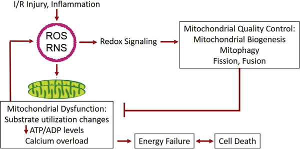
Mitochondria are key to the cellular response to energetic demand, but are also vital to reactive oxygen species signaling, calcium hemostasis, and regulation of cell death. Cardiac surgical patients with diabetes, heart failure, advanced age, or cardiomyopathies may have underlying mitochondrial dysfunction or be more sensitive to perioperative mitochondrial injury. Mitochondrial dysfunction, due to ischemia/reperfusion injury and an increased systemic inflammatory response due to exposure to cardiopulmonary bypass and surgical tissue trauma, impacts myocardial contractility and predisposes to arrhythmias. Strategies for perioperative mitochondrial protection and recovery include both well-established cardioprotective protocols and targeted therapies that remain under investigation.
Keywords: Cardiac surgery; Cardioprotection; Cardiopulmonary bypass; Inflammation; Ischemia/reperfusion injury; Mitochondria; Myocardial metabolism.
Copyright © 2019 Elsevier Inc. All rights reserved.
Figures
Similar articles
-
Mitochondrial Dysfunction in Cardiac Surgery.Anesthesiol Clin. 2025 Jun;43(2):357-375. doi: 10.1016/j.anclin.2025.02.004. Epub 2025 Mar 20. Anesthesiol Clin. 2025. PMID: 40348547 Review.
-
Humanin exerts cardioprotection against cardiac ischemia/reperfusion injury through attenuation of mitochondrial dysfunction.Cardiovasc Ther. 2016 Dec;34(6):404-414. doi: 10.1111/1755-5922.12210. Cardiovasc Ther. 2016. PMID: 27434747
-
Cardioprotection against ischemia/reperfusion injury by QiShenYiQi Pill® via ameliorate of multiple mitochondrial dysfunctions.Drug Des Devel Ther. 2015 Jun 15;9:3051-66. doi: 10.2147/DDDT.S82146. eCollection 2015. Drug Des Devel Ther. 2015. PMID: 26109848 Free PMC article.
-
High-dose Humanin analogue applied during ischemia exerts cardioprotection against ischemia/reperfusion injury by reducing mitochondrial dysfunction.Cardiovasc Ther. 2017 Oct;35(5). doi: 10.1111/1755-5922.12289. Cardiovasc Ther. 2017. PMID: 28726291
-
Mitochondrial transplantation in cardiac surgical patients: optimism, caveats, and outstanding questions.Curr Opin Anaesthesiol. 2023 Feb 1;36(1):5-10. doi: 10.1097/ACO.0000000000001202. Epub 2022 Oct 20. Curr Opin Anaesthesiol. 2023. PMID: 36550601 Review.
Cited by
-
Frailty as a predictor of mortality and morbidity after cholecystectomy: A systematic review and meta-analysis of cohort studies.Langenbecks Arch Surg. 2024 Nov 18;409(1):352. doi: 10.1007/s00423-024-03537-z. Langenbecks Arch Surg. 2024. PMID: 39557689
-
Signaling Pathways (TNF-α-NF-κB, TLR2-TLR4 as well as ROS-MDA) and Cardiac Damages during Cardiac Surgeries (Coronary Stenting, Permanent Pacemaker Implantations, Radiofrequency Ablations).Curr Top Med Chem. 2025;25(2):196-208. doi: 10.2174/0115680266314899240919081451. Curr Top Med Chem. 2025. PMID: 39350416
-
Comparison of Machine Learning Models Including Preoperative, Intraoperative, and Postoperative Data and Mortality After Cardiac Surgery.JAMA Netw Open. 2022 Oct 3;5(10):e2237970. doi: 10.1001/jamanetworkopen.2022.37970. JAMA Netw Open. 2022. PMID: 36287565 Free PMC article.
-
Targeted Mitochondrial Delivery to Hepatocytes: A Review.J Clin Transl Hepatol. 2022 Apr 28;10(2):321-328. doi: 10.14218/JCTH.2021.00093. Epub 2021 Oct 19. J Clin Transl Hepatol. 2022. PMID: 35528979 Free PMC article. Review.
-
Efficacy of Bioenergetic Health Index to Predict Delirium After Major Abdominal Surgery in Elderly Patients: A Protocol for a Prospective Observational Cohort Study.Front Med (Lausanne). 2022 Apr 25;9:809335. doi: 10.3389/fmed.2022.809335. eCollection 2022. Front Med (Lausanne). 2022. PMID: 35547218 Free PMC article.
References
-
- Westerhof N Cardiac work and efficiency. Cardiovasc Res 2000;48(1):4–7. - PubMed
-
- Schaper J, Meiser E, Stammler G. Ultrastructural morphometric analysis of myocardium from dogs, rats, hamsters, mice, and from human hearts. Circ Res 1985;56(3):377–91. - PubMed
-
- Stanley WC, Recchia FA, Lopaschuk GD. Myocardial substrate metabolism in the normal and failing heart. Physiol Rev 2005;85(3):1093–129. - PubMed
-
- Razeghi P, Young ME, Alcorn JL, et al. Metabolic gene expression in fetal and failing human heart. Circulation 2001;104(24):2923–31. - PubMed
Publication types
MeSH terms
Grants and funding
LinkOut - more resources
Full Text Sources
Medical
