

Silicon substituted hydroxyapatite/VEGF scaffolds stimulate bone regeneration in osteoporotic sheep
- PMID: 31678741
- PMCID: PMC7616907
- DOI: 10.1016/j.actbio.2019.10.033
Silicon substituted hydroxyapatite/VEGF scaffolds stimulate bone regeneration in osteoporotic sheep
Abstract
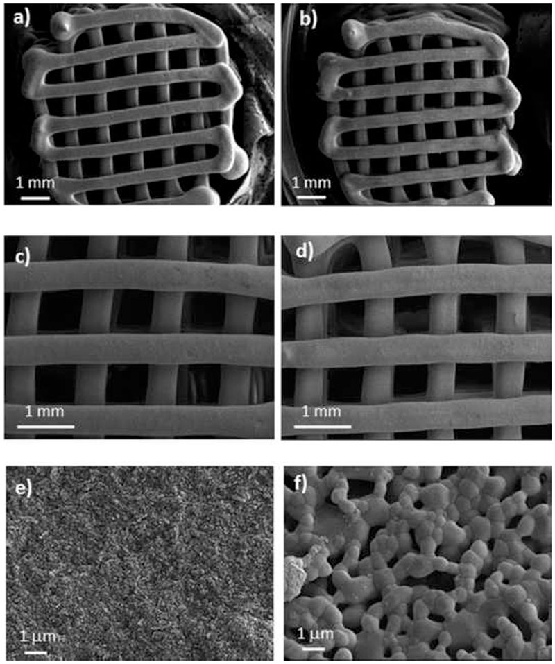
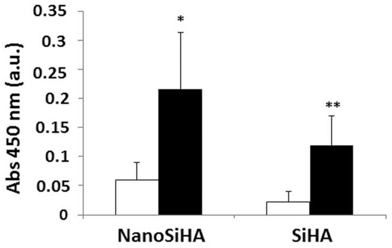
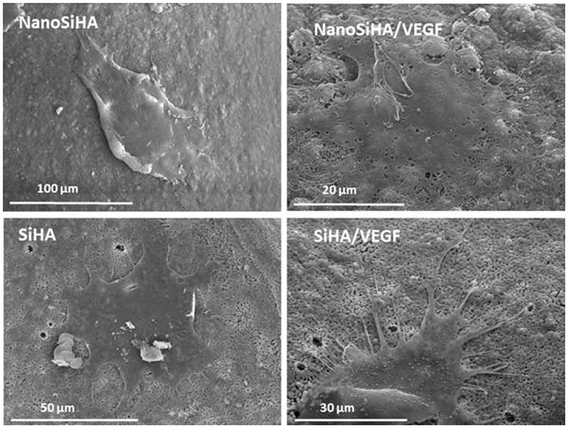
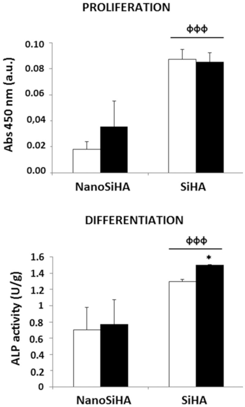
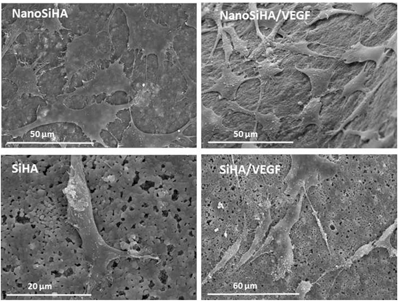
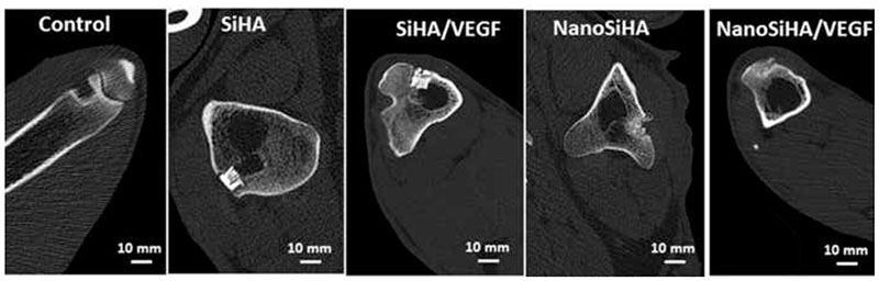
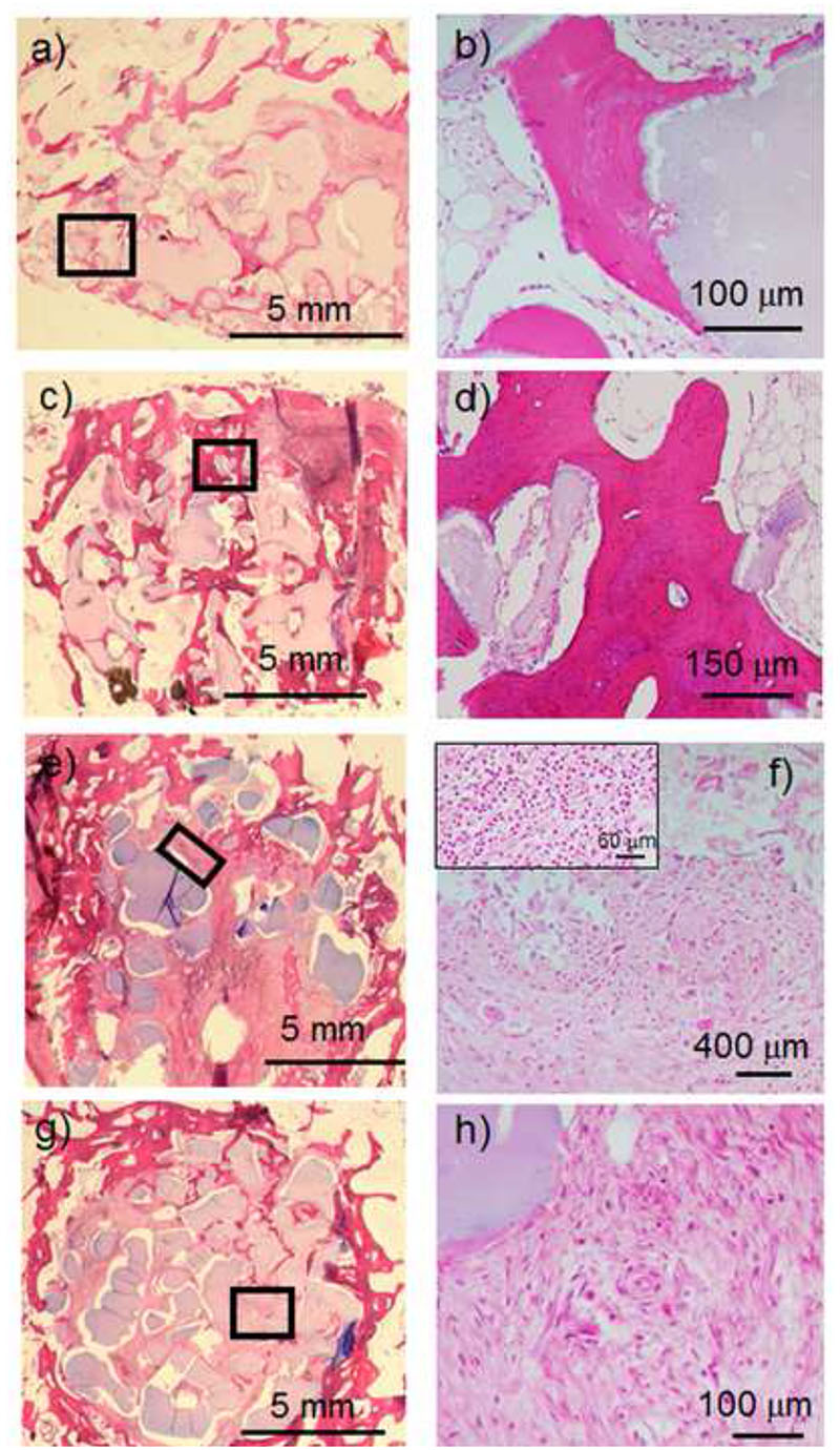
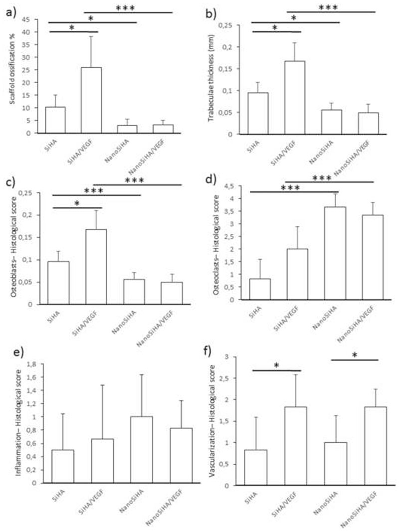
Silicon-substituted hydroxyapatite (SiHA) macroporous scaffolds have been prepared by robocasting. In order to optimize their bone regeneration properties, we have manufactured these scaffolds presenting different microstructures: nanocrystalline and crystalline. Moreover, their surfaces have been decorated with vascular endothelial growth factor (VEGF) to evaluate the potential coupling between vascularization and bone regeneration. In vitro cell culture tests evidence that nanocrystalline SiHA hinders pre-osteblast proliferation, whereas the presence of VEGF enhances the biological functions of both endothelial cells and pre-osteoblasts. The bone regeneration capability has been evaluated using an osteoporotic sheep model. In vivo observations strongly correlate with in vitro cell culture tests. Those scaffolds made of nanocrystalline SiHA were colonized by fibrous tissue, promoted inflammatory response and fostered osteoclast recruitment. These observations discard nanocystalline SiHA as a suitable material for bone regeneration purposes. On the contrary, those scaffolds made of crystalline SiHA and decorated with VEGF exhibited bone regeneration properties, with high ossification degree, thicker trabeculae and higher presence of osteoblasts and blood vessels. Considering these results, macroporous scaffolds made of SiHA and decorated with VEGF are suitable bone grafts for regeneration purposes, even in adverse pathological scenarios such as osteoporosis. STATEMENT OF SIGNIFICANCE: For the first time, the in vivo behavior of scaffolds made of silicon substituted hydroxyapatites (SiHA) has been evaluated under osteoporosis conditions. In order to optimize the bone regeneration properties of these bioceramics, 3D macroporous scaffolds have been manufactured by robocasting and implanted in osteoporotic sheep. Our experimental design shed light on the important issue of the biological response of nano-sized bioceramics vs highly crystalline bioceramics, as well as on the importance of coupling vascularization and bone growth processes by decorating SiHA scaffolds with vascular endothelial growth factor.
Keywords: In vivo test; Macroporous scaffold; Osteoporosis; Silicon substituted hydroxyapatite; VEGF.
Copyright © 2019. Published by Elsevier Ltd.
Conflict of interest statement
☒ The authors declare that they have no known competing financial interests or personal relationships that could have appeared to influence the work reported in this paper.
☐ The authors declare the following financial interests/personal relationships which may be considered as potential competing interests:
Figures
References
-
- Ruys AJ. Silicon-doped hydroxyapatite. J Aust Ceram Soc. 1993;29:71–80.
-
- Carlisle EM. Silicon: a possible factor in bone calcification. Science. 1970;167:279–80. - PubMed
-
- Hench LL, Hench JW, Greenspan DC. Bioglass: a short history and bibliography. J Aust Ceram Soc. 2004;40:1–42.
-
- Gibson IR, Best SM, Bonfield W. Chemical characterization of silicon-substituted hydroxyapatite. J Biomed Mater Res. 1999;44:422–428. - PubMed
-
- Langstaff S, Sayer M, Smith Tj, Pugh SM, Hesp SA, Thompson WT. Resorbable bioceramics based on stabilized calcium phosphates. Part I: rational design, sample preparation and material characterization. Biomaterials. 1999;20:1727–1741. - PubMed
Publication types
MeSH terms
Substances
Grants and funding
LinkOut - more resources
Full Text Sources
Medical
Research Materials
