Review
. 2020 Feb;45(2):108-122.
doi: 10.1016/j.tibs.2019.10.003. Epub 2019 Nov 1.

Evasins: Tick Salivary Proteins that Inhibit Mammalian Chemokines

Affiliations
Review

Evasins: Tick Salivary Proteins that Inhibit Mammalian Chemokines

Ram Prasad Bhusal et al. Trends Biochem Sci. 2020 Feb.

Abstract

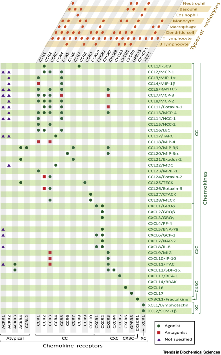
Ticks are hematophagous arachnids that parasitize mammals and other hosts, feeding on their blood. Ticks secrete numerous salivary factors that enhance host blood flow or suppress the host inflammatory response. The recruitment of leukocytes, a hallmark of inflammation, is regulated by chemokines, which activate chemokine receptors on the leukocytes. Ticks target this process by secreting glycoproteins called Evasins, which bind to chemokines and prevent leukocyte recruitment. This review describes the recent discovery of numerous Evasins produced by ticks, their classification into two structural and functional classes, and the efficacy of Evasins in animal models of inflammatory diseases. The review also proposes a standard nomenclature system for Evasins and discusses the potential of repurposing or engineering Evasins as therapeutic anti-inflammatory agents.

Keywords: Evasin; anti-inflammatory; binding protein; chemokine; protein family.


Figures

None
Figure I. Chemokines, Chemokine Receptors, and Leukocytes. The selectivity of human chemokines (listed on right in green) for conventional chemokine receptors (listed at top and bottom in black) and atypical chemokines receptors (listed at bottom left in black) is shown in the green and gray grid. Chemokine–ligand pairs are categorized as listed in the International Union of Basic and Clinical Pharmacology (IUPHAR) Guide to Pharmacology (http://www.guidetopharmacology.org/) (agonists, green circles; antagonists, red squares; not specified, purple triangles). The expression of conventional chemokine receptors on different types of leukocytes (listed top right in orange) is shown in the orange and gray grid; further details, including expression patterns on subtypes of leukocytes (especially T cells) and non-hematopoietic cells, are presented in . Abbreviations for chemokine nonsystematic names: BCA, B cell-attracting chemokine; BRAK, breast- and kidney-expressed chemokine; CTACK, cutaneous T cell-attracting chemokine; ENA, epithelial cell-derived neutrophil activating peptide; GCP, granulocyte chemotactic protein; GRO, growth-regulated oncogene; HCC, hemofiltrate CC chemokine; IL, interleukin; IP, interferon γ-induced protein; ITAC, interferon-inducible T cell α chemoattractant; LEC, liver-expressed chemokine; NAP, neutrophil-activating peptide; MCP, monocyte chemotactic protein; MDC, macrophage-derived chemokine; MECK, mucosae-associated epithelial chemokine; MIG, monokine induced by γ-interferon; MIP, macrophage inflammatory protein; MPIF, myeloid progenitor inhibitory factor 1; PF, platelet factor; RANTES, regulated on activation, normal T cell expressed and secreted; SCM, single cysteine motif; SDF, stromal cell-derived factor; TARC, thymus- and activation-regulated chemokine; TECK, thymus-expressed chemokine.
Figure 1
Figure 1
Sequence Alignments, Phylogenetic Tree, and Pairwise Identity Matrix of Class A Evasins. (A) Sequence alignment of all validated Class A Evasins with proposed nomenclature. The consensus sequence (above alignment) and a graphical representation of the amino acid conservation (sequence logo; below alignment) show that eight cysteine residues (green) are conserved (except for two missing Cys residues in the bottom three sequences) and two glycine residues are completely conserved across the family. The secondary structure of EVA-1 [Protein Database (PDB) ID: 3FPR] and EVA-1 residues forming hydrogen bond interactions with CCL3 (in PDB ID: 3FP), analyzed by PDBSum, are presented at the top of the alignment. The alignment was performed using MAFFT, with default parameters, in the program DNASTAR Navigator 15 (DNASTAR, Madison, USA). Amino acid residues are color coded by physicochemical properties (aromatic, light yellow; acidic, medium salmon; basic, medium blue; nonpolar aliphatic, medium orange; polar neutral, medium green). (B) Phylogenetic tree (left side) and pairwise identity matrix of all validated Class A Evasins. Pairwise identities between sequences were calculated using MAFFT and are color coded on a continuous scale from rose (high identity) to blue (low identity). The phylogenetic tree was generated on FigTree v1.4.3 (http://tree.bio.ed.ac.uk/software/figtree/) using the alignment data from MAFFT.
Figure 2
Figure 2
Structure of EVA-1. (A) Ribbon representation of one protomer of nonglycosylated EVA-1 [Protein Database (PDB) ID: 3FPR, resolution 1.70 Å] showing the base of the structure formed of the third β-sheet (β5, β6, and β7 strands) and the α-helix (blue); the first β-sheet (β1 and β2 strands) and second β-sheet (β3 and β4 strands) (green); the N-terminal region (black); the C-terminal region (gray); and cysteine residues and disulfide bonds (sticks) (yellow). (B) Topology diagram corresponding to (A) showing β-strands as arrows and the α-helix as a cylinder, with coloring the same as in (A), except that disulfide bond connectivity is shown as yellow-orange lines. (C) Dimer of nonglycosylated EVA-1 (PDB ID: 3FPR; left) and trimer of glycosylated EVA-1 (PDB ID: 3FPT, resolution 2.70 Å; right). Each protomer is represented in a different color, with cysteine residues and disulfide bonds (sticks) in yellow. (D) Ribbon representation of one protomer of glycosylated EVA-1 (PDB ID: 3FPT) showing confirmed glycosylated residue Asn-19 (magenta sticks); other potential N-glycosylation sites Asn-34 and Asn-42 (orange sticks); residues corresponding to predicted glycosylation sites in other Evasins (cyan sticks); and putative sulfation site Tyr-23 (red sticks). The two conserved glycine residues and their interacting residues are shown in space-filling representation with Gly colored by atom type (C, green; H, white; O, red; N, blue), Cys in yellow, and other residues in gray. Gly-68 of EVA-1 is positioned adjacent to the disulfide bond linking strand β5 to strand β7 such that no l-amino acid side chain can be accommodated in this closely packed region of the structure. Gly-73 of EVA-1 is located on the β6–β7 turn with its CH2 group packed closely against the side chains of residue Lys-30 (on β3) and Thr-39 (on β4). This interaction may be important to define the relative positions of the β3–β4 sheet and the β5–β6–β7 sheet.
Figure 3
Figure 3
Structure of the Complex between EVA-1 and CCL3. (A) EVA-1 [α-helix and third β-sheet (blue); the first β-sheet and second β-sheet (green); the N-terminal region (black); the C-terminal region (gray); and cysteine residues and disulfide bonds (sticks) (yellow)] bound to CCL3 [magenta but highlighting the N terminus (orange), N-loop and β3 strand (cyan), and conserved cysteine residues with side chain sticks (yellow)] [Protein Database (PDB) ID: 3FPU, 1.9 Å resolution]. (B) The complex (PDB ID: 4RWS) between viral chemokine vMIP-II, colored the same as CCL3 in panel (A), and chemokine receptor CXCR4 (gray); the N-terminal 22 residues of vMIP-II were not defined in this structure. (C) Stick (left) and space-filling (right) representations showing the four Cys residues of CCL3 (magenta carbon backbone and labels) forming two conserved disulfide bonds and the residues of EVA-1 (green carbon backbone and labels) with which they directly interact. The first disulfide bond of EVA-1 (Cys-12 to Cys-33) is also shown. Side chains of EVA-1 Phe-14 and Leu-15 are omitted for clarity. Two hydrogen bonds from CCL3 Cys-11 to EVA-1 Phe-14 are indicated as broken lines; these extend the first β-sheet of EVA-1 by a very short β-strand (β0). The orientation of the CC motif relative to the first β-sheet of EVA-1 is further constrained by hydrophobic interactions of each CCL3 disulfide bond with EVA-1 side chains (Val-16, Pro-24, and Pro-13), which are conserved or substituted by other hydrophobic residues in most other Evasin sequences. The interaction is further stabilized by an edge to face π–π interaction between the side chains of EVA-1 Phe-14 and CCL3 Phe-13 (inset). It appears that the insertion of an additional residue within the CC motif, as found in CXC chemokines, cannot readily be accommodated while retaining these key interactions.
Figure 4
Figure 4
Sequence Alignments, Phylogenetic Tree, and Pairwise Identity Matrix of Class B Evasins. (A) Sequence alignment of all validated Class B Evasins. The consensus sequence (above alignment) and sequence logo (below alignment) show that six cysteine residues (green) and one glycine residue are completely conserved across the family. The secondary structure of EVA-3 [Protein Database (PDB) ID: 6I31] is presented at the top. The alignment was performed using MAFFT, with default parameters, in the program DNASTAR Navigator 15 (DNASTAR, Madison, USA). Amino acid residues are color coded by physicochemical properties (aromatic, light yellow; acidic, medium salmon; basic, medium blue; nonpolar aliphatic, medium orange; polar neutral, medium green). (B) Phylogenetic tree (left side) and pairwise identity matrix of all validated Class B Evasins, indicating subclasses B(I) and B(II). Pairwise identities between sequences were calculated using MAFFT and are color coded on a continuous scale from rose (high identity) to blue (low identity). The phylogenetic tree was generated on FigTree v1.4.3 (http://tree.bio.ed.ac.uk/software/figtree/) using the alignment data from MAFFT.
Figure 5
Figure 5
Structure of EVA-3 Showing the ‘Knottin’ Cystine Knot Topology. (A) Ribbon representation of one protomer of EVA-3 [Protein Database (PDB) ID: 6I31]. Pairs of cysteines that form disulfide bonds (Cys-21 and Cys-37; Cys-26 and Cys-39; Cys-33 and Cys-50) are shown in different colors (yellow, green, and red, respectively). Other residues within the macrocycle created by the first two disulfide bonds are shown in gray. (B) EVA-3 structure, colored as in (A), showing the protein backbone and Cys side chains. This view highlights the third (red) disulfide bond passing through the macrocycle created by the first two (yellow and green) disulfide bonds. (C) Topology diagram showing β-strands as arrows and the α-helix as a cylinder, with coloring corresponding to (A) and (B).

References

    1. Murphy P.M. Chemokines and chemokine receptors. In: Rich R.R., editor. Clinical Immunology E-Book: Principles and Practice. 5th edn. Elsevier Health Sciences; 2018. pp. 157–170.
    1. Zlotnik A., Yoshie O. The chemokine superfamily revisited. Immunity. 2012;36:705–716. - PMC - PubMed
    1. Burns J.M. Comprehensive mapping of poxvirus vCCI chemokine-binding protein: expanded range of ligand interactions and unusual dissociation kinetics. J. Biol. Chem. 2002;277:2785–2789. - PubMed
    1. Bahar M.W. Structure and function of A41, a vaccinia virus chemokine binding protein. PLoS Pathog. 2008;4:e5. - PMC - PubMed
    1. Xue X. Structural basis of chemokine sequestration by CrmD, a poxvirus-encoded tumor necrosis factor receptor. PLoS Pathog. 2011;7:e1002162. - PMC - PubMed

MeSH terms

LinkOut - more resources