. 2019 Nov 18;51(4):421-430.e3.
doi: 10.1016/j.devcel.2019.09.017. Epub 2019 Oct 31.

An Ancient CFTR Ortholog Informs Molecular Evolution in ABC Transporters

Affiliations

An Ancient CFTR Ortholog Informs Molecular Evolution in ABC Transporters

Guiying Cui et al. Dev Cell. .

Abstract

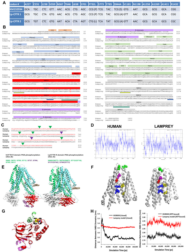
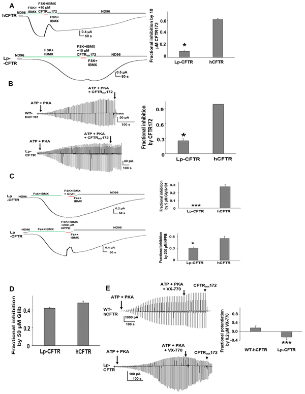
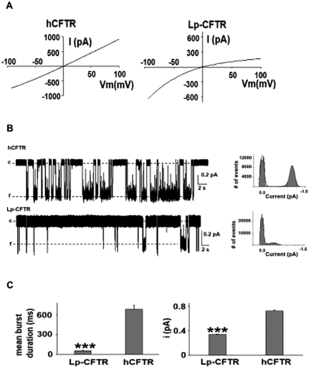
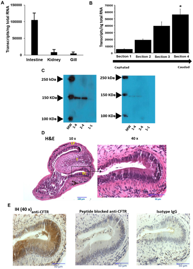
The cystic fibrosis transmembrane conductance regulator (CFTR) is a chloride channel central to the development of secretory diarrhea and cystic fibrosis. The oldest CFTR ortholog identified is from dogfish shark, which retains similar structural and functional characteristics to the mammalian protein, thereby highlighting CFTR's critical role in regulating epithelial ion transport in vertebrates. However, the identification of an early CFTR ortholog with altered structure or function would provide critical insight into the evolution of epithelial anion transport. Here, we describe the earliest known CFTR, expressed in sea lamprey (Petromyzon marinus), with unique structural features, altered kinetics of activation and sensitivity to inhibition, and altered single-channel conductance compared to human CFTR. Our data provide the earliest evolutionary evidence of CFTR, offering insight regarding changes in gene and protein structure that underpin evolution from transporter to anion channel. Importantly, these data provide a unique platform to enhance our understanding of vertebrate phylogeny over a critical period of evolutionary expansion.

Keywords: ABC transporters; CFTR; channel; lamprey; molecular evolution; phosphorylation; vertebrates.


Figures

Figure 1:
Figure 1:. Lamprey CFTR (Lp-CFTR) demonstrates key similarities and differences in sequence and structure compared to human CFTR (hCFTR)
A: SNPs identified in Lp-CFTR. The Lp-CFTR gene was cloned from lamprey cDNA, sequenced, and compared to a published putative sequence (KM232934). Eighteen SNPs were identified with 2 amino acid changes between the three Lp-CFTR sequences. B: Amino acid sequence of Lp-CFTR aligned with that of hCFTR. Key major domains are delineated above alignment. Lp-CFTR has an extended N-terminus, a leucine at the position corresponding to F508 in hCFTR (delineated by red letter and underline), intact Walker A domains (yellow underline) and Walker B domains (green underline) in both NBD1 and NBD2, but conserved signature sequence (blue lines) in NBD1 only. C: Lp-CFTR has a reduced number of consensus PKA phosphorylation sites. The R-domains of Lp-CFTR and hCFTR were aligned and predicted phosphorylation sites were identified for activation (green arrowheads) and inhibition (purple arrowheads), reflecting a reduced number of consensus PKA phosphorylation sites in Lp-CFTR. D: Similar hydropathy predicted from lamprey and human CFTR sequences. CFTR sequences were compared using the Kyte-Doolittle scale for hydropathy via ExPasy, with increased hydrophilicity as negative values and increased hydrophobicity as positive values. E: Structural homology models of Lp-CFTR. Lp-CFTR was modeled using the cryo-EM structures of CFTR in the closed state, left, and in the ATP-bound nearly open state, right. Domains colored as in Figure 1B. The segment of the R-domain visible in the closed state (purple) is not shown in the nearly open state. F: Structure of the pore domain in the homology models of Lp-CFTR. Lp-CFTR was modeled using CFTR cryo-EM structures in closed (left) and ATP-bound nearly open (right) conformations, demonstrating development of a pore; residues 1101-1153 deleted in order to reveal the inner pore. Of note, F337 (L360 in Lp-CFTR, purple) occludes the pore in the closed state and S341 (S364, red) is at the narrowest spot in the pore interior although not yet rotated into the pore axis. R347 interacts with D924 (R370 and D927, upper blue-yellow pair) in both states, while R352 interacts with D993 (R375 and D996, bottom blue-yellow pair) only in the nearly open state. Of note, a key interaction between D110 in ECL1 and K892 in ECL4 observed in hCFTR in the closed state is not enabled in Lp-CFTR (N134 with R896, green pair, partially occluded in the view of the ATP-bound near-open state). G: NBD1 of Lp-CFTR bears a key substitution. Lp-CFTR modeled using the CFTR cryo-EM structures demonstrated folding of NBD1 similar to that of human CFTR. NBD1 is noted in red ribbon and a portion of the R-domain is in purple ribbon. The lamprey substitution for phenylalanine 508, leucine 525, is noted in dashed box region and is positioned to interact with intracellular loop 4 (green ribbon). H: NBD domains are more distant from each other in Lp-CFTR compared to the human ortholog: Comparison of distances between the centers of mass of NBD1 and NBD2 of human (black) and lamprey (red) CFTR conformations obtained from 100 ns MD simulations. (A) Closed hCFTR (5uak.pdb) and the corresponding Lp-CFTR homology model. (B) ATP-bound nearly-open hCFTR (6msm.pdb) and the corresponding Lp-CFTR homology model. See also Data Figure S1 and Figure S1
Figure 2:
Figure 2:. Lp-CFTR has limited chloride conduction and is poorly responsive to both CFTR potentiators and inhibitors
A: Whole oocyte characterization of Lp-CFTR function. Like hCFTR, Lp-CFTR can be activated by 10 μM Forskolin (FSK) with 1 mM isobutylmethylxanthine (IBMX) but exhibits distinctly slower activation. CFTRinh172 significantly blocked hCFTR (upper panel), with markedly diminished effect on Lp-CFTR (lower panel). Summary data are shown (right panel), data shown as mean±SEM; *= p< 0.05 via unpaired t-test, n=10 for hCFTR; n=4 for Lp-CFTR. B: Macropatch characterization of Lp-CFTR. Representative inside-out macropatch currents of hCFTR (upper panel) and Lp-CFTR (lower panel). Channels were activated by 127.6 U/mL PKA and 1 mM ATP until currents reached plateau. A voltage-ramp protocol was applied every 5 s. CFTR currents were blocked with 10 μM CFTRinh172. Summary data are shown in right panel, data shown as mean±SEM. *= p<0.05 compared to hCFTR via unpaired t-test; n=4 for each group. C: Lp-CFTR is not inhibited by GlyH-101 and is poorly sensitive to NPPB. Lp-CFTR demonstrates reduced sensitivity to inhibition by the CFTR pore blockers GlyH-101 (GlyH, 5uM, upper panel) and NPPB (200 μM) in whole oocyte recordings. Summary data are shown in right panels, data shown as mean±SEM. *** P<0.001; * P<0.05 compared to hCFTR via unpaired t-test. n=4 for Lp-CFTR, n=7 for hCFTR. D: Inhibition of Lp-CFTR by glibenclamide. Glibenclamide (50 μM) blocked hCFTR and Lp-CFTR to a similar extent in inside-out macropatches; no statistical difference between groups via unpaired t-test. Data shown as mean±SEM; n=6 for each group. E: Lp-CFTR is insensitive to potentiation by VX-770. Representative currents of WT-hCFTR (top panel) and Lp-CFTR (bottom panel) were recorded in inside-out macropatches in presence of 1 mM ATP and 127.6 U/mL PKA with or without 0.2 μM VX-770. A voltage-ramp protocol was applied every 5 s. CFTR currents were blocked with 10 μM CFTRinh172. Summary data for fractional potentiation are shown in right panel (Fractional potentiation by VX-770 = (I(ATP + PKA + VX-770)/ I(ATP + PKA) −1), data shown as mean±SEM; n = 4 –7 each. ***, p < 0.001 compared to hCFTR via unpaired t-test. See also Figure S2
Figure 3:
Figure 3:. Lamprey CFTR inwardly rectifies and has altered single channel kinetics
A: Notable inward rectification of lamprey CFTR. Representative current-voltage (IV) curves of hCFTR (left panel) and Lp-CFTR (right panel) recorded in inside-out macropatches in symmetrical chloride conditions demonstrated inward rectification of Lp-CFTR current; similar results seen in n=5 for hCFTR and n=7 for Lp-CFTR. B: Single channel recordings exhibit reduced channel stability. (Left) Traces of Lp-CFTR and hCFTR (at VM = −100 mV, activated with 1 mM MgATP and 127.6 U/mL PKA) demonstrated significant differences in opening frequency (c=closed, f=open). (Right) Representative all points histograms over the recording periods show reduced open conductance in Lp-CFTR. Solid lines in the histograms represent fits to a Gaussian function. C: Lamprey CFTR demonstrates altered single channel behavior. Lp-CFTR exhibit shorter open burst duration (left) and lower single channel amplitude (right) compared to hCFTR in inside-out patches. Data shown as mean±SEM. ***=p< 0.001 vs. hCFTR, n= 6 for Lp-CFTR. hCFTR data (n=10) were cited from a previous publication (Cui et al., 2014). VM = −100 mV.
Figure 4:
Figure 4:. Lamprey CFTR is highly expressed in the distal gastrointestinal (GI) tract of the lamprey
A: CFTR transcripts are found in various organs in the lamprey. RNA was isolated from intestine, kidney, and gill from seven different larval lampreys and quantified for Lp-CFTR via SYBR-green RT-qPCR (each lamprey organ run in triplicate), data shown as mean+SD; p<0.001 via ANOVA for all groups, p<0.001 for intestine vs. kidney and p<0.001 for intestine vs. gill via Tukey’s post-test. Of note, no difference was observed in 40S rRNA between tissues. B: CFTR transcripts are increased in the distal GI tract. RNA was isolated from different sections of the GI tract (cephalad (Section 1) to caudad (Section 4)) from 7 different larval lamprey and quantified for Lp-CFTR via SYBR-green RT-qPCR (each lamprey section run in triplicate), data shown as mean+SD. p<0.01 via ANOVA for all groups, * p<0.01 Section 4 vs. Section 1 via Tukey’s post-test. Of note, no difference was observed in 40S rRNA between sections. C: CFTR expression in lamprey GI tract. Representative Western blot (from six separate lampreys) with rabbit anti-lamprey CFTR antibody. Blot of Section 4 GI tract tissue from two separate lampreys (numbers 1-4 and 2-4), and a representative GI tract tissue of the Section 1 of a lamprey (number 1-1), demonstrating predicted ~150 kDa band at higher density in Section 4 vs. Section 1 (left panel). Antibody depletion with competing peptide demonstrating loss of 150 kDa band in all samples (right panel). D Transverse section of lamprey GI tract. Image of the caudal section (Section 4) representative of four separate lampreys. Lampreys were dissected transversally and stained with H&E. At 10x magnification, the lamina propria (LP) and typhosole (T) were identified with lining epithelial cells (EC). At 40x, the ECs exhibit features of simple columnar epithelium with darkly stained nuclei and brush border. E: CFTR is expressed in lamprey GI tract. Image of the caudal region of the intestine (Section 4) representative of images from four separate lampreys. Lampreys were dissected transversely and stained with either anti-CFTR antibody (1:3000 dilution), isotype control IgG (1:3000 dilution), or peptide blocked anti-CFTR antibody (1:3000 dilution), and imaged at 40x over a region of epithelial cells. Of note, there is significant Lp-CFTR staining of intestinal epithelia that is greatly reduced with peptide block and not observed with isotype IgG control.

References

    1. Aghaallaei N, Gruhl F, Schaefer CQ, Wernet T, Weinhardt V, Centanin L, Loosli F, Baumbach T, and Wittbrodt J (2016). Identification, visualization and clonal analysis of intestinal stem cells in fish. Development 143, 3470–3480. - PMC - PubMed
    1. Bessadok A, Garcia E, Jacquet H, Martin S, Garrigues A, Loiseau N, Andre F, Orlowski S, and Vivaudou M (2011). Recognition of sulfonylurea receptor (ABCC8/9) ligands by the multidrug resistance transporter P-glycoprotein (ABCB1): functional similarities based on common structural features between two multispecific ABC proteins. J Biol Chem 286, 3552–3569. - PMC - PubMed
    1. Bozoky Z, Ahmadi S, Milman T, Kim TH, Du K, Di Paola M, Pasyk S, Pekhletski R, Keller JP, Bear CE, et al. (2017). Synergy of cAMP and calcium signaling pathways in CFTR regulation. Proc Natl Acad Sci U S A 114, E2086–E2095. - PMC - PubMed
    1. Clemens JD, Nair GB, Ahmed T, Qadri F, and Holmgren J (2017). Cholera. Lancet 390, 1539–1549. - PubMed
    1. Cotten JF, and Welsh MJ (1999). Cystic fibrosis-associated mutations at arginine 347 alter the pore architecture of CFTR. Evidence for disruption of a salt bridge. J Biol Chem 274, 5429–5435. - PubMed

Publication types

MeSH terms

Substances

LinkOut - more resources